Correlation of Myocardial Interstitial Collagen in the Right Ventricular
Septum with Ventricular Function of Patients with Ischemic
Cardiomyopathy
Marcelo Westerlund Montera
1,2,3, Cantídio Drumond
5, Cristina Takiya
6, Cláudio Tinoco Mesquita
3, Hans Fernando R.
Dohmann
4, Charles Mady
1Universidade de São Paulo, Instituto do Coração (InCor), São Paulo, SP1, Hospital Pró-Cardíaco - Centro de Insuficiência Cardíaca2 - Laboratório de Medicina Nuclear3 e Laboratório de Cateterismo Cardíaco4, Rio de Janeiro, RJ; Santa Casa de Misericórdia - Departamento de Cardiologia, Rio de Janeiro, RJ5, Universidade Federal do Rio de Janeiro - Departamento de Histologia, Rio de Janeiro, RJ6 - Brazil
Summary
Background:Myocardial collagen content influences ventricular relaxation, contraction, and morphology. Its relationship with ventricular function in patients (Pts) with ischemic cardiomyopathy (ICMP) has not yet been fully studied in humans.
Objective: To assess the relationship between interstitial collagen content in non-infarcted areas of the right ventricular septum and ventricular function in ICMP.
Methods:3 pts with coronary artery disease were divided into four groups as follows:The control group consisted of 7 pts with normal left (LVEF) and right (RVEF) ventricular ejection fraction (group C); Group : 5 patients with RVEF < 40%; Group 2: 9 pts with LVEF < 40%; and Group 3, 0 pts with biventricular dysfunction. RVEF and LVEF were measured by radionuclide angiography. For quantitative analysis of interstitial collagen volume fraction (CVF), endomyocardial biopsy specimens were taken from the right ventricle and stained with picrosirius red.
Results:Mean CVF was significantly higher in group 3, compared with the control group and with groups and 2 (30.2 ± 7.9% vs. 6.8 ± 3.3% vs. 5.8 ± 4.% vs. 7.5±7.7%, respectively; p =0.000). It was also significantly higher in patients belonging to group 2, compared with those in the control group (7.5 ± 7.7% vs. 6.8 ± 3.3%, p =0.000). CVF was inversely correlated with RVEF (r = - 0.50, p = 0.003) and LVEF (r = -0.70, p = 0.000).
Conclusion: In ICMP, CVF is elevated in non-infarcted areas of the right ventricular septum and inversely correlated with right and left ventricular function. (Arq Bras Cardiol 2009;92():52-60)
Key words:Myocardial ischemia; cardiac output, low; ventricular function, right; ventricular function, left; collagen.
Mailing address: Marcelo Westerlund Montera •
Rua Conde de Irajá, 513 – Ipanema - 22271-020 – Rio de Janeiro, RJ - Brazil E-mail: [email protected], [email protected]
Manuscript received November 29, 2007; revised manuscript received February 18, 2008; accepted March 05, 2008
Introduction
Heart failure (HF) is a highly prevalent syndrome that affects approximately 1% to 1.5% of the adult population. It is estimated that 4.7 million people in the United States have heart failure, with 400,000 new cases being diagnosed every year1-3. The majority of cases result from heart muscle diseases, known as cardiomyopathies, of which ischemic cardiomyopathy (ICM) is the most common, accounting for 40% to 80% of all cardiomyopathies1,4. Ischemic cardiomyopathy carries a worse prognosis than idiopathic dilated cardiomyopathy4-7, probably due to the presence of areas of myocardial ischemia and fibrosis from prior infarction, which may predispose to arrhythmias and extensive ventricular remodeling.
The activation of ventricular remodeling is related to the extent of myocardial necrosis, fibrosis, and ischemia in the infarcted area. However, it has been demonstrated that morphological changes in the remote non-infarcted myocardium play a major role in the development of ventricular remodeling8,9. These include diffuse perivascular fibrosis, interstitial fibrosis, and diffuse atrophy of cardiac myocytes10.Changes in myocardial matrix at sites distant from myocardial scarring account for approximately two-thirds of all cardiac fibrous tissue and are regarded as the primary component of ventricular remodeling in ischemic cardiomyopathy11,12.
The morphological and morphometric changes that occur in the myocardium following myocardial infarction, with the development of left ventricular (LV) systolic and diastolic dysfunctions, are well described in the literature 8,13-16. However, the relationship between thesechanges in the
The aim of this study is to assess the relationship between interstitial collagen content, measured by right ventricular septal biopsy, and right and left ventricular systolic function in ICM patients with varying degrees of ventricular dysfunction.
Methods
From January to December 2003, thirty-one patients with angiographically documented coronary artery disease were consecutively included in the study for diagnostic evaluation of precordial pain or left ventricular dysfunction. They were divided into four groups according to left (LVEF) and right (RVEF) ventricular ejection fractions measured by radionuclide angiography as follows: Control group (Group C), seven patients with preserved LV and RV systolic function; Group 1, five patients with isolated RV systolic dysfunction (RVEF < 40%); Group 2, nine patients with isolated LV systolic dysfunction (LVEF < 40%); and Group 3, ten patients with right and left ventricular systolic dysfunction. Patients with pulmonary hypertension, chronic obstructive pulmonary disease (COPD), infiltrative or restrictive myocardial diseases, and collagen diseases were excluded from the study, as were those with suspected myocarditis, myocardial infarction with septal involvement or right ventricular dysfunction secondary to right ventricular cardiomyopathy. The study protocol was approved by the Institutional Ethics Committee, and an informed consent was obtained from all patients.
All patients underwent biochemical tests, including renal function tests, electrocardiography (ECG), and transthoracic echocardiography (ECHO). Exclusion criteria were septal myocardial infarction on ECG and ECHO, significant valvular heart disease, systemic hypertension (blood pressure > 140x90 mmHg), serum creatinine above 1.4 mg/dL, atrial fibrillation, clinical instability of less than two weeks, and chronic obstructive pulmonary disease.
Cardiac catheterization
All patients underwent coronary angiography via the femoral approach using Sones’ technique. Coronary artery disease was defined as stenosis greater than 50% in at least one major epicardial coronary artery. Ventriculography was also performed in all subjects.
Analysis of ventricular function
Both right and left ventricular functions were assessed by equilibrium and first-pass radionuclide angiography. Left and right ventricular ejection fractions were calculated using the standard method recommended by the American Society of Nuclear Cardiology17, with the lower limit of normal set at 40% for both ventricles. Right ventricular ejection fraction (RVEF) was measured by first-pass radionuclide angiography using a DIACAM Siemens gamma camera connected to an ICON computer. The study protocol was validated by the Cardiovascular Nuclear Imaging Laboratory of the Yale University School of Medicine18. Tc-99m was injected at rest in all patients to assess ventricular function. Images were acquired in the supine position, with the detector in the right anterior oblique (RAO) projection (20° to 30°). First-pass images were analyzed independently by two nuclear medicine physicians,
using two regions of interest. Mean values for right and left ventricular functions were used for statistical analysis. Beats with diastolic counts below 50% of the maximum end-diastolic count were excluded, as were premature ventricular beats and post-premature ventricular contraction (PVC) beats. For statistical analysis purposes, normal right ventricular ejection fraction was considered to be equal to or greater than 40%.
LVEF was measured by planar equilibrium radionuclide angiography with the modified technique for in-vivo labeling, using 2 to 3 mg of stannous pyrophosphate 15 minutes before injecting 20 mCi of Tc-99m–pertechnetate19. Images were acquired in the supine position with the detector in the left anterior oblique (LAO) projection at 45 degrees. Left ventricular systolic dysfunction was defined as LVEF ≤ 40%.
Endomyocardial biopsy
Right ventricular endomyocardial biopsies were performed percutaneously according to Mason’s technique20 using a Cordis bioptome, and four tissue samples were obtained from the interventricular septum of each patient.
Histological study and quantiication of interstitial collagen
Biopsy specimens were fixed in 10% buffered formalin and embedded in paraffin. Serial, 5-�m-thick sections were�m-thick sections werem-thick sections were mounted on glass slides and stained with hematoxylin-eosin, Masson’s trichrome, and Sirius Red (0.1% Sirius red F3BA dissolved in saturated picric acid, pH 2.0). Ten fields per section were analyzed at a magnification of x320. Collagen content was measured by quantitative morphometric analysis of sirus-red-stained sections, with an automatic image analyzer (Image-Pro Plus 4.5.1 software, Media Cybernetics, Silver Spring, MD, USA). Images were captured using a Coolpix 900 digital camera attached to an Eclipse E400 light microscope (both manufactured by Nikon, Japan). Each histological section was viewed at a magnification of x40. Collagen fibers were stained blue under direct light, and images were digitized. Interstitial collagen volume fraction (CVF) in each field was calculated as the sum of all connective tissue area divided by the sum of all connective tissue and cardiac myocyte areas, as demonstrated previously in a number of studies21-24.
Statistical analysis
Results
Patients’ characteristics
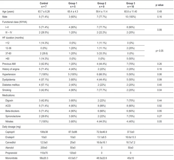
Clinical characteristics of the patients are summarized in Table I. No significant difference was found in clinical and demographic variables among the four groups, except for lower incidence of smoking in group 3. Most patients were on angiotensin-converting enzyme inhibitors and beta-blockers, with no significant differences among groups regarding drug regimen.
Coronary artery disease
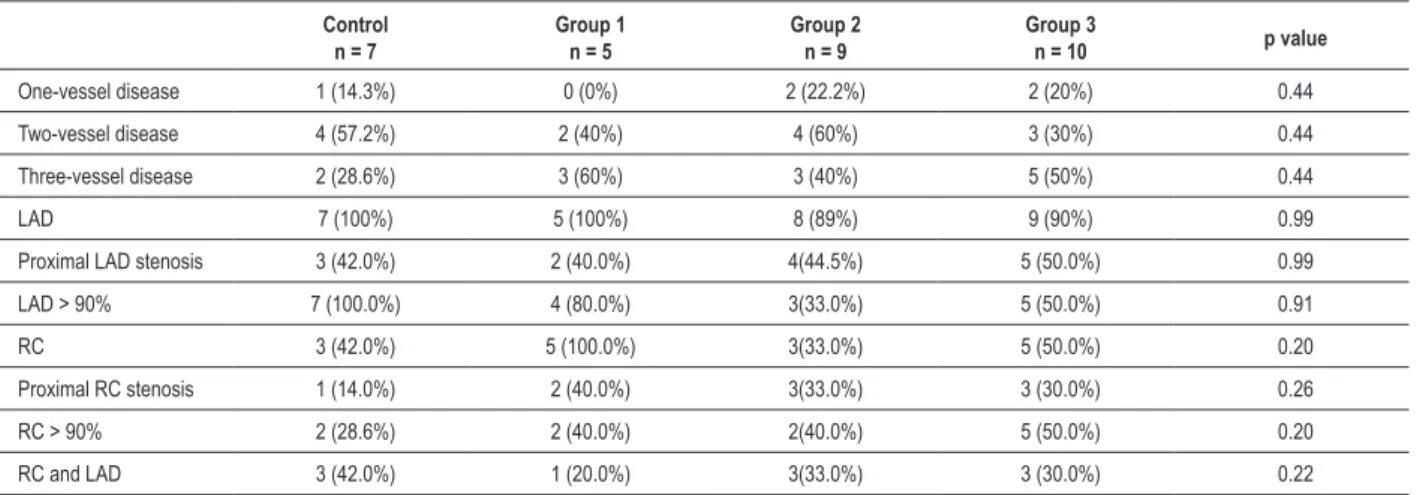
The majority of patients (84%) had two- or three-vessel disease, the left anterior descending artery (LAD) being the most frequently affected. There was no difference among the
Table 1 – Baseline clinical characteristics of the study population
Control n = 7
Group 1 n = 5
Group 2 n = 9
Group 3
n = 10 p value
Age (years) 63.7 ± 6.26 65.4 ± 4.3 59.4 ± 11.4 60.8 ± 11.40 0.49 Male 5 (71.4%) 3 (60%) 7 (77.7%) 10 (100%) 0.16 Functional class (NYHA)
I–II 5 (71.4%) 4 (80%) 7 (77.7%) 8 (80%)
0.99 III – IV 2 (28.5%) 1 (20%) 2 (22.2%) 2 (20%)
HF duration (months)
<12 1 (14.3%) 0 (0%) 1 (11.1%) 0 (0%)
p> 0.05 12-36 0 (0%) 1 (20%) 1 (11.1%) 2 (20%)
37-60 2 (28.6) 1 (20%) 3 (33.3%) 0 (0%) >60 1 (14.3%) 0 (0%) 0 (0%) 5 (50%)
Previous AMI 3 (42.8%) 1 (20%) 4 (44.4%) 7 (70%) 0.26 History of angina 5 (71.4%) 4 (40%) 2 (22%) 2 (20%) 0.16 Hypertension 7 (100%) 5 (100%) 6 (66.5%) 5 (50%) 0.06 Dyslipidemia 4 (57.1%) 3 (60%) 4 (44.4%) 5 (50%) 0.99 Diabetes mellitus 4 (57.1%) 2 (40%) 2 (22%) 2 (20%) 0.40 Smoking 3 (42.8%) 4 (80%) 7 (77.7%) 2 (20%) 0.04 Medications:
Digoxin 3 (42.8%) 3 (60%) 2 (22%) 7 (70%) 0.44 ACEI 5 (71.4%) 4 (80%) 8 (89%) 8 (80%) 0.92 Beta-blockers 5 (71.4%) 4 (80%) 6 (66%) 6 (60%) 0.95 Spironolactone 2 (28.6%) 3 (60%) 2 (22%) 7 (70%) 0.27 Nitrates 7 (100%) 3 (60%) 4 (44.5%) 4 (40%) 0.05 Daily dosage (mg)
Captopril 108±38 87.5±88 72.9±46.9 37.5±0 Enalapril 10±0 10±0 13.1±8.5 16.6±13.3 Carvedilol 12.5±0 25±0 16.6±16.1 18.7±7.2
Atenolol 200±0 50±0 0 50±0
Propranolol 110±70 120±0 0 0
Mononitrate 58±20.3 43.5±5.7 48.5±22.6 45±10
ACEI – angiotensin-converting enzyme inhibitor; HF - heart failure; AMI - acute myocardial infarction; NYHA - New York Heart Association classiication.
four groups with regard to the presence of lesions involving the proximal left anterior descending and right coronary (RC) arteries (p = 0.91) Table 2.
Analysis of ventricular function
Table 2 - Distribution of coronary stenoses among groups
Control n = 7
Group 1 n = 5
Group 2 n = 9
Group 3
n = 10 p value
One-vessel disease 1 (14.3%) 0 (0%) 2 (22.2%) 2 (20%) 0.44 Two-vessel disease 4 (57.2%) 2 (40%) 4 (60%) 3 (30%) 0.44 Three-vessel disease 2 (28.6%) 3 (60%) 3 (40%) 5 (50%) 0.44
LAD 7 (100%) 5 (100%) 8 (89%) 9 (90%) 0.99
Proximal LAD stenosis 3 (42.0%) 2 (40.0%) 4(44.5%) 5 (50.0%) 0.99 LAD > 90% 7 (100.0%) 4 (80.0%) 3(33.0%) 5 (50.0%) 0.91 RC 3 (42.0%) 5 (100.0%) 3(33.0%) 5 (50.0%) 0.20 Proximal RC stenosis 1 (14.0%) 2 (40.0%) 3(33.0%) 3 (30.0%) 0.26 RC > 90% 2 (28.6%) 2 (40.0%) 2(40.0%) 5 (50.0%) 0.20 RC and LAD 3 (42.0%) 1 (20.0%) 3(33.0%) 3 (30.0%) 0.22
LAD - left anterior descending artery; RC - right coronary artery
dysfunction and isolated left ventricular dysfunction (28.2 ± 2.7 vs. 22.0 ± 6.5%, p = 0.10).
Analysis of collagen volume fraction
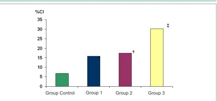
Mean collagen volume fraction (CVF) is shown in figure 1. Collagen content was higher in the group with biventricular dysfunction, compared with the control group and groups with isolated right or left ventricular dysfunction (30.2 ± 7.9
vs. 6.8 ± 3.3, 15.8 ± 4.1, and 17.5 ± 7.7%, respectively; p
= 0.0001). Patients with isolated left ventricular dysfunction had higher CVF than the control group (17.5 ± 7.7 vs. 6.8 ± 3.3%, p = 0.0001). The group with isolated right ventricular dysfunction showed a tendency to higher CVF, compared with the control group (15.8 ± 4.1 vs. 6.8 ± 3.3, p = 0.08) (Figure 2).
Correlation between collagen volume fraction and ventricular function
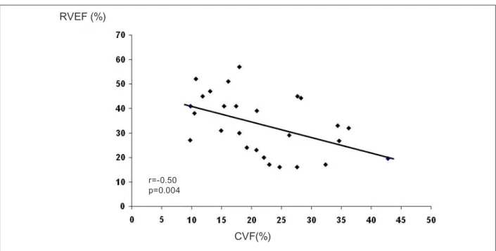
There was a significant inverse linear relationship between collagen fraction and RVEF (r = -0.50; p = 0.003, n = 31) and LVEF (r = -0.70; p = 0.0001, n = 27), demonstrating that the higher the collagen fraction the lower the RVEF and LVEF, as shown in Figures 3 and 4.
ROC curve analysis
The best cut-off point for CVF for the association with LVEF ≤ 40% was 18.3%, with sensitivity of 70.5%, specificity of 90%, and area under the ROC curve of 0.87 (95% CI: 0.7- 0.9; p = 0.0001) (Figure 5 a). For the association with RVEF ≤ 40%, the cut-off point was 12.9%, with sensitivity of 82%, specificity of 64.2%, and area under the ROC curve of 0.77 (CI 95%: 0.59-0.9; p = 0.0001) (Figure 5 b).
Discussion
This is the first human study published in the literature to demonstrate a direct relationship between interstitial collagen content in the right ventricle and right and left ventricular
dysfunction in patients with ischemic cardiomyopathy. A linear correlation was observed between CVF in the non-infarcted myocardium and the degree of right and left ventricular dysfunction. Interstitial collagen fraction was 4.4 times higher in patients with biventricular dysfunction, 2.5 times higher in patients with isolated left ventricular dysfunction, and 2.3 times higher in patients with isolated right ventricular dysfunction, as compared with those with preserved ventricular function, showing an inverse correlation between collagen content and LVEF and RVEF. The association between ventricular dysfunction and the degree of interstitial collagen proliferation was reinforced by the demonstration of an inverse linear correlation between collagen content and RVEF and LVEF (Figures 3 and 4). The analysis of the relationship between several volume fractions of interstitial collagen and ventricular function allowed us to establish a cut-off point of 12.9% and 18%, respectively, for the prognosis of right ventricular dysfunction and left ventricular dysfunction, with high sensitivity (82.0% and 70.5%, respectively) and specificity (64.2% and 90.0%, respectively), and areas under the ROC curve of 0.87 and 0.77, respectively, indicating that these cut-off points are strongly correlated with CVF for discriminating the presence of ventricular dysfunction (Figures 5 a and b).
Figure 2 - Total interstitial collagen content among groups. Histological analysis with picrosirius red; a. Control group; b. Group 1; c. Group 2; d. Group 3 Figure 1 - Interstitial collagen fraction (CVF) among groups; p - 0.08 vs. control group; † p - 0.0001 vs. control group; ‡ p - 0.0001 vs. all groups
Group Control
Group 1
Group 2
Group 3
The difficulty in confirming the association between the degree of right or left ventricular dysfunction and interstitial collagen content lies in the lack of human in vivo studies analyzing collagen and the degree of ventricular dysfunction, particularly right ventricular dysfunction. This may be partly due to a failure of current methods to accurately assess right
ventricular systolic and diastolic function.
Figure 3 - Correlation between interstitial collagen fraction (CVF) and right ventricular ejection fraction (RVEF).
CVF(%)
RVEF (%)
r=-0.50 p=0.004
Figure 4 - Correlation between interstitial collagen fraction (CVF) and left ventricular ejection fraction (LVEF).
CVF(%)
LVEF (%)
r=-0.70 p=0.001
Interstitial collagen content is also increased in non-ischemic dilated cardiomyopathy30, but to a lesser extent than in ischemic cardiomyopathy12,13. This increased collagen content in ischemic cardiomyopathy results not only from ventricular remodeling induced by myocardial infarction, but also from the trophic stimulation of myocardial ischemia31,32.
Figure 5 - a - ROC curve for estimating the probability of left ventricular ejection fraction ≤ 40 relative to interstitial collagen fraction (CVF); b - ROC curve for
estimating the probability of right ventricular ejection fraction ≤ 40 relative to interstitial collagen fraction (CVF); AROC: Area under the ROC curve; CI: Conidence interval
related to the clinical severity of heart failure, the degree of hemodynamic impairment, hyponatremia, and need for heart transplantation37. In contrast, hypertensive patients treated with angiotensin-converting enzyme inhibitors or angiotensin receptor blockers have shown a reduction in myocardial fibrosis and ventricular hypertrophy, regardless of the decrease in blood pressure, with improvement in systolic and diastolic ventricular function, cardiac arrhythmias, and clinical symptoms, as well as reduced ventricular mass38-40.
This strongly suggests that increased interstitial collagen in ischemic cardiomyopathy is a major determinant of ventricular dysfunction, and not only a marker of the degree of ventricular remodeling. In patients with ischemic cardiomyopathy, collagen content was found to be increased not only in the infarcted, but specially in the non-infarcted myocardium8,12,16, a finding that was challenged by Marijianowski et al41 but corroborated by others37,42.
Our findings have important implications for clinical practice, since they demonstrate that, in less severe forms of ventricular dysfunction, there is a significant increase in collagen content and that its progression is directly related to worsening ventricular dysfunction and the degree of ventricular remodeling. They also suggest that in less advanced phases of ventricular dysfunction, the inhibition of collagen production with angiotensin-converting enzyme inhibitors, angiotensin receptor blockers, and aldosterone antagonists
may contribute to reduce both the progression of ventricular remodeling and mortality, as was demonstrated in advanced ventricular dysfunction43-47.
Study limitations
Finally, the presence of myocardial ischemia, which might have influenced the degree of collagen content, was not evaluated in our study, although the extent of coronary artery disease, as documented by coronary angiography, was found to be similar among groups.
Conclusion
Concluding, in patients with ischemic cardiomyopathy, there is an increase in collagen content in the non-infarcted region of the right ventricular septum, which is inversely correlated to the degree of right and left ventricular systolic dysfunction.
Potential Conflict of Interest
No potential conflict of interest relevant to this article was reported.
Sources of Funding
There were no external funding sources for this study.
Study Association
This article is part of the thesis of Doctoral submitted by Marcelo Westerlund Montera, from Universidade de São Paulo.
References
1. Gheorghiade M, Bonow RO. Chronic heart failure in the United States: a manifestation of coronary artery disease. Circulation. 1998; 97: 282-9. 2. Hunt SA, Baker DW, Chin MH, Cinquegrani MP, Feldman AM, Francis GS,
et al. ACC/AHA guidelines for the evaluation and management of chronic heart failure in the adult: executive summary. A report of the American College of Cardiology / American Heart Association Task Force on Practice Guidelines (Committee to Revise the 1995 Guidelines for the Evaluation and Management of Heart Failure): developed in Collaboration with the International Society for Heart and Lung Transplantation; endorsed by the Heart Failure Society of America. Circulation. 2001; 104: 2996-3007. 3. Levy D, Kenchaiah S, Larson MG, Benjamin EJ, Kupka MJ, Ho KK, et al.
Long-term trends in the incidence of and survival with heart failure. N Engl J Med. 2002; 347: 1397-402.
4. Felker GM, Thompson RE, Hare JM, Hruban RH, Clemetson DE, Howard DL, et al. Underlying causes and long-term survival in patients with initially unexplained cardiomyopathy. N Engl J Med. 2000; 342: 1077-84. 5. Likoff MJ, Chandler SL, Kay HR. Clinical determinants of mortality in chronic
congestive heart failure secondary to idiopathic dilated or to ischemic cardiomyopathy. Am J Cardiol. 1987; 59: 634-8.
6. Franciosa JA, Wilen M, Ziesche S, Cohn JN. Survival in men with severe chronic left ventricular failure due to either coronary heart disease or idiopathic dilated cardiomyopathy. Am J Cardiol. 1983; 51: 831-6.
7. Bart BA, Shaw LK, McCants CB Jr, Fortin DF, Lee KL, Califf RM, et al. Clinical determinants of mortality in patients with angiographically diagnosed ischemic or nonischemic cardiomyopathy. J Am Coll Cardiol. 1997; 30: 1002-8.
8. Volders PGA, Willems IE, Cleutjens JP, Arends JW, Havenith MG, Daemen MJ. Interstitial collagen is increased in the noninfarcted myocardium after myocardial infarction. J Mol Cell Cardiol. 1993; 25: 1317-23.
9. Wei S, Chow LT, Schum IO, Chin L, Sanderson JE. . Left and right ventricular collagen type I/III ratio and remodeling post-myocardial infarction. J Card Fail. 1999; 5: 117-26.
10. Cooper GIV. Basic determinants of myocardial hypertrophy: a review of molecular mechanisms. Annu Rev Med. 1997; 48: 13-23.
11. Iwai N, Shimoike H, Kinoshita M. Genes up-regulated in hypertrophied ventricle. Biochem Biophys Res Commun. 1995; 209: 527-34.
12. Beltrami CA, Finato N, Rocco M, Feruglio GA, Puricelli C, Cígola E, et al. Structural basis of end-stage failure in ischemic cardiomyopathy in humans. Circulation. 1994; 89: 151-63.
13. Schaper J, Froede R, Hein ST, Buck A, Hashizume H, Speiser B, et al. Impairment of the myocardial ultrastructure and changes of the cytoskeleton in dilated cardiomyopathy. Circulation. 1991; 83: 504-14.
14. Weber KT, Brilla CG. Pathological hypertrophy and cardiac interstitium. Circulation. 1991; 83: 1849-65.
15. Litwin SE, Litwin CM, Raya TE, Warner AL, Goldman S. Contractility and stiffness of noninfarcted myocardium after coronary ligation in rats: effects of chronic angiotensin converting enzyme inhibition. Circulation. 1991; 83: 1028-37.
16. Hare JM, Walford GD, Hruban RH, Hutchins GM, Deckers JW, Baughman KL. Ischemic cardiomyopathy: endomyocardial biopsy and ventriculographic evaluation of patients with congestive heart failure, dilated cardiomyopathy and coronary artery disease. J Am Coll Cardiol. 1992; 20: 1318-25. 17. Port SC. Imaging guidelines for nuclear cardiology procedures. Part II.
American Society of Nuclear Cardiology. J Nucl Cardiol. 1999; 6: G47-84. 18. Berger HJ, Gottschalk A, Zaret BL. First-pass radionuclide angiocardiography
for evaluation of right and left ventricular performance: computer applications and technical considerations. In: Sorenson JA (ed). Nuclear cardiology: selected computer aspects. New York: Society of Nuclear Medicine; 1978. p. 29-44.
19.Callahan RJ, Froelich JW, Mckusick KA, Leppo J, Strauss HW. A modified method for the in vivo labeling of red blood cells with Tc-99m: concise communication. J Nucl Med. 1982; 23: 315-8.
20. Mason JW. Techniques for right and left ventricular endomyocardial biopsy. Am J Cardiol. 1978; 41: 887-92.
21. Brilla CG, Janicki JS, Weber KT. Impaired diastolic function and coronary reserve in genetic hypertension: role of interstitial fibrosis and medial thickening of intramyocardial coronary arteries. Circ Res. 1991; 69: 107-15.
22. Brilla CG, Pick R, Tan LB, Janicki JS, Weber KT. Remodeling of the rat right and left ventricles in experimental hypertension. Circ Res. 1990; 67: 1355-64. 23. Doering CW, Jalil JE, Janicki JS, Pick R, Aghili S, Abrahams C, et al. Collagen
network remodeling and diastolic stiffness of the rat left ventricle with pressure overload hypertrophy. Cardiovasc Res. 1988; 22: 686-95.
24. Brilla CG, Janicki JS, Weber KT. Cardioreparative effects of lisinopril in rats with genetic hypertension and left ventricular hypertrophy. Circulation. 1991; 83: 1771-9.
25. Pauschinger M, Knopf D, Petschauer S, Doerner A, Poller W, Schwimmbeck PL, et al. Dilated cardiomyopathy is associated with significant changes in collagen type I/III ratio. Circulation. 1999; 99: 2750-6.
26.Mukherjee D, Sen S. Alteration of collagen phenotypes in ischemic cardiomyopathy. J Clin Invest. 1991; 88: 1141-6.
27. Weber KT, Anversa P, Armstrong PW, Brilla CG, Burnett JC Jr, Cruickshank JM, et al. Remodeling and reparation of the cardiovascular system. J Am Coll Cardiol. 1992; 20: 3-16.
29.Bishop JE, Greenbaum R, Gibson DG, Yacoub M, Laurent GJ. Enhanced deposition of predominantly type I collagen in myocardial disease. J Mol Cell Cardiol. 1990; 22: 1157-65.
30.Brooks A, Schinde V, Bateman AC, Gallagher PJ. Interstitial fibrosis in the dilated non-ischaemic myocardium. Heart. 2003; 89: 1255-6.
31.Karsch KR, Kreuzer H, Neuhaus KL. Myocardial function of the right ventricle in coronary disease. Z Kardiol. 1976; 65 (11): 972-82.
32.La Vecchia L, Luca Spadaro G, Paccanaro M, Favero L, Zanolla L, Vincenzi P, et al. Predictors of right ventricular dysfunction in patients with coronary artery disease and reduced left ventricular ejection fraction. Coron Artery Dis. 2002; 13 (6): 319-22.
33. Hess OM, Schneider J, Koch R, Bamert C, Crimm J, Krayenbuehl HP. Diastolic function and myocardial structure in patients with myocardial hypertrophy: special reference to normalized viscoelastic data. Circulation. 1981; 63: 360-71.
34. Pelouch V, Dixon IMC, Golfman L, Beamish RE, Dhalla NS. Role of extracellular matrix proteins in heart function. Mol Cell Biochem. 1993; 129: 101-20.
35. Smits JFM, Van Krimpen C, Schoemaker RG, Cleutjens JP, Daemen MJ. Angiotensin II receptor blockade after myocardial infarction in rats: effects on hemodynamics, myocardial DNA synthesis, and interstitial collagen content. J Cardiovasc Pharmacol. 1992; 20: 772-8.
36. Pfeffer JM, Pfeffer MA, Fishbein MC, Frohlich ED. Cardiac function and morphology with aging in the spontaneously hypertensive rat. Am J Physiol. 1979; 6: H461-8.
37. Klappacher G, Franzen P, Haab D, Mehrabi M, Binder M, Plesch K, et al. Measuring extracellular matrix turnover in the serum of patients with idiopathic or ischemic dilated cardiomyopathy and impact on diagnosis and prognosis. Am J Cardiol. 1995; 175 (14): 913-8.
38. Brilla CG, Funck RC, Rupp H. Lisinopril-mediated regression of myocardial fibrosis in patients with hypertensive heart disease. Circulation. 2000; 102: 1388-93.
39. Dahlof B, Pennert K, Hansson L. Reversal of left ventricular hypertrophy in hypertensive patients: a metanalysis of 109 treatment studies. Am J Hypertens. 1992; 5: 95-110.
40. Diez R, Querejeta R, Lopez B, Gonzalez A, Larman M, Martinez Ubago JL. Losartan-dependent regression of myocardial fibrosis is associated with reduction of left-ventricular chamber stiffness in hypertensive patients. Circulation. 2002; 105: 2512-7.
41. Marijianowski MMH, Teeling P, Becker A. Remodeling after myocardial infarction in humans is not associated with intersticial fibrosis of noninfarcted myocardium. J Am Coll Cardiol. 1997; 30: 76-82.
42. Volders PGA, Daemen MJAP. Interstitial fibrosis and angiotensin-converting enzyme inhibition in patients with end-stage myocardial infarction. (letter). J Am Coll Cardiol. 1998; 32: 552.
43. Pfeffer MA, Braunwald E, Moye LA, Basta L, Brown EJ Jr, Cuddy TE, et al. Effect of captopril on mortality and morbidity in patients with left ventricular dysfunction after myocardial infarction: results of the Survival and Ventricular Enlargement Trial. N Engl J Med. 1992; 327: 669-77.
44. Effect of enalapril on mortality and the development of heart failure in asymptomatic patients with reduced left-ventricular ejection fractions: the SOLVD Investigators. N Engl J Med. 1992; 327: 685-91.
45. Pitt B, Zanna F, Remm WJ, Cody R, Castaigne A, Perez A, et al. for The Randomized Aldactone Evaluation Study Investigators. The effect of spironolactone on morbidity and mortality in patients with severe heart failure. N Engl J Med. 1999; 341: 709-17.
46. Pitt B, Remme W, Zannad F, Neaton J, Martinez F, Roniker B, et al. The Eplerenone Post–Acute Myocardial Infarction Heart Failure Efficacy and Survival Study Investigators. Eplerenone, a selective aldosterona blocker in patients with left ventricular dysfunction after myocardial infarction. N Engl J Med. 2003; 348: 1309-21.
47. Hayashi M, Tsutamoto T, Wada A, Tsutsui T, Ishii C, Ohno K, et al. Immediate administration of mineralocorticoid receptor antagonist spironolactone prevents postinfarct left ventricular remodeling associated with suppression of a marker of myocardial collagen synthesis in patients with first anterior acute myocardial infarction. Circulation. 2003; 107: 2559-65.
48. Pickering JG, Boughner DR. Fibrosis in the transplanted heart and its relation to donor ischemic time. Circulation. 1990; 80: 949-58.