Coronary Flow Reserve in Sickle Cell Anemia
José Leão de Souza Júnior, Ana Clara Tude Rodrigues, Paula Cássia Buck, Sandra Fátima Menosi Guallandro*,
Charles Mady
Instituto do Coração do Hospital das Clínicas – FMUSP, *Disciplina de Hematologia e Hemoterapia do HC – FMUSP - São Paulo, SP - Brazil
Summary
Background: Patients with sickle cell anemia (SCA) frequently present with episodes of chest pain, alterations in the resting electrocardiogram, and changes in cardiac structure and functions.
Objective: To evaluate the effect of recurrent episodes of vaso-occlusion on the coronary microcirculation.
Methods:&RURQDU\IORZYHORFLW\DQGFRURQDU\IORZUHVHUYH&)5RIVWDEOHSDWLHQWVZLWK6&$Q IHPDOHV
years) were measured in the anterior descending coronary artery with transesophageal echocardiogram at baseline and after intravenous adenosine-induced maximum hyperemia, and compared to those of patients with sickle cell trait (TRA,
Q IHPDOHV\HDUVLURQGHILFLHQF\DQHPLD,52Q IHPDOHV\HDUVDQGFRQWUROJURXS 125Q IHPDOHV\HDUV
Results: 7KH 6&$ JURXS SUHVHQWHG LQFUHDVHG GLDVWROLF FRURQDU\ IORZ YHORFLWLHV S DW EDVHOLQH DQG GXULQJ
PD[LPXPK\SHUHPLDDQGFPVUHVSHFWLYHO\ZKHQFRPSDUHGZLWKWKHRWKHUWKUHHJURXSV75$ DQGFPV,52DQGFPVDQG125DQGFPV +RZHYHU&)5ZDVQRUPDOLQWKH6&$JURXSDQGFRPSDUDEOHS WRWKHRWKHUJURXSV²75$ ,52DQG125
Conclusion: Despite the higher coronary flow velocities already observed at baseline and also during maximum hyperemia, CFR is normal in SCA, which suggests preserved coronary microcirculation. The episodes of vaso-occlusion are not responsible for the cardiologic findings in this disease.
Key words: Anemia; sickle cell; microcirculation; echocardiography.
0DLOLQJDGGUHVV-RVp/HmRGH6RX]D-~QLRU
Rua Manoel da Nóbrega, 405/11 – 04001-082 – São Paulo, SP - Brazil E-mail: [email protected]
Manuscript received October 16, 2006; revised manuscript received Octo-ber 16, 2006; accepted NovemOcto-ber 24, 2006.
Introduction
Sickle cell disease is the most prevalent hereditary blood disorder worldwide. The sickle cell anemia (SCA) genotype represents its most severe clinical form, with an estimated incidence of 1:300 to 1:600 births of black children1.
However, because of the constant miscegenation between peoples, this disease is not restricted to this race any longer. Since the first year of life, patients with SCA present hemolytic anemia and recurrent episodes of microvascular occlusion that result in tissue ischemia and subsequent chronic organ damage. Thus, SCA progressively reduces quality of life and life expectancy2.
Cardiovascular diseases are the most frequent clinical manifestation of SCA and contribute to increase the morbidity and mortality of these patients. Cardiovascular alterations in SCA are more intense and persistent than those produced by other forms of anemia3. During childhood, all SCA patients
already present cardiac hypertrophy, and a progressive dilatation of the heart chambers starts, especially of the left
ventricle4,5. Additionally, histological studies have demonstrated
the presence of degenerative lesions in cardiac myofibrils and fibromuscular dysplasia of coronary arterioles6,7.
The cardiac alterations found in SCA have been attributed to the chronic presence of anemia, and volume overload, as well as high output syndrome, play a secondary role. On the other hand, recurrent occlusions of the coronary microcirculation due to sickling episodes may also characterize a decisive event in the cardiac findings of the disease. Several studies have pointed to the presence of myocardial ischemia in SCA, both in children and adults with no risk factors for atherosclerosis8-10. For this reason, a sickle cell cardiomyopathy
has already been proposed11. However, up to this moment,
no studies evaluating the functional integrity of the coronary microcirculation in SCA are available.
Coronary flow reserve (CFR) assessed by transesophageal Doppler echocardiography (TDE) has been widely used in the study of the integrity of coronary microcirculation in several diseases12-14. TDE is a safe, extensively validated
monitoring, pulse oximetry and non-invasive arterial pressure measurement were continuously performed during the tests, with a non-invasive automatic monitoring instrument. M-mode transthoracic echocardiography guided by the two-dimensional mode was used to determine the cardiac structure and functions, and the parameters were recorded according to the recommendations of the American Society of Echocardiography15. Because of the small body surface
area (BSA) of the SCA and IRO groups, the echocardiographic measurements were indexed. Diastolic function measurements were performed with pulsed Doppler on the transmitral flow and in pulmonary veins, according to recommendations of the Canadian Consensus on Echocardiography16.
The method validated by Denenberg et al17 was used
to assess the left ventricular function, so as to minimize the influence of the loading conditions imposed to the heart, which are altered in SCA; this method consists in the ratio between left ventricular end-systolic wall stress (LVESS) and the left ventricular end-systolic volume index (iESV). LVESS was estimated by a noninvasive method, according to Reichek et al’s formula18.
After the end of the transthoracic examination, TDE was performed to determine coronary flow velocities. Introduction of the probe into the esophagus was preceded by local anesthesia of the oropharynx with xylocaine spray and intravenous administration of midazolam, at a maximum dose of 15 mg, for mild sedation. The probe was positioned at the basal short-axis section of the aortic valve, with identification of the anterior descending coronary artery (ADA). The pulsed Doppler signal was then oriented over the proximal portion of the ADA, maintaining an ultrasound beam angulation not higher than 30º; systolic and diastolic coronary flow velocities were identified and continuously recorded in baseline conditions (CFVbas) and after maximum hyperemia (CFVmax) obtained after intravenous infusion of adenosine, at a dose of 140 µg/kg/min for six minutes. After adenosine administration, the records of the highest flow velocities observed were selected for analysis. Coronary arterial flow reserve (CFR) was calculated as the ratio between peak hyperemic diastolic coronary flow velocity and peak baseline diastolic coronary flow velocity199DOXHVZHUHFRQVLGHUHGUHSUHVHQWDWLYH
of a CFR reduction.
Statistical analysis - Results were expressed as mean ± standard deviation. The Kolmogorov-Smirnov test was used to evaluate the normal distribution in all quantitative variables. The Kruskal-Wallis test was used for determining the difference between the four groups. When differences were found between them, a pairwise comparison was made using the Tukey – HSD (Honest Significant Differences) test. Pearson’s correlation coefficient was used to verify the correlation between CFR and clinical, hematological and echocardiographic variables. P values < 0.05 were considered statistically significant.
Results
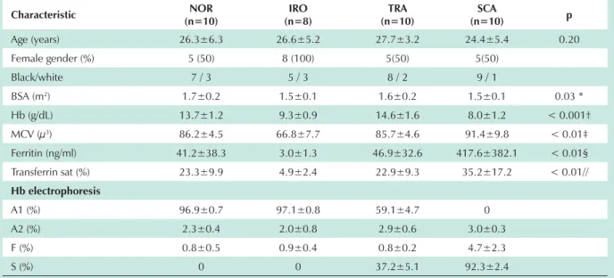
Clinical characteristics - Clinical and demographic characteristics of the individuals participating in the study are shown in Table 1. Mean BSA of the SCA and IRO groups
Methods
A total of 38 individuals of both genders with age above 18 years (ranging from 19 to 39) were prospectively studied. Ten patients with SCA and 10 patients with sickle cell trait (TRA) were randomly selected from the outpatient clinic of the Discipline of Hematology and Hemotherapy of Hospital das Clínicas da Faculdade de Medicina da Universidade de São Paulo. Patients were clinically stable, with no pain crises, hospitalizations or blood transfusion in the three months prior to the beginning of the study. No patients received cytotoxic drugs such as hydroxyurea. Patients with TRA were asymptomatic and had no laboratory alterations such as anemia or morphologic alterations of erythrocytes. Diagnosis of both diseases was confirmed with hemoglobin electrophoresis.
Eight female patients with chronic iron deficiency anemia (IRO) and 10 healthy individuals (NOR) were consecutively selected from the Blood Bank of Hospital das Clínicas da Faculdade de Medicina da Universidade de São Paulo. IRO was confirmed by the presence of hemoglobin (Hb) < 12 g/dL, erythrocyte mean corpuscular volume (MCV) < 82 µ3,
serum ferritin < 9 ng/mL and transferrin saturation < 16%. Patients with IRO had had symptoms attributable to anemia for more than a year, and were chronically adapted to it. Anemia resulted from hypermenorrhea secondary to uterine myoma in five patients and nutritional deficiency in the remaining women. The NOR group was comprised of asymptomatic volunteer blood donors who had donated for the last time more than three months prior to admission to the study.
Patients with systemic and pulmonary hypertension, diabetes, dyslipidemia, coronary artery disease or history of chest pain, asthma or obstructive pulmonary disease, esophageal disease, renal failure, stroke, pregnancy, presence of antibodies against human immunodeficiency virus, alcohol consumption and smoking were not included in the study.
The study was conducted according to the principles of the Declaration of Helsinki and approved by the Research Ethics Committee of Instituto do Coração (Heart Institute). Awritten consent form was obtained from all participants in the study.
Clinical assessment - Complete history taking and physical examination, as well as 12-lead electrocardiogram, pulse oximetry, urinalysis, chest radiograph, hemoglobin electrophoresis, and blood tests for hematological and biochemical profiles were performed for all participants in the study.
were lower than those observed in the TRA and NOR groups (p=0.03). No significant differences were observed in the mean Hb level between the SCA and IRO groups (p=0.17), and between the TRA and NOR groups (p=0.39). However, both SCA and IRO groups had Hb levels lower than TRA and NOR groups (p<0.01). The IRO group was comprised only of females. Mean age and race distribution were similar between the groups.
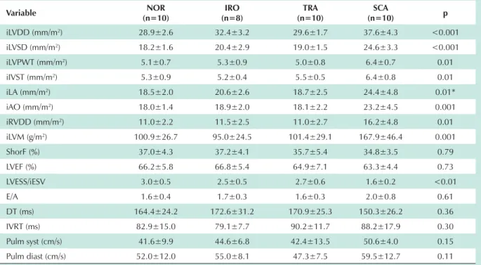
Cardiac structure and functions - As shown in Table 2, patients with SCA had higher indexes of left ventricular systolic and diastolic diameter, left ventricular wall and interventricular septal thickness, right ventricular diameter, left atrial diameter, aortic diameter, and left ventricular mass with statistical significance when compared with TRA, IRO and NOR groups – p<0.05. Left ventricular systolic and diastolic function parameters were similar between the groups studied, except for the lower LVESS / iESV ratio in the SCA group, which was statistically significant in comparison with TRA, IRO and NOR groups – p<0.01. These results corroborate previous data from the literature4,5,20,21.
Coronary flow velocity and coronary flow reserve - At baseline, the SCA group had higher systolic and diastolic CFVbas in comparison with TRA and NOR groups (p<0.01). When compared with the IRO group, the SCA group had higher systolic and diastolic CFVbas; however, the systolic CFVbas difference did not reach statistical significance (p=0.12). As shown in Table 3, during adenosine infusion the SCA group also showed higher values of systolic and diastolic CFVmax when compared with the TRA, IRO and NOR groups, with a statistically significant difference – p<0.01. However, a proportional increase of CFVmax was verified
in all groups, on average of three times the baseline values. Thus, CFR, which corresponds to the ratio between diastolic CFVmax and diastolic CFVbas was normal in the four study groups, with no statistical difference between them (p=0.70), although it was 14% lower in the SCA group – Figure 1. No statistically significant correlation was observed between CFR and clinical, hematological, cardiac structure, and coronary flow velocity variables.
Discussion
The present study found no alterations of CFR in SCA, and proved the functional integrity of the coronary microcirculation in this disease. Despite a higher CFVbas in SCA, the vasodilatation capacity of coronary resistance vessels is preserved and allows an adequate increase in CFVmax.
Several factors described in the literature could suggest, in an initial analysis, results different from those found in this study. First, the bare presence of anemia has been pointed as the responsible for a reduction in CFR19,22,23. Tissue hypoxia
observed in these syndromes determines a reduction in coronary arteriolar resistance, with a subsequent increase in baseline coronary flow. Thus, part of the microvascular vasodilation capacity would already be used at baseline, thus limiting the capacity of coronary flow increase during maximum hyperemia. Our results show that even with increased CFVbas, anemia observed in the SCA and IRO groups did not result in this reduction of CFVmax, with a proportional increase occurring in comparison with the groups without anemia, TRA and NOR.
Although higher values of CFVbas in the SCA and IRO Table 1 - Clinical characteristics of the four study groups
Characteristic Q 125 IRO (n=8)
TRA
Q Q SCA p
Age (years) 26.3±6.3 26.6±5.2 27.7±3.2 24.4±5.4 0.20 Female gender (%) 5 (50) 8 (100) 5(50) 5(50)
Black/white 7 / 3 5 / 3 8 / 2 9 / 1
BSA (m2) 1.7±0.2 1.5±0.1 1.6±0.2 1.5±0.1 0.03 *
Hb (g/dL) 13.7±1.2 9.3±0.9 14.6±1.6 8.0±1.2 < 0.001† MCV (µ3) 86.2±4.5 66.8±7.7 85.7±4.6 91.4±9.8 < 0.01‡
Ferritin (ng/ml) 41.2±38.3 3.0±1.3 46.9±32.6 417.6±382.1 < 0.01§ Transferrin sat (%) 23.3±9.9 4.9±2.4 22.9±9.3 35.2±17.2 < 0.01//
+EHOHFWURSKRUHVLV
A1 (%) 96.9±0.7 97.1±0.8 59.1±4.7 0 A2 (%) 2.3±0.4 2.0±0.8 2.9±0.6 3.0±0.3 F (%) 0.8±0.5 0.9±0.4 0.8±0.2 4.7±2.3
S (%) 0 0 37.2±5.1 92.3±2.4
Values are expressed as mean±standard deviation. BSA - body surface area; Hb - hemoglobin; MCV - erythrocyte mean corpuscular volume; Transferrin sat - transferrin saturation. * SCA vs125²S ,52vs125²S 6&$vs,52S 125vs75$S Â,52vs NOR, TRA and
6&$²S6&$vs125,52DQG75$²S,52vs125²S ,52vs75$²S ,52vs6&$²S,52vs NOR and TRA
groups corroborate the results of hemodynamic studies in anemias24,25, the difference in velocities found between
the two groups claims attention, since both present similar hemoglobin concentration – 8.0±1.2 and 9.3±0,9 g/dL, respectively – p=0.17. The SCA group had a mean CFBbas approximately 1.6 times higher than that of the IRO group, with a statistically significant difference – p< 0.01. As previously mentioned, the most important factor determining
the increase in coronary flow velocity in anemia is the decrease in coronary vascular resistance resulting from the reduction in arterial oxygen content. Previous data suggest that SCA with a similar Hb level in comparison with IRO and other forms of anemia causes a higher degree of tissue hypoxemia25.
Second, cardiac hypertrophy leads to alterations in coronary flow patterns with reduction of CFVmax26. In light
of the eccentric hypertrophy found in SCA, a lower CFR Table 2 - Values of cardiac structure and functions in the four study groups
Variable Q 125 IRO
(n=8)
TRA
Q Q SCA p
iLVDD (mm/m2) 28.9±2.6 32.4±3.2 29.6±1.7 37.6±4.3 <0.001
iLVSD (mm/m2) 18.2±1.6 20.4±2.9 19.0±1.5 24.6±3.3 <0.001
iLVPWT (mm/m2) 5.1±0.7 5.3±0.9 5.0±0.8 6.4±0.7 0.01
iIVST (mm/m2) 5.3±0.9 5.2±0.4 5.5±0.5 6.4±0.8 0.01
iLA (mm/m2) 18.5±2.0 20.6±2.6 18.7±2.5 24.4±4.8 0.01*
iAO (mm/m2) 18.0±1.4 18.9±2.0 18.1±2.2 23.2±4.5 0.001
iRVDD (mm/m2) 11.0±2.2 11.5±2.5 11.0±2.7 16.2±4.8 0.01
iLVM (g/m2) 100.9±26.7 95.0±24.5 101.4±29.1 167.9±46.4 0.001
ShorF (%) 37.0±4.3 37.2±4.1 35.7±5.4 34.8±3.5 0.79 LVEF (%) 66.2±5.8 66.8±5.4 64.9±7.1 63.3±4.4 0.73 LVESS/iESV 3.0±0.5 2.5±0.5 2.7±0.6 1.6±0.2 <0.01 E/A 1.6±0.4 1.7±0.3 1.6±0.3 2.0±0.8 0.61 DT (ms) 164.4±24.2 172.6±31.2 170.9±25.3 150.3±26.2 0.36 IVRT (ms) 82.9±15.0 79.1±7.7 90.2±11.7 88.2±17.9 0.30 Pulm syst (cm/s) 41.6±9.9 44.6±6.8 42.4±13.5 50.6±4.0 0.15 Pulm diast (cm/s) 52.0±12.0 55.0±8.1 47.3±7.5 59.5±12.7 0.11
Values are expressed as mean±standard deviation. iLVDD - left ventricular end-diastolic diameter index; iLVSD - left ventricular end-systolic diameter index; iLVPWT - left ventricular posterior wall thickness index; iIVST - interventricular septal thickness index; iLA - left atrium index; iAO - aortic root index; iRVDD - right ventricular end-diastolic diameter index; iLVM - left ventricular mass index; ShorF - left ventricular shortening fraction; LVEF - left ventricular ejection fraction; LVESS/iESV - left ventricular end-systolic stress/left ventricular end-systolic volume index ratio; E/A - mitral flow E wave / A wave ratio; DT - deceleration time; IVRT - isovolumetric relaxation time; Pulm syst - pulmonary systolic wave; Pulm diast - pulmonary diastolic wave. * SCA vs,52²S
7DEOH9DOXHVRIFRURQDU\IORZYHORFLWLHVDQGRIFRURQDU\IORZUHVHUYHLQWKHVWXG\JURXSV
Variable Q 125 IRO
(n=8)
TRA
Q Q SCA p
Baseline
CFV diast (cm/s) 38.1±10.0 42.4±10.4 34.4±11.9 67.3±14.0 <0.001 CFV syst (cm/s) 16.7±6.0 21.8±6.6 18.5±7.3 29.0±6.8 0.005*
+\SHUHPLD
CFV diast (cm/s) 126.8±24.6 141.0±18.7 114.7±36.4 198.2±37.9 <0.001 CFV syst (cm/s) 51.2±10.5 63.2±14.9 49.7±20.2 95.1±24.5 <0.001 CFR 3.4±0.8 3.5±1.2 3.4±0.8 3.0±0.7 0.70
1. Serjeant GR. Sickle cell disease. Lancet. 1997;350:725-30.
2. Stuart MJ, Nagel RL. Sickle cell disease. Lancet. 2004;364:1343-60.
3. Covitz W, Espeland M, Gallagher D, Hellenbrand W, Leff S. The heart in sickle cell anemia: the cooperative study of sickle cell disease. Chest. 1995;108:1214-9.
4. Martins WA, Mesquita ET, Cunha DM, Pinheiro LAF, Romeo Filho LJM, Pareto Jr RC. Estudo ecoDopplercardiográfico em adolescentes e adultos jovens portadores de anemia falciforme. Arq Bras Cardiol. 1999;73:463-8.
5. Lamers L, Ensing G, Pignatelli R, Goldberg C, Bezold L, Ayres N, et al. Evaluation of left ventricular systolic function in pediatric sickle cell anemia patients using the end systolic wall stress velocity of circumferential fiber shortening relationship. J Am Coll Cardiol. 2006;47:2283-8.
6. Tap SM, Mete UO, Kaya M. Ultrastructural alterations in the myocardium of patients with sickle cell anemia. J Submicrosc Cytol Pathol. 2001;33:151-6.
7. James TN, Riddick L, Massing GK. Sickle cell and sudden death: morphologic abnormalities of the cardiac conduction system. J Lab Clin Med. 1994;124:507-20.
References
could also be expected. Left ventricular hypertrophy and dilatation develop as a compensatory measure in response to volume overload, so as to reduce the ventricular wall stress and normalize the cardiac performance27. However, growth
of the coronary microcirculation is not proportional to the increase in the cardiac mass, with an increased minimum coronary vascular resistance and decreased blood flow to the endocardium being observed. In addition, interstitial fibrosis and vascular media hypertrophy also contribute to the reduction of vasodilation capacity28,29. Comparatively, in
diseases such as idiopathic dilated cardiomyopathy30 or in
chronic aortic regurgitation31, both with eccentric hypertrophy,
a reduction in CFR is observed, and its presence characterizes a subgroup of patients with increased mortality, regardless of the degree of left ventricular dysfunction30. However, our
results do not corroborate these reports. Also, in addition to a normal CFR, no correlation between CFR and left ventricular mass index was observed in the SCA group .
Some experimental studies have demonstrated evidences of increased capillary density in hypertrophies secondary to volume overload32,33, thyroxin stimulation34, and also
following physical training35. In these situations, a proportional
growth would occur between the coronary microvasculature and myocyte hypertrophy. Additionally, in humans, a result
similar to that of the present study was observed in athletes with left ventricular hypertrophy36. These data suggest that
the specificity of the inducing stimulus is important to the development of cardiac hypertrophy, and some of them are also able to induce a physiologic adaptation of the coronary microvasculature. In support of this hypothesis, some of the stimuli able to produce microvascular growth such as tissue hypoxia, increased blood flow velocity, and microvascular vasodilation are known to be present in anemias37. Additionally, the presence of serum markers of
angiogenesis has been reported in patients with SCA, both at baseline and during painful crises38.
Third, although the heart muscle has a high rate of oxygen extraction from the arterial blood, the heart does not have a sinuous microcirculation or reduced pH, which are factors that favor red cell sickling and its adherence to the vascular endothelium. The hemoglobin S polymerization process does not occur instantaneously. There is a period of time, called delay time, during which the cascade of events leading to HbS aggregation is activated. This period is attributed to the process of red cell nucleation, in which HbS forms small clusters, however without modifying the inner red cell viscosity yet. When these clusters grow to form a substantial mass, a rapid addition of new HbS units occurs, thus forming polymers that distort the red cells and lead to the characteristic conformation of a sickle39. Delay time is
estimated at 10 to 15 sec, and is inversely proportional to the intracellular concentration of HbS in the erythrocytes40.
If the HbS is reoxygenated within this period of time, the erythrocyte will not present significant structural alterations and will not adhere to the endothelium. Therefore, increased coronary blood flow velocity may act as a protective factor, thus favoring a rapid transit time and erythrocyte oxygenation in adequate time. This action may prevent sickled cells from binding to the coronary vascular endothelium.
Conclusion
Thus, we conclude that despite higher coronary flow velocities at baseline and also during maximum hyperemia, CFR is normal in SCA, which suggests integrity of the coronary microcirculation. This study did not find evidence that the lesions produced by coronary resistance vessels are responsible for the cardiac alterations found in this disease, so such alterations result from the chronic presence of anemia.
Fig. 1 - Individual distribution of coronary flow reserve in the four study groups
Groups
NOR IRO TRA SCA p=0.70
C
o
ro
n
a
ry
r
e
s
e
rv
8. Montalembert M, Maunoury C, Acar P, Brousse V, Sidi D, Lenoir G. Myocardial ischaemia in children with sickle cell disease. Arch Dis Child. 2004;89:359-62.
9. Berezowski K, Mautner GC, Roberts WC. Scarring of the left ventricular papillary muscles in sickle cell disease. Am J Cardiol. 1992;70:1368-70.
10. Tanner MA, Westwood MA, Pennell DJ. Myocardial infarction following sickle cell chest syndrome. Br J Haematol. 2006;134:2.
11. Lester LA, Sodt PC, Hutcheon N, Arcilla RA. Cardiac abnormalities in children with sickle cell anemia. Chest. 1990;98:1169-74.
12. Marcus ML, Doty DB, Hiratzka LF, Wright CB, Eastham CL. Decreased coronary reserve: a mechanism for angina pectoris in patients with aortic stenosis and normal coronary arteries. N Engl J Med. 1982;307:1362-7.
13. Zehetgruber M, Mundigler G, Christ G, Mortl D, Probst P, Baumgartner H, et al. Estimation of coronary flow reserve by transesophageal coronary sinus Doppler measurements in patients with syndrome X and patients with significant left coronary artery disease. J Am Coll Cardiol. 1995;25:1039-45.
14. Memmola C, Iliceto S, Napoli VF, Cavallari D, Santoro G, Rizzon P. Coronary flow dynamics and reserve assessed by transesophageal echocardiography in obstructive hypertrophic cardiomyopathy. Am J Cardiol. 1994;74:1147-51.
15. Sahn DJ, DeMaria A, Kisslo J, Weyman A. Recommendations regarding quantitation in M-mode echocardiography: results of a survey of echocardiographic measurements. Circulation. 1978;58:1072-83.
16. Rakowski H, Appleton C, Chan KL, Dumesnil JG, Honos G, Jue J, et al. Canadian consensus recommendations for the measurement and reporting of diastolic dysfunction by echocardiography. J Am Soc Echocardiogr. 1996;9:736-60.
17. Denenberg BS, Criner G, Jones R, Spann JF. Cardiac function in sickle cell anemia. Am J Cardiol. 1983;51:1674-8.
18. Reichek N, Wilson J, Sutton MSJ, Plappert TA, Goldberg S, Hirshfeld JW. Noninvasive determination of left ventricular end-systolic stress: validation of the method and initial application. Circulation. 1982;65:99-108.
19. Hoffman JIE. Maximal coronary flow and the concept of coronary vascular reserve. Circulation. 1984;70:153-9.
20. Gerry JL Jr, Bulkley BH, Hutchins GM. Clinicopathologic analysis of cardiac dysfunction in 52 patients with sickle cell anemia. Am J Cardiol. 1978;42:211-6.
21. Falk RH, Hood WB Jr. The heart in sickle cell anemia. Arch Intern Med. 1982;142:1680-4.
22. Nitenberg A, Antony I. Coronary vascular reserve in humans: a critical review of methods of evaluation and of interpretation of the results. Eur Heart J. 1995;16 (Suppl 1): 7-21.
23. Roy SB, Bathia ML, Mathur VS, Virmani S. Hemodynamic effects of chronic severe anemia. Circulation. 1963;28:346-56.
24. Bhatia ML, Manchanda SC, Roy SB. Coronary haemodynamic studies in
chronic severe anaemia. Br Heart J. 1969;31:365-74.
25. Leight L, Snider TH, Clifford GO, Hellems HK. Hemodynamic studies in sickle cell anemia. Circulation. 1954;10:653-62.
26. Pichard AD, Smith H, Holt J, Meller J, Gorlin R. Coronary vascular reserve in left ventricular hypertrophy secondary to chronic aortic regurgitation. Am J Cardiol. 1983;51:315-20.
27. Grossman W, Jones D, McLaurin LP. Wall stress and patterns of hypertrophy in the human left ventricle. J Clin Invest. 1975;56:56-64.
28. Krayenbuehl HP, Hess OM, Schneider J, Turina M. Physiologic or pathologic hypertrophy. Eur Heart J. 1983;4 (Suppl A): 29-34.
29. Opie LH, Commerford PJ, Gersh BJ, Pfeffer MA. Controversies in ventricular remodelling. Lancet. 2006;367:356-67.
30. Rigo F, Gherardi S, Galderisi M, Pratali L, Cortigiani L, Sicari R, et al. The prognostic impact of coronary flow-reserve assessed by Doppler echocardiography in non-ischemic dilated cardiomyopathy. Eur Heart J. 2006;27:1319-23.
31. Nitenberg A, Foult JM, Antony I, Blanchet F, Rahali M. Coronary flow and resistance reserve in patients with chronic aortic regurgitation, angina pectoris and normal coronary arteries. J Am Coll Cardiol. 1988;11:478-86.
32. Tomanek RJ. Response of the coronary vasculature to myocardial hypertrophy. J Am Coll Cardiol. 1990;15:528-33.
33. Chen Y, Torry RJ, Baumbach GL, Tomanek RJ. Proportional arteriolar growth accompanies cardiac hypertrophy induced by volume overload. Am J Physiol. 1994;267:H2132-7.
34. Chilian WM, Wangler RD, Peters KG, Tomanek RJ, Marcus ML. Thyroxine-induced left ventricular hypertrophy in the rat: anatomical and physiological evidence for angiogenesis. Circ Res. 1985;57:591-8.
35. Cohen MV. Coronary vascular reserve in the greyhound with left ventricular hypertrophy. Cardiovasc Res. 1986;20:182-94.
36. Hildick-Smith DJR, Johnson PJ, Wisbey CR, Winter EM, Shapiro LM. Coronary flow reserve is supranormal in endurance athletes: an adenosine transthoracic echocardiographic study. Heart. 2000;84:383-9.
37. Rakusan K, Cicutti N, Kolar F. Effect of anemia on cardiac function, microvascular structure, and capillary hematocrit in rat hearts. Am J Physiol. 2001;280:H1407-14.
38. Duits AJ, Rodriguez T, Schnog JJ. Serum levels of angiogenic factors indicate a pro-angiogenic state in adults with sickle cell disease. Br J Haematol. 2006;134:116-9.
39. Francis RB Jr, Johnson CS. Vascular occlusion in sickle cell disease: current concepts and unanswered questions. Blood. 1991;77:1405-14.