Heart Institute, Hospital das Clínicas - FMUSP
Mailing address: William Azem Chalela - Incor - Serviço de Radioisótopos Av. Dr. Enéas C. Aguiar, 44-AB - 05403-000 - São Paulo, SP - Brazil
Objective - To compare single-photon-emission
com-puted tomography (SPECT) imaging scans using 201Tl and 99mTc-MIBI in detection of viable myocardium, in regions
compromised by infarction.
Methods - Thirty-two (59.3±9.8 years old and 87%
male) myocardial infarction patients were studied. All had Q waves on the ECG and left ventricle ejection frac-tion of <50%. They underwent coronary and left ventricle angiographies and SPECT before (including 201Tl
reinjec-tion) and after coronary artery bypass surgery (CABG). Improvement in perfusion observed after surgery was considered the gold standard for myocardial viability.
Results - Among 102 studied regions of the heart, there
were 40 (39.2%) areas of transient perfusion defects in the conventional protocol with 201Tl and 52 (51.0%) after
rein-jection. Therefore, 12/62 (19.4%) more viable regions were identified by reinjection. Using 99mTc-MIBI, only 14 (13.7%)
regions with transient defects were identified, all of which were seen also in 201Tl protocols. After surgery, 49 of a total of
93 regions analyzed (52.7%) were viable. Sensitivity, spe-cificity, accuracy, positive and negative prediction values were, respectively, 201Tl SPECT scans - 65.3%, 90.9%, 77.4%,
88.9% and 70.2%, reinjection protocol with 201Tl scans
-81.6%, 81.8%, 81.7%, 83.3% and 80.0%; 99mTc-MIBI SPECT
scans - 20.4%, 90.9%, 53.8%, 71.4% and 50.6%. Logistic regression demonstrated that the reinjection protocol with
201Tl was the best predictor of viability (P<0.001).
Conclusion - Our data suggest the election of 201Tl for
viability studies, especially when using the reinjection protocol.
Keywords: myocardial viability, coronary artery disease, myocardial perfusion scintigraphy
Arq Bras Cardiol, volume 72 (nº 5), 535-545, 1999
Willliam A. Chalela, Paulo J. Moffa, José A. F. Ramires, Aguinaldo P. Moraes, José Soares Jr, José C. Meneghetti
São Paulo, SP - Brazil
Detection of the Viable Myocardium. A Perfusion
Scintigraphic Study, Before and After Coronary Bypass
Surgery in Myocardial Infarction Patients
Ischemic cardiomyopathy is a frequent cause of hos-pitalization and death. In addition, patients with severe is-chemic heart disease have high mortality, when treated me-dically, with about a 31% two-year survival rate 1. Cardiac
transplantation is sometimes indicated for these patients 2,3.
Ischemic cardiomyopathy can be due to ventricular dysfunction caused by large areas of infarction or by hypoki-netic regions caused by chronic hypoperfusion without ne-crosis. The presence of viable myocardium could favor the indication of myocardial revascularization, thus halting the evolution to larger necrotic areas with the consequent ven-tricular dysfunction and the need for heart transplantation 4.
The correct identification of the state of myocardial via-bility constitutes an important element in the choice of the therapy subsequently adopted: myocardial revasculariza-tion, resection of aneurysms, or heart transplantation.
Often, clinical data, the electrocardiogram (ECG), coro-nary angiography and the analysis of regional wall motion at rest, are not enough for the distinction of fibrosis from via-ble myocardium.
Initially, postextrasystolic potentiation and infusion of inotropic agents were used to identify viable myocardium based upon the observation of the recovery of myocardial contraction using sensors placed directly on the epicardium under experimental conditions, or by ventricular wall motion analysis in conventional ventriculograms. Recently, echo-cardiography and gated radionuclide angiography with pharmacological stress (low dose dobutamine) were used for the same purpose.
536
Chalela et al
Scintigraphy in the detection of viable myocardium
Arq Bras Cardiol volume 72, (nº 5), 1999
Cardiac imaging techniques using myocardial perfu-sion scintigraphy (MPS), based on the study of myocardial perfusion and cellular membrane integrity, have achieved substantial success in the assessment of myocardial viability 5.
Thus, the present work aimed to investigate the pre-sence of viable muscle in myocardial infarction areas using single-photon-emission computed tomography (SPECT). For that purpose, both thallium-201 chloride (201Tl) and
2-methoxi-isobuthyl-isonitrile labeled with technetium-99m (99mTc-MIBI) were used. Improvement in perfusion of these
areas after coronary artery bypass graft surgery (CABG) was used in the statistical calculations.
201Tl is the radionuclide most often used as a
myocar-dial perfusion marker in the assessment of myocarmyocar-dial viability. 99mTc-MIBI is frequently used for the diagnosis of
coronary artery disease; however, there are insufficient data about its utility in the detection of the viable myocardium.
Methods
Thirty-two patients with a history of MI (of a total of 113 patients referred for viability studies) were selected. The patients fulfilled the following inclusion criteria: MI between 3 and 12 months prior; presence of Q waves (<40ms) on the ECG; angina and heart failure functional class 6 < III; global left ventricle ejection fraction (LVEF)
<50%; no absolute contraindications to the stress testing (ST). Patients were excluded for: clinical instability;the use of thrombolytic agents; coronary bypass surgery during an acute MI; bundle branch block on ECG; uncontrolled hypertension; valve disease, pericardial disease or other cardiac diseases.
All patients underwent cardiac catheterization and both rest and stress 201Tl and 99mTc-MIBI scintigraphies, up
to six months before CABG. Complete revascularization was confirmed 7, and the patients repeated the radionuclide
eva-luation after approximately three months and the ca-theterization up to six months after the procedure. Thallium and technetium studies were performed a minimum of three and a maximum of seven days apart. Patients were under the same medication and doses during the follow-up.
This study was approved by the scientific committee of the Heart Institute and also by the ethics committee of our hospital. All patients agreed to participate in the study after informed consent.
ECG tracings were conventionally obtained on the sa-me days of the scintigraphies. Myocardial regions affected by MI were classified as: septal, anterior, inferior, dorsal and lateral, according to the literature 8.
Coronary lesions were classified according to the per-centage of obstruction: occluded (100% of the lumen), seve-re lesions (>90% and <100%); critical lesion from 70% to 90%; and noncritical lesions (<70%). The presence of colla-teral circulation (Cc) was assessed as well as its origin and intensity 9. Obstructed arteries were considered
infarct-related arteries depending on the anatomical location of the
affected region, i. e., left anterior descending artery (LAD) for septal, anterior and apical regions; right coronary (RC) for inferior region; and circumflex (Cx) arteries for lateral and dorsal regions. When RC and Cx had a similar degree of obstruction, the dominant artery assumed to be responsi-ble for the inferior region was the RC and for the dorsal region the Cx branch.
Ventricular wall motion analysis was performed using left ventriculography, in right anterior oblique (RAO) pro-jection. Ventricular wall motion abnormalities were classified as follows: hypokinesis - discrete, moderate or accentuated diminished motion; akinesis - total absence of motion; dyskinesis - paradoxal systolic expansion of a given LV segment. Wall motion was graded arbitrarily ranging from zero to five, according to LV motion (0 = normal, 1 to 3 = discrete to accentuated hypokinesis; 4 = akinesis and 5 = dyskinesis). A change of at least one point was considered an improvement, when comparing parameters before and after the procedure.
Global LVEF was calculated by Dodge’s uniplanar method 10 modified by Kennedy 11.
Stress tests were conventionally performed 12, using
the treadmill and the Ellestad’s protocol 13, with a 12-lead
simultaneous recording.
The protocol of the 201Tl scintigraphy included three
phases: 1st, imaging was acquired immediately after
radio-nuclide injection at peak exercise; the dose injected was 111 MBq (3mCi) and tomographic images obtained with the patient in the supine position; 2nd, after 4h the redistribution
images, similarly acquired; 3rd, reinjection, performed after
24h, with an additional dose of thallium administered at rest and SPECT images acquired after 4h.
For 99mTc-MIBI studies, the entire procedure was
per-formed in one day. Initially, images were obtained at rest, with a 296 to 370 MBq (8 to 10mCi) intravenous infusion, followed by the stress images. The radionuclide [888 to 1110 MBq (24 to 30mCi)] was injected at peak exercise, three ti-mes the resting dose. To acquire tomographic images, a Sie-mens scintigraphic camera, model Orbiter ZLC-Digitrac 750, coupled to a Maxdelta (Microvax - 3300) computer was used. Analog imaging was converted to digital in a 64x64, 64 projections with 20 s duration each, over a 180o arch,
begin-ning from 45o RAO to 45o left posterior oblique LPO (step/
shoot acquisition mode). The filter used was a Butterworth, with a cutoff frequency of 0.4 Ny (Ny-Nyquist frequency) for 201Tl and 0.5 Ny for 99mTc-MIBI.
con-centration (0 = normal perfusion; 1 = mild perfusion defect, 3 = severe perfusion defect or tracer absence). A change of at least one point in the grade of a given region was considered as transient perfusion defects and was clas-sified as partial when zero was not observed in the redistri-bution and/or reinjection images. Maintenance of perfusion values was indicative of persistent perfusion defects (fibrosis).
In this study, we analyzed only LV regions compromi-sed by MI, as shown on the ECG. The apical region was considered as an extension of anterior and/or inferior involvement, when graded with values greater than zero. Areas of improvement in myocardial perfusion observed on the resting images were considered as true viable ones.
To accomplish the goals of this study, descriptive tables were constructed. Continuous variables were pre-sented descriptively as mean and standard deviations. Comparisons before and after surgery were performed with Wilcoxon’s (nonparametric) or Student (paired) t tests 14.
Agreement between methods for myocardial viability was analyzed using Kappa statistics 15 (K). Values >0.75
repre-sent excellent agreement, between 0.40 and 0.75 good agree-ment, and <0.40, poor agreement. Sensitivity (S), specificity
(E), accuracy (A), positive (PPV) and negative (NPV) predic-tive values were calculated. Viability confirmed after surge-ry was adjusted using a stepwise logistic regression model in which the other protocols were considered predictive factors.
The significance level used was 0.05. Calculations were made using the SAS system 16. Intra and interobserver
variability analysis was calculated by reevaluating a sample of 10 cases after at least 30 days.
Results
Studied population data are found in table I. The mean age was 59.3±9.8 years (ranging from 42 to 75 years), 28 (87.6%) males and 4 (12.4%) females. Patients were studied on average 8.14±3.29 months after MI.
Seventy-eight myocardial regions of fibrosis were studied. They were divided into: 23 (29.5%) septal, 23 (29.5%) anterior, 21 (26.9%) inferior, 10 (12.8%) lateral and 1 (1.3%) dorsal regions.
Catheterization (data are displayed in table II) was performed a mean of 2.79±2.73 months before surgery. Single vessel disease occurred in one (3.1%) patient; double
Table I - Population Data
No Name Age Gender Weight Height BMI HBP DM HC HT S FH ECG - areas Time FC FC
(years) (kg) (Cm) (kg/m2) S A I L P (months) A P HF
1 GAL 61 M 88 1.75 28.7 YES NO NO NO YES NO S A I 3.8 I III
2 CABG 42 M 79 1.70 27.3 YES NO NO NO YES YES S A L 10.3 III III
3 MIS 61 M 102 1.78 31.9 NO YES YES NO NO YES I 12.0 III III
4 OVA 47 M 92 1.79 28.7 YES NO NO NO YES YES S A I 3.8 III II
5 ACGD 49 M 63 1.62 24.0 YES YES YES NO NO NO S A I 6.8 II III
6 SS 65 M 67 1.65 24.6 YES YES NO NO NO NO I L 12.0 II II
7 TJ 55 M 68 1.70 23.5 YES NO YES NO YES NO S A I 4.3 III III
8 AOA 74 M 63 1.72 21.3 NO NO NO NO YES NO S A I 6.9 III III
9 PSS 72 M 86 1.72 29.1 YES YES NO NO YES YES S A L 11.7 III III
10 P L 65 M 60 1.60 23.4 NO NO NO NO YES NO I L 11.7 I III
11 CP 48 M 86 1.74 28.4 NO NO NO NO YES YES S A L 4.6 I II
12 MTM 67 M 81 1.66 29.4 NO YES NO NO YES NO I 3.1 II II
13 OF 63 M 70 1.70 24.2 NO NO YES NO YES YES S A 12.0 III II
14 BM 72 M 65 1.80 20.1 NO YES NO NO YES NO S A 3.3 II III
15 DCA 63 M 68 1.73 22.7 NO YES NO NO NO NO S A I 11.6 I III
16 PAAG 65 M 79 1.77 25.2 YES YES NO NO YES NO S A 11.1 I III
17 JMP 54 F 51 1.58 20.4 YES NO YES NO YES NO S A 12.0 III II
18 NCP 65 F 66 1.56 27.1 YES YES YES NO NO YES S A I 4.2 III II
19 JBS 56 M 73 1.54 30.8 YES NO NO NO YES NO S A I 8.7 I II
20 JMB 74 M 75 1.60 29.3 YES NO NO NO NO YES I L P 11.7 I III
21 FP 56 M 66 1.55 27.5 YES YES YES NO NO NO I L 11.8 III I
22 NGS 42 F 52 1.46 24.4 NO YES YES NO NO NO S A I 5.5 III III
23 JB 61 M 70 1.70 24.2 YES NO NO NO YES YES i 11.6 III III
24 GMS 63 M 70 1.70 24.2 NO NO NO NO YES NO S A I L 12.0 I III
25 SAC 67 M 72 1.72 24.3 NO NO NO NO YES YES I L 11.1 III III
26 JMC 52 M 85 1.73 28.4 YES YES NO NO YES YES S A I 12.0 I III
27 SAA 44 M 71 1.58 28.4 NO NO YES NO YES NO S A 3.4 I III
28 GDS 75 M 81 1.72 27.4 NO NO NO NO YES NO I L 5.7 II III
29 AM 65 M 82 1.71 28.0 YES YES YES NO NO NO S A 3.3 III III
30 JCNN 45 M 78 1.80 24.1 YES YES NO NO YES NO S A I 3.3 III III
31 GTB 62 M 56 1.63 21.1 NO NO NO NO NO NO S A 11.9 III III
32 MFL 49 F 90 1.60 35.2 YES NO YES NO NO YES S A I 3.7 III III
BMI - body mass index kg/m2; HBP- high blood pressure; DM- diabetes mellitus; HC- hypercholesterolemia, HT- hipertriglyceridemia; S- smoking;
5
3
8
Chalela et al
Scintigraphy in the detection of viable myocardium
Arq Bras Cardiol
volume 72, (nº 5), 1999
Table II - Coronary angiographic data before coronary artery bypass graft
No ∆ CABG Coronary Angiography LV
RC LDA DB LCx ME LC Reg. Mot. EF
Cc Cc Cc Cc Cc Cc
Months Dom. Lesion Source Grade Lesion Source Grade Lesion Source Grade Lesion Source Grade Lesion Source Grade Lesion Source Grade Ant. Inf. Ap
% % % % % %
1 0.63 RC 95 - - - 99 - - - 80 - - - 70 - - - A HA A 35
2 5.70 RC - - - - 100 - - - 80 - LDA +++ 100 - - - HM HD HM 47
3 1.53 RC 80 - - - 100 - - - 100 - - - D HM HM 18
4 0.70 RC 100 LDA LCx ++ 99 - - - 100 - - - HM HM D 45
5 3.57 RC 100 - - - 100 - - - 70 - - - 30 - - - 100 - LCx +++ 40 - - - A HA A 20
6 3.13 RC 90 - - - 80 - - - 100 - LCx +++ - - - - N HD HD 48
7 1.07 RC 100 LDA LCx ++ 100 - RC +++ - - - - 40 - - - 80 - - - HA HA HA 48
8 2.33 RC 100 - RC + 100 - LDA + 30 - - - 70 - - - A HM A 21
9 4.00 RC 100 - RC +++ 80 - - - 70 - - - 40 - - - HM HD HM 39
10 1.87 RC 100 - LCx +++ 100 - RC +++ - - - 100 - LCx +++ - - - - A HA A 12
11 5.23 RC 80 RC LCx +++ 95 - RC ++ 80 - - - 100 - - - 40 - - - 40 - - - A HM A 46
12 0.23 RC 100 - RC +++ 100 RC LDA +++ - - - - 80 - - - 80 - - - A HA D 17
13 2.23 RC 100 RC LCx +++ 100 - LDA +++ 70 - - - 40 - - - 80 - - - D HM D 28
14 5.53 LCx 90 - - - 90 - - - 40 - - - HM HM HA 40
15 3.40 RC 100 LDA LCx ++ 100 RC LDA +++ - - - - 100 - - - HM HA A 36
16 5.67 RC 100 - LCx ++ 100 - LCx ++ - - - A HD A 20
17 5.37 RC 100 - - - 100 RC LCx ++ - - - 80 - - - HA HÁ A 39
18 4.70 RC 100 - RC + 100 - - - 75 - - - D HA D 35
19 4.80 RC 100 - RC + 100 - LDA + - - - - 75 - - - D HA D 35
20 5.70 RC 100 - LDA +++ 95 - - - 90 - - - 100 - - - HM HA HD 30
21 2.50 RC 100 - - - 100 - - - 90 - - - 100 - 3 + 40 - - - HA HA HA 43
22 5.70 RC 100 RC LDA + 100 - LDA ++ - - - HD HM A 33
23 2.10 RC 100 - - - 90 - - - 95 - - - 90 - - - HM HM A 47
24 0.43 Bal. 90 - - - 99 - - - 90 - - - 100 - - - A HM A 30
25 3.47 RC 100 - - - 80 - - - 100 - - - HM HÁ A 40
26 4.37 RC 100 - - - 100 - LDA ++ - - - - 90 - - - 80 RC LCx +++ - - - - HA HD D 42
27 1.50 RC 70 - - - 95 - - - A HD D 38
28 6.0 RC 100 LDA LCx ++ 80 RC LCx +++ - - - - 100 - - - 40 - - - HM HM HA 35
29 0.23 RC 30 - - - 95 - - - 90 - - - 30 - - - 30 - - - 30 - - - HA HM HA 35
30 0.50 RC 100 - LCx +++ 95 - - - 70 - - - HA HD HD 26
31 4.17 RC 80 - - - 100 RC LDA ++ 80 - - - 70 - - - A HM A 27
32 0.87 RC 100 - LDA ++ 95 - - - 40 - - - 70 - - - HD HM HD 30
∆ - time interval; CABG- coronary artery bypass graft; LV - left ventriculography; Dom. - dominance; RC- right coronary; LAD - Left anterior descending artery; DB -diagnonals branches; LCx - Left Circumflex artery, ME- marginal branches; LCA- left coronary artery; LC - left coronary; Reg. Mot.- regional motility; EF- global ejection fraction; Cc- collateral circulation; Ant - anterior; Inf. inferior; Ap- apical; Bal.- balanced; N- normal; HD- mild hypokinesis; HM - moderate hypokinesis; HA - severe hypokinesis; A - akinesis; D - dyskinesis;
vessel disease in 7 (21.9%) , most frequently the LAD and RC combination (71.4%); triple vessel disease in 24 (75.0%). Collateral circulation was observed in 24 (75.0%) patients. Regional motion could be studied in the anterior, inferior and apical LV wall, making a total of 96 regions. Changes found were: 61 (63.4%) regions with hypokinesis, 23 (24%) with akinesis and 11 (11.6%) with dyskinesis. Global LVEF observed in all patients was 34.9±10.2%. The 3 patients who died in the immediate postoperative (PO) period had LVEF of 20%, 30% and 43%.
There were no significant differences in the main clini-cal, electrocardiographic, hemodynamic and metabolic stress test variables. Mean maximal heart rates were: 132±20.6 bpm and 130.5±18.6 bpm; systolic blood pressure, 159.5±30.5 mmHg and 153.7±23.2 mmHg; and double pro-duct 21159.2±5676.1 and 20063.0±4125.9 (p=ns). The tracers were injected in similar conditions during both SPECT studies.
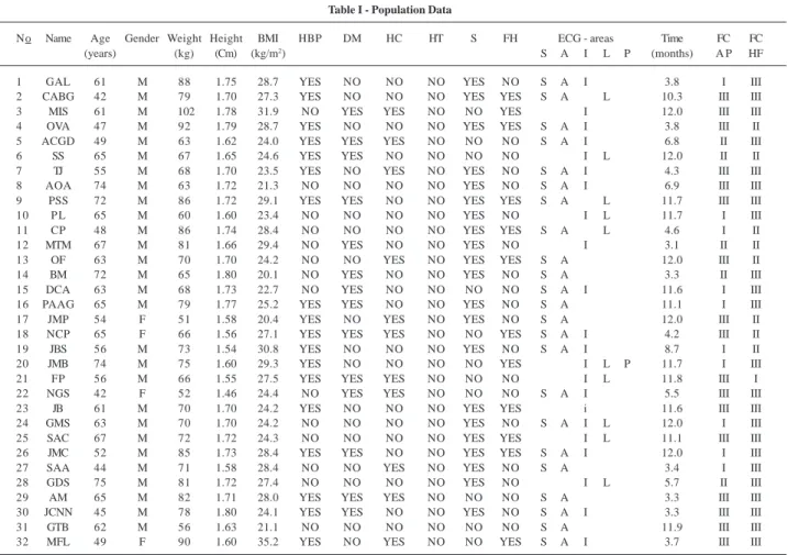
Every artery with lesions >50% was bypassed. Follow-up period after MR surgery was 15.16±10.17 months. Three (9.4%) patients died soon after operation due to cardiogenic shock. Another patient died suddenly after four months. Angina and CHF symptoms were evaluated in twenty-nine (90.6%) patients to determine functional class before and after CABG (fig. 1), and statistical analyses showed signi-ficant (p<0.001) improvement in both parameters analyzed. Coronary angiographic studies after surgery could be performed in 23 patients (4.98±1.09 months postoperative-ly). All grafts were patent. There was wall motion improve-ment in 48 (69.6%) regions and no change in 21 (30.4%) myo-cardial regions, when compared with the preoperative stu-dies (p<0.001). Mean global LVEF also increased (46.4± 13.5%; p<0.001; fig. 2).
For the detection of viable myocardium using scinti-graphy, 102 myocardial regions affected by MI were analy-zed. 201Tl, conventional protocol (comparing stress and
4h redistribution imaging) showed 40 (39.2%) reversible
regions and 62 (60.8%) nonviable ones (fixed defect); the reinjection protocol (stress and 24 h reinjection imaging) showed 52 (51.0%) and 50 (49%) myocardial regions to be reversible and irreversible, respectively. Therefore, rein-jection identified 12 additional reversible regions (19.4%) initially classified as irreversible by the conventional stu-dy. With the conventional 99mTc-MIBI protocol (stress
and rest imaging) there were 14 (13.7%) reversible regions and the remaining 88 (86.3%) were considered nonviable (tab. III).
After surgery, 29 of 32 patients were able to undergo scintigraphy, except for the three cases of death in an early postoperative phase. Therefore, 93/102 (91.2%) regions were analyzed: 21 (22.6%) septal, 21 (22.6%) anterior, 22 (23.7%) apical, 19 (20.4%) inferior and 10 (10.7%) latero-dorsal. 49 (52.7%) regions were classified as truly viable, in 41 (44.1%) there was agreement between scintigraphies and
Fig. 1 - Functional Class, before and after Coronary Artery Bypass Graft Surgery. N = 29 patients; CABG = Coronary Artery Bypass Graft Surgery.
ANGINA PECTORIS
p < 0.0001
Heart Failure
p < 0.0001
Before CABG After CABG Before CABG After CABG Fig. 2 - Left Ventricle ejection fraction distribution, before and after Coronary Artery Bypass Graft. N = 23 patients; EF = Global Left Ventricle Ejection Fraction; CABG = Coronary Artery Bypass Graft Surgery
EF (%)
5
4
0
Chalela et al
Scintigraphy in the detection of viable myocardium
Arq Bras Cardiol
volume 72, (nº 5), 1999
Table III - Myocardial Perfusion Imaging results before Coronary Artery Bypass Graft
Before CABG After CABG
Conventional 201Tl Reinjection 201Tl Conventional 99mTc - MIBI Rest - 201Tl Rest - 99mTc - MIBI
No ∆ CABG Myocardial Segments Myocardial Segments ∆ CABG Myocardial Segments ∆ CABG Myocardial Segments ∆ CABG Myocardial Segments
Months S A A P I L P S A A P I L P Months S A A P L I P Months S A A P I L P Months S A A P L I P
1 1.6 I I I I - - R R R R - - 1.4 I I I - I - 3.1 NV NV NV NV - - 3.2 NV NV NV - -
-2 5.2 I I I - I - R R R - R - 5.1 I I I I - - 2.7 V V V - V 2.8 V V V V -
-3 1.4 - - - I - - - I - - 1.3 - - - - I - 3.0 - - - NV 3.1 - - - - V
-4 2.4 R R R R - - R R R R - - 2.2 I I I - I - 3.0 V V V V - - 3.2 V V V - -
-5 3.9 R R R R - - R R R R - - 3.7 I I I - I - 3.2 V V V V - - 3.3 V V V - V
-6 1.3 - - I I I - - I - I I - 1.4 - - I I I - 3.0 - - NV NV NV - 3.1 - NV - NV NV
-7 0.8 I I I I - - I I I I - - 0.6 I I I - I - 3.0 V V V V V - 3.1 NV NV NV - NV
-8 1.7 I I I I - - I I I I - - 1.5 I I I - I - 3.0 NV NV NV NV NV - 3.0 NV NV NV - NV
-9 3.5 R R R - R - R R R - R - 3.6 I I I I - - 3.0 V V V - NV - 3.1 V V V V -
-10 0.7 - - - I I - - - - I I - 0.7 - - - I I - 3.1 - - / NV - - 3.2 - - - NV NV
-11 0.8 I I I - I - I I I - I - 0.7 I I I I - - 3.0 NV NV NV - NV - 3.1 NV NV NV NV -
-12 0.3 - - - R - - - R - - 0.4 - - - - R - 3.3 - - - NV - - 3.2 - - - - NV
-13 0.1 R R R - - - R R R - - - 0.2 R R R - - - 3.1 V V V - - - 3.1 V V V - -
-14 3.8 R R R - - - R R R - - - 3.9 I I I - - - 3.3 V V V - - - 2.9 V V V - -
-15 0.4 I I I I - - R R R R - - 0.3 I I I - I - 3.0 NV NV NV NV - - 3.0 V V V - V
-16 4.7 I I I - - - I I I - - - 4.5 I I I - - -
-17 0.6 I I I - - - I I I - - - 0.8 I I I - - - 3.1 NV NV NV - - - 3.3 NV NV NV - -
-18 0.8 I I I I - - I I I I - - 0.6 I I I - I - 2.8 NV NV NV NV - - 2.9 NV NV NV - NV
-19 0.4 R R R - - - R R R - - - 0.2 R R R - - - 3.2 NV NV NV - - - 3.1 NV NV NV -
-20 3.4 - - - I I I - - - I I I 3.2 - - - I I I 2.8 - - - NV NV NV 3.0 - - - NV NV NV
21 1.7 - - - I I - - - - I I - 1.5 - - - I I - 3.0 - - -
-22 2.4 R R R R - - R R R R - - 2.2 R R R - R - 2.9 V V V V - - 2.7 V V V - V
-23 1.4 - - - I - - - I - - 1.5 - - - - I - 2.7 - - - NV - - 2.9 - - - - NV
-24 4.5 I I I I I - I I I I I - 4.7 I I I I I - 2.8 V V V V V - 3.1 V V V V V
-25 3.4 - - - I I - - - - I I - 3.6 - - - I I - 2.6 - - - NV NV - 2.7 - - - NV NV
-26 0.3 I I I I - - I I I I — - 0.2 I I I - I - 2.9 NV NV NV NV - - 3.0 NV NV NV - NV
-27 0.4 I I I - - - I I I - - - 0.2 I I I - - - 3.1 NV NV NV - - - 3.0 NV NV NV - -
-28 2.2 - - - I I - - - - I I - 1.9 - - - I I - 3.1 - - - NV NV - 3.2 - - - NV NV
-29 0.1 R R R - - - R R R - - - 0.2 R R R - - - 3.0 V V V - - - 3.1 V V V - -
-30 0.1 R R R R - - R R R R - - 0.2 I I I - I - 3.1 V V V V - - 3.2 V V V - V
-31 4.6 R R R - - - R R R - - - 4.4 I I I - - - 2.8 V V V - - - 2.9 V V V - -
-32 0.4 R R R R - - R R R R - - 0.2 I I I - I - - -
of the remaining 8.4 (4.3%) were viable according to 201Tl
and 4 (4.3%) according to 99mTc-MIBI; the other 44 (47.3%)
regions were truly nonviable (tab. III). There was agreement in 85 (91.4%) regions, which is significant (p<0.001) with excellent reproducibility (k = 0.828).
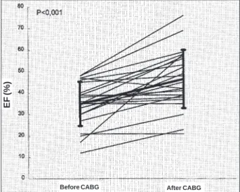
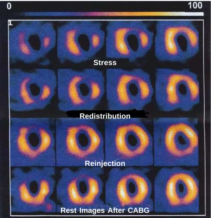
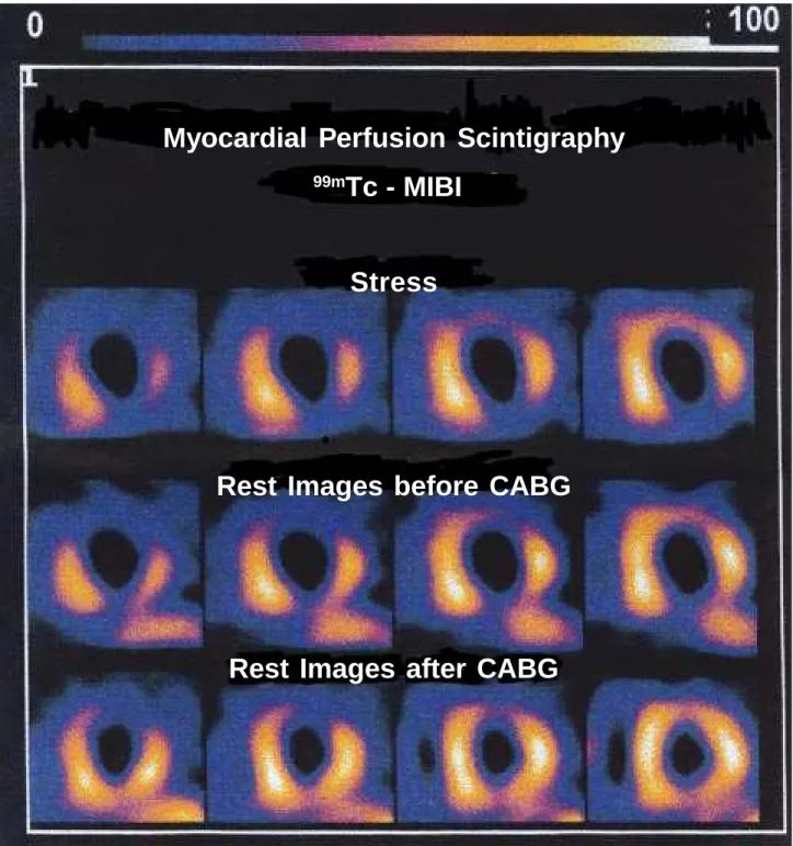
Figures 3 and 4 show an MPS example with tomogra-phic slices of the oblique axis of a patient with MI and viable muscle.
Calculations of operating variables S, E, A, PPV and NPV in the entire population were, respectively: 65.3%, 90.9%, 77.4%, 88.9% and 70.2% for the conventional
proto-col with 201Tl; and 81.6%, 81.8%, 81.7%, 83.3% and 80.0% for reinjection one; 20.4%, 90.9%, 53.8%, 71.4% and 50.6% for the conventional protocol with 99mTc-MIBI. It is con-cluded that there is an underestimation of viable myocar-dium by the conventional protocol with 99mTc-MIBI. The
in-creased perfusion, observed after revascularization of myo-cardial regions with MI and fibrosis but still viable areas, was shown better by 201Tl SPECT, notably with the
rein-jection protocol. It could be confirmed, using logistic re-gression analyses, that the reinjection protocol was the best predictor of viability (p<0.001).
Fig. 3 - Myocardial perfusion scintigraphy with 201Tl and tomographic slices in the oblique plan. Redistribution images suggest viability in the anterior and lateral regions; reinjection suggest viability of the inferior wall.
Stress
Redistribution
Reinjection
542
Chalela et al
Scintigraphy in the detection of viable myocardium
Arq Bras Cardiol volume 72, (nº 5), 1999
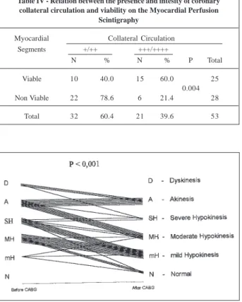
It was observed that, through severe lesions (obstruc-tion >90% lumen), coronary and Cc in the same territory were responsible for perfusion of 24/49 (49.0%) viable re-gions and of 26/44 (59.0%) nonviable ones (p=ns). Howe-ver, the semiquantified analysis of collateral circulation in-tensity of +++/++++ was more frequent in viable regions (60%) and of +/++ in nonviable ones (78.6%). These diffe-rences were significant (p=0.004, tab. IV).
Forty-nine myocardial regions (of a total of 69) evalua-ted after revascularization were MI relaevalua-ted. There was
regio-nal wall motion improvement in 36/49 (73.5%) after CABG (p<0.001, fig. 5).
The relationship between perfusion and systolic func-tion recovery could be observed in 49 myocardial regions, 34 of which (69.4%) were in agreement, and 25 (51.%) improved both perfusion and regional motility, but 9 (18.4%) did not. The re-sults were contradictory in 15 (30.6%) regions. There was an im-provement in motion in 11 (22.4%) and in perfusion in 4 (8.2%). However, the improvement in regional perfusion was signifi-cantly related (p=0.011) to the improvement in wall motion. Fig. 4 - Myocardial perfusion scintigraphy with 99mTc-MIBI and tomographic slices in the oblique plan. Conventional protocol imaging shows persistent defects in the anterior,
inferior and lateral regions. After CABG, images confirmed that these persistent defects contained viable muscle. CABG - Coronary artery bypass graft.
Myocardial Perfusion Scintigraphy
99m
Tc - MIBI
Stress
Rest Images before CABG
Discussion
In patients with CAD and LV dysfunction, CABG sur-gery has been shown to have a positive impact on survival. In the present study, survival was 96.6% during a follow-up period of 15.2 ± 10.2 months. Only one of 29 patients discharged (3.4%) died suddenly.
The presence of viable myocardium is not the only factor to indicate the need for CABG, but also the size of the area at risk, which once recovered, will lead to changes both in the clinical condition and in the natural history after re-vascularization. In this sense, surgical benefits could be confirmed by symptoms of angina and heart failure.
The study has also shown an in-hospital survival of 90.6%. No patient with EF <20% died in this phase. 19 patients had EF >20% and <40%, of whom two died in the immediate postoperative period. Therefore, hospital survi-val for these patients was 89.5% (17/19 patients), similar to that observed in the literature. Hochberg et al 17 studied 466
patients with CAD and LVEF <40% who underwent CABG and followed them for 36 months. Hospital survival (up to 30 days after surgery) for patients with LVEF >20% and <30% was 89%. The CASS study 18 observed significant surgical
benefit in a subgroup of patients with CAD in the three main coronary vessels and LVEF between 35% and 50%. They also observed increased survival after surgery in patients with LV dysfunction, particularly those with LVEF <25%, concluding that the presence of severe heart failure does not constitute a specific contra indication for surgery.
These data of increased survival, and the clinical im-provement observed, favor the indication for surgery in pa-tients with different degrees of left ventricular dysfunction and CAD. However, the improvement observed after MR represents a complex interaction between compensatory mechanisms, coronary anatomy, surgical results and patient selection.
The later the evaluation - two to three months after surgery - the more correct the interaction between recovery of perfusion and function, perioperative injury and graft patency will be.
In the conventional protocol, there were perfusion scans with 201Tl showing 40/102 (39.2%) regions of
reversi-bility. After reinjection 52/102 (51.0%) regions were viable. Therefore, from 62/102 irreversible regions by the conventio-nal protocol, 12/62 (19.4%) regions were shown to be viable after reinjection. This figure is smaller than that in the literature. Several studies show that the reinjection protocol, as compared with conventional scans with 201Tl, lowers the
number of defects considered fixed by about 31 to 49% 19-21.
In the present study, the smaller overestimation of the degree of fibrosis could be justified by the method used, because the redistribution images of the conventional pro-tocol were performed later than in the studies quoted above. Overestimation of the degree of fibrosis (persistent perfusion defects) by the conventional protocol, when compared with that of 201Tl reinjection, is dependent on the
redistribution phenomenon. Soon after the initial 201Tl
uptake by the myocardium, there is a continuous exchange of this univalent ion with the blood that determines its redistribution. 201Tl is continuously released from normal
myocardium and replaced by recirculating 201Tl, from
resi-dual activity in the blood. Although its initial biodistribu-tion is similar to K+, compartment redistribubiodistribu-tion presents a different velocity of exchange between the intra- and extra-cellular milieu. 201Tl uptake by the cells is dependant on
regi-onal availability, as well as on membrane and Na+/K+ pump integrity. However, ischemia-related metabolic changes alter the extraction rate and the exchange with the extra-cellular medium. In contrast to fibrotic cells, viable myocar-dium maintains a slow metabolism that preserves the basic cellular structure, making possible the recovery of its origi-nal functions. This complex protective mechanism may be detected by nuclear medicine and allows observation, using the dynamic biodistribution of the radionuclide in cardiac muscle. The smaller accumulation and slow washout of
201Tl in the ischemic areas result in the perfusion defects.
This process, based upon redistribution, is only possible if myocardium is viable, with an intact cellular membrane 22-24.
The redistribution phenomenon is, however, highly influenced by serum concentration of the tracer and by cellular washout. Redistribution may occur later in situa-tions in which there is resting low blood flow due to severe coronary stenosis, absence of hyperemia after stress, long-lasting metabolic abnormalities and low serum concentra-tion of the tracer 22,25. Slow redistribution, observed in
ischemic (viable) areas, makes the 3 to 4 hour images depen-Fig. 5 - Regional motility before and after revascularization of the regions affected by
myocardial infarction. D- dyskinesis; A- akinesis; HA- accentuated hypokinesis; HM- moderate hypokinesis; HD- discrete hypokinesis; N- normal.
Table IV - Relation between the presence and intesity of coronary collateral circulation and viability on the Myocardial Perfusion
Scintigraphy
Myocardial Collateral Circulation
Segments +/++ +++/++++
N % N % P Total
Viable 10 40.0 15 60.0 25
0.004
Non Viable 22 78.6 6 21.4 28
544
Chalela et al
Scintigraphy in the detection of viable myocardium
Arq Bras Cardiol volume 72, (nº 5), 1999
dent on limitations in the differentiation of viable from fibrotic myocardium.
The conventional protocol using 99mTc-MIBI showed
only 14/102 (13.7%) viable regions; 201Tl conventional
scans identified 26 (25.5%) additional viable regions; and after reinjection, 38 (37.3%) more regions, when compared with the 99mTc-MIBI scans. Therefore, protocols with 201Tl
were superior in detecting reversible defects in areas affec-ted by MI. This is similar to findings in the literature.
Cuocolo et al 26 studied 20 patients with CAD and LV
dysfunction (EF of 30%±8%) using 201Tl and 99mTc-MIBI
scans, comparing conventional protocols with reinjection protocols. 122 regions had permanent defects in the con-ventional protocol with 201Tl; 57 (47%) of them had
tran-sient defects in the reinjection protocol. Using the protocol with 99mTc-MIBI, there were 22 (18%) regions of transient
defects. Thus, 99mTc-MIBI did not identify 35 (29.0%)
regions when compared with the reinjection protocol. Dilsizian et al 27, using similar methodology, observed an
underestimation in the protocol with 99mTc-MIBI of 36%
when compared with the reinjection protocol. Reinjection imaging with 201Tl identifies a greater number of viable
myocardial regions when compared with 99mTc-MIBI.
Clinical and experimental observations showed that, in normal tissue, soon after initial uptake, 99mTc-MIBI fixes in
the myocardial cell, having minimal redistribution later. When depolarization of cell membranes and mitochondria occurs, as during ischemia, there is some difficulty in the storage and uptake of the tracer 28,29. Possibly, more intense
metabolic changes diminish the passive transport of 99m
Tc-MIBI. On the other hand, Na+/K+ATPase activity, an impor-tant factor in cell survival, would hardly interfere with 201Tl
extraction (active transport).
The statistical analysis demonstrated that the reinjec-tion protocol was the best approach for detecting viable myocardium, and there was an important depreciation of
99mTc-MIBI when compared with 201Tl for myocardial
viability identification.
Positron emission tomography 30 (PET) is considered
the most sensitive noninvasive technique for identifying viable myocardium and has been used for comparison with other methods. Several studies 20,31 showed that 201Tl
positivity after reinjection is as sensitive as PET imaging with 18-F-deoxyglucose (FDG) in viability detection. Agreement between methods was 88% in the 1st study and
85% in 2nd. Other studies 19,29,32-34 showed similar predictive
values, using recovery of wall motion observed after revascularization as the gold standard: PPV of 78% to 85% for PET and 73% to 87% for the reinjection protocol; NPV of 80% to 92% for PET and 75% to 100% for the reinjection protocol. In the present study the reinjection protocol had
similar predictive values: positive of 83.3% and negative of 80.0%, using perfusion recovery assessed by greater myocardial uptake after surgical revascularization.
A significant relationship was found between myocar-dial perfusion recovery and regional wall motion recovery (p=0.011) after revascularization in myocardial infarction areas.
These data are relevant to the observation that the me-tabolic abnormalities due to inappropriate perfusion reco-ver after coronary flow restoration. The presence of chro-nic myocardial contractile dysfunction is not enough to indicate revascularization. The extent of the area potentially recoverable - viable myocardium - allied to proper coronary anatomy, could lead to change in the clinical condition of a patient after revascularization.
Probably due to the lack of other more objective and precise methods for CAD identification, coronary angio-graphy, despite its limitations, has been used to evaluate the diagnostic precision of the methods used for studying myocardial ischemia. Angiography also has a number of problems related to the possibility of underestimation or overestimation of coronary lesions 35. Quantitative
corona-ry angiography 36 seems to be more objective than visual
estimation but also there are divergences among the several methods used (i.e. density against geometric method), espe-cially when analyzing unstable and eccentric plaques. There is the possibility that the degree of anatomic stenosis does not express the real coronary flow in specific situations such as during cardiac stress 37,38.
Evaluations of perfusion and LV contractile function have some limitations, especially because of the absence of a precise and of a practical quantitative method. The best quantification method currently available in practice is the semiquantitative scoring system. Therefore, the method is observer-dependant, causing intra and interobserver varia-bility common to most diagnostic methods.
In this study, intra and interobserver variability were, respectively, 4.6% and 7% for coronary angiography; 3% and 4% for left ventriculography; 2% and 4% for the stress test; and 4% and 5.1% for the myocardial perfusion scinti-graphy.
Despite these limitations, intra and interobserver varia-bilities were acceptable. Myocardial perfusion scintigraphy and coronary angiography with visual quantification are accepted and performed routinely in most medical centers.
Acknowledgments
References
1. Franciosa JA, Wilen M, Ziesche S, Cohn JN. Survival in men with severe chronic left ventricular failure due to either coronary heart disease or idiopathic dilated cardiomyopathy. Am J Cardiol 1983; 51: 831-6.
2. Primo G, Le Clerc JL, Goldstein JP, De Smet JM, Joris MP. Cardiac transplantation for the treatment of end-stage ischemic cardiomyopathy. Adv Cardiol 1988; 36: 293-7 .
3. Kriett JM, Kaye MP. The registry of the international society for heart trans-plantation: seventh official report ñ 1990. J Heart Transplant 1990; 9: 323-30. 4. Iskandrian AS, Shelbert HR. Myocardial viability assessment. J Nucl Med
1994a ; 35 (suppl): 1S-3S.
5. Lucas JR, Botvinick EH, Dae MW. Myocardial viability; evidence provided by analysis of left ventricular systolic function. Coron Artery Dis 1993; 4: 485-94. 6. New York Heart Association: the criteria committee. Inc: Diseases of the heart and blood vessels. Nomenclature and criteria for 6th ed. Boston Little Brown, 1964. 7. Braunwald E. Heart Disease. A Textbook of Cardiovascular Medicine. 5th ed.
Philadelphia: WR Saunders Co, 1996.
8. Tranchesi J. Eletorcardiograma Normal e PatolÛgica. NoÁies de Vectorcardio-grafia. 6a ed. São Paulo: Editora Athenas, 1983: 724.
9. Arie S. Circulação colateral com fator de proteção do miocárdio em portadores de insuficiência coronária crônica (Master thesis). São Paulo: Faculdade de Medicina da Universidade de São Paulo, 1977:53.
10. Dodge HT, Sandler H, Bellew BW, Lord Jr JD, Seatle W. The use of biplane angiography for the measurement of left ventricular volume in man. Am Heart J 1960; 60: 762.
11. Kennedy JW, Baxley WA, Figley MN. Quantitative angiography. The normal left ventricle in man. Circulation 1966, 34:272.
12. Consenso Nacional de Ergometria. Florianópolis, SC. Arq Bras Cardiol 1995; 65: 189-211.
13. Ellestad MA. Stress Testing. Principles and Practice. 3rd ed. Philadelphia:
Daves Company, 1986: 526.
14. Rosner B. Fundamentals of Biostatistics - 2nd ed. Boston: PWS Publishers,
1986: 584.
15. Fleiss JL. Statistical Methods for Rates and Proportions. New York: John Wiley & Sons, 1981: 325.
16. SAS Institute Inc., SAS/STAT User’s Guide, Version 6, 4th ed, vol.2, Cary, NC:
SAS Institute. 1989: 1686.
17. Hochberg MS, Parsonnet V, Gielschinsky I, Hussain SM. Newark NJ. Coronary artery bypass grafting in patients with ejection fractions below forty percent. J Thorac Cardiovasc Surg 1983; 86: 519-27.
18. Coronary Artery Surgery Study (CASS): a randomized trial of coronary artery bypass surgery: survival data. Circulation 1983; 68: 939-50.
19. Dilsizan V, Rocco T, Freedman N, Martins BL, Bonow RO. Enhanced direction of ischemic but viable myocardium by the reinjection of thallium after stress-redistribution imaging. N Engl J Med 1990; 323: 141-6.
20. Tamaki N, Ohtani H, Yamashita K, et al. Metabolic activity in the areas of new fill-in after thallium-201 refill-injection: comparison with positron emission tomogra-phy using fluorine-18-deoxyglucose. J Nucl Med 1991; 32: 673-8. 21. Hendel RC. Single photon perfusion imaging for the assessment of myocardial
viability. J Nucl Med 1994; 35: 235-315.
22. Strauss HW, Harrison K, Langan JK, Lebowitz E, Pitt B. Thallium 201 for myocardial imaging: relation to thallium-201 to regional myocardial perfusion. Circulation 1975; 51: 641-5.
23. Weich HF, Strauss W, Pitt B. The extraction of thallium-201 by the myocardium. Circulation 1977; 56: 188-97.
24. Pohost GM, Alpert NM, Ingwall JS, Strauss W. Thallium-201 redistribution: mechanism and clinical utility. Semin Nucl Med 1980; 10: 70-93.
25. Camici P, Araujo LI, Spinks T, et al. Increase uptake of 18F-fluorodeoxyglucose in post-ischemic myocardium in patients with exercise induced angina. Circulation 1986; 74: 81-6.
26. Cuocolo A, Pace L, Ricciardelli B, Chiariello M, Trimarco B, Salvatore M. Identification of viable myocardium in patients with chronic coronary artery disease: comparison of thallium-201 scintigraphy with reinjection and technetium-99m methoxyisobutyl. J Nucl Med 1992; 33: 505-11.
27. Dilsizian V, Arrighi JA , Diodati JG, et al. Myocardial viability in patients with chronic coronary artery disease: comparison of Tc-99m MIBI with Thallium reinjection and F-18 fluorodeoxyglucose. Circulation 1994; 89: 578-87. 28. Okada RD, Glover D, Gaffney T, Williams S. Myocardial kinetics of
technetium-99m hexakis-2-methoxy-2-methypropyl-isonitrile. Circulation 1988; 77: 491-8. 29. Sinusas AJ, Watson DD, Cannon JM, Jr ME, Beller GA. Effect of ischemia and postischemic dysfunction on myocardial uptake of technetium- 99 m labeled methoxyisobutyl isonitrile and thallium-201. J Am Coll Cardiol 1989; 14: 1785-93.
30. Gropler RJ, Bergmann SR - Myocardial viability ñ what is the definition?J Nucl Med 1991; 32: 10-2.
31. Bonow RO, Dilsizian V, Cuocolo A, Bacharach SL. Identification of viable myocardium in patients with coronary artery disease and left ventricular dysfunction: comparison of thallium scintigraphy with reinjecton and PET imaging with 18F-fluorodeoxyglucose. Circulation 1991; 83: 26-37. 32. Tillisch J, Brunken R, Marshall R, et al. Reversibility of cardiac wall-motion
ab-normalities predicted by position tomography. N Engl J Med 1986; 314: 884-8. 33. Tamaki N, Yonekura Y, Yamashita K, Saji H, Magata Y, Senda M. Position emission. Position emission tomography using fluorine-18-deoxglucose in evaluation of coronary artery bypass grafting. Am J Cardiol 1989; 64: 860-5. 34. Ohtani H, Tamaki N, Yonekura Y, et al. Value of thallium-201 reinjection after
delayed SPECT imaging for predicting reversible ischemia after coronary artery bypass grafting. Am J Cardiol 1990; 66: 394-9.
35. Fleming RM, Kiekeeide RL, Gould KL. Patterns in visual interpretation of coronary arteriograms as detected by quantitative coronary angiography. J. Am Coll Cardiol 1991; 18: 945-51.
36. Rodrigues AS, Santaera O, Fernandez M, et al. ñ Digital coronaryangiography: a new approach in the analysis of atherosclerotic plaque. Medicine 1991; 51: 209-16.
37. Meyer Sl, Dousk GCM, Twieg DB ñ Influence of dobutamine on hemodynamics and coronary blood flow in patients with and without coronary artery disease. Am J Cardiol 1976; 38: 103-8.