Comparison of central corneal edema
and visual recovery between liquefaction
and conventional phacoemulsification
in soft cataracts
Comparação do edema de córnea central
e da recuperação visual após facoemulsificação
por liquefação e convencional em cataratas moles
Celso Takashi Nakano1
,Wilson Takashi Hida1
,Newton Kara-Jose Junior3
, Antonio Francisco Pimenta Motta1
, Alexandre Reis2
, Mauricio Pamplona2
, Reinaldo Fujita2
, Iris Yamane1
, Ricardo Holzchuh1
, Amaryllis Avakian3
A
BSTRACTPurpose: The aim of the present study is to assess central corneal edema and visual recovery after cataract surgery performed according to two technologies: conventional ultrasonic and liquefaction (Aqualase®
). Methods: This is a prospective contralateral study in wich 20 patients with comparable preoperative conditions were submitted to cataract surgery were evaluated. Preoperative assessment involved complete ophthalmological examination and the study included patients with bilateral cataracts up to grade 2, according to the Lens Opacity Classification System II. The same cristaline fracture technique was used in all cases, and surgical procedures were performed by the same experienced surgeon, using two technologies: liquefaction or conventional phacoemulsification. Postoperative central corneal edema was measured by corneal optical pachymetry (Orbscan II®
) on the 1st
, 3rd
, 7th
and 10th
postoperative days. Results:
None of the 20 patients submitted to surgery was lost during the postoperative follow-up or excluded from the analysis. On the first postoperative, the visual acuity average was 0.031 logMAR in the Aqualase®
group and 0.043 logMAR in the conventional surgery group. No statistical difference was detected in the assessment of visual acuity throughout the postoperative period. Central corneal pachymetry varied from 543.93 + 34.69 preoperatively to 545.08 ± 25.67 on the last day of follow-up in the Aqualase®
group, and from 543.13 + 30.62 to 536.08 + 34.89 in the conventional technique group, without statistical significance. Conclusion: This study suggests that both techniques are equally effective for surgery on lenses with grade I or II cataract, and that they provide similar results in terms of visual recovery and central corneal edema.
Keywords: Phacoemulsification/methods; Cataract extraction/methods; Techniques, measures, measurement equipment; Diagnostic techniques, ophthalmological; Biometry/ instrumentation
1
Estagiário do Setor de Catarata do Departamento de Oftalmologia do Hospital das Clínicas da Universidade de São Paulo – USP – São Paulo (SP), Brasil;
2
Medico Residente do Departamento de Oftalmologia do Hospital das Clínicas da Universidade de São Paulo – USP – São Paulo (SP), Brasil;
3
Chefe do Setor de Catarata do Departamento de Oftalmologia do Hospital das Clínicas da Universidade de São Paulo – USP – São Paulo (SP), Brasil.
I
NTRODUCTIONS
ince the 1970’s, the importance of preserving the corneal endothelium has been established as a factor in the recovery from post-operatory edema(1-3). Durovic et al. has suggested that corneal edema is one of the main causes of low visual acuity in the immediate postoperative period after intraocular surger (4)
.
As a rule, candidates to cataracts surgery aim for early visual recovery after the procedure, so that they can resume their routine activities (5,6)
. Surgery performed by phacoemulsification with small incision induces little corneal edema, and provides early visual recovery (7,9)
. The main causes of corneal endothelium edema after phacoemulsification surgery is believed to be: the thermal energy released by ultrasonic frequency vibration of the tip and turbulent flow of fluid and particles within the anterior chamber (4,10,11)
.
In order to reduce endothelial injury, the following alternatives have been developed: viscoelastic substances (12-15)
; special surgical techniques such as, for example, nuclear prefracture; and new modalities of tip vibration at ultrasonic frequency, such as the White-star®
, Neosonix®
, dynamic rising time and torsional systems
(1,2,5,6,16-23)
.
By the year 2000, an alternative technology was proposed for cataracts surgery (5,16,17)
. This modality, named Aqualase®
, consists in the liquefaction of the lens core by jets of balanced saline solution (BSS) without tip vibration. Therefore, the thermal energy release component is eliminated as a causal factor of postoperative corneal edema. This method is assumed to provide for safe and effective emulsification of lenses with cataracts up to grade 2, based on the brunescence criterion of the Lens Opacities Classification System, LOCS II (1,5,16,17)
. Some findings suggest that liquefaction could not be a good option for harder cataract nuclei (16,17)
. Several studies have been performed comparing different surgical techniques and modes of modulating the physical parameters of the phacoemulsifier, with the objective of demonstrating the benefits of reducing conventional phacoemulsification-induced corneal edema, but very few studies have compared the technique of conventional phacoemulsification with liquefaction of the lens core by Aqualase® (2,3,7,8,11,15-19)
.
The purpose of this study is to assess central corneal edema and visual recovery after cataract surgery performed according to two approaches: conventional phacoemulsification and liquefaction by Aqualase®
.
M
ETHODSThis randomized clinical trial was conducted according to established ethical standards for clinical research and the Institutional Review Board of University of São Paulo Medical School General Hospi-tal (HC-FMUSP) has approved the protocol. In order to maintain the study blinding, physicians conducting postoperative evaluation did not have access to the randomization codes or the patients’ medical records.
Twenty patients, with mean age of 65.13 ± 6.34 years (ranging from 58 to 72) underwent cataract surgery. The inclusion criteria for the study were bilateral cataract, up to grade 2 of nuclear opalescence (Lens Opacities Classification System, LOCS II) in both eyes
(1)
, with normal age-matched endothelial count (more than 2000 cells/mm2
), and no other ocular surgery or illness. Preoperative ocular examinations included Snellen visual acuity, detailed biomicroscopic examination, Goldmann tonometry, and axial length measurement (AL) with ultrasonic technology. Written informed consent was obtained from each patient.
Both eyes of each patient was included in the study. For the first surgery, the eyes were randomized to Aqualase®
or conventional phacoemulsification. The fellow eye was submitted to the surgery 30 days after the first, with the opposite technique.
All surgeries were performed between March 2005 and July 2006 by the same surgeon (C.T.N.), with the same technique and preoperative peribulbar anesthesia. Cyclopentolate 1% and phenylephrine 2.5% eye drops were used three times for obtaining mydriasis, 1 hour before the surgery. A three step clear corneal tunnel self-sealing incision was made with a 2.75 mm disposable metal blade on the steepest axis and a side port incision. After inject dispersive and cohesive ophthalmic viscosurgical devices (Celoftal and Provisc, Alcon Laboratories, Fort Worth, Texas, USA) into the anterior chamber using the soft shell technique (20)
, uttrata forceps was used to grasp the capsule and perform the capsulorhexis (2)
. Karate pre chop technique with Akahoshi’s pre chopper to make the initial fracture and endocapsular phacoemulsification was performed in all eyes by using Infiniti®
Vision System Phacoemulsification machine (Alcon Laboratories, Fort Worth, Texas, USA) using both technologies, Aqualase®
devices were removed. During the postoperative time, eye drops were prescribed, including the use of topic fourth generation quinolones 4 times a day, for 7 days, and steroids (dexamethasone 1%) 4 times a day, tapered over a 30-day period. Intraoperative parameters settled in the machine, for conventional phacoemulsification were 70% linear ultrasonic power with 40% time on and 40cc/min aspiration rate with 500 mmHg vacuum and for Aqualase were 70% linear liquefaction power with 40% time on and 40cc/min aspiration rate with 500 mmHg vacuum. The in the conventional phacoemulsification were 49.12 ± 11.93 seconds and Aqualase time were 65.51 ± 12.19 seconds. All number and magnitude were listed and recorded at the end of the surgery, the ultrasonic/liquefaction elapsed time, mean power time for conventional phacoemulsification and pulses magnitude for Aqualase.
Postoperative corneal edema was not clinically significant at biomicroscopic examination, no descemet’s folds or loss of tranparency was noted. Measurement of the central edema was done using corneal optical pachymetry (Orbscan II®
) on the 1st
, 3rd
, 7th
and 10th
postoperative days. This is diagnostic system that scans the surface of the eye and acquires over 9000 data points in 1.5 seconds to map the entire corneal surface (11mm), analyzing the elevation and the curvature measurements on both the anterior and posterior surfaces of the cornea. Some trials have shown excellent correlation, 4 weeks afer surgery, between this system and ultrasonic measures and good repeatability(3-6)
.
Data were analyzed using the Statistical Program Statistica, version 5.1. Statistical analysis of the results were performed by Mann-Whitney testing for analysis of variance. A p value less than 0.05 was considered statistically significant.
R
ESULTSNone of the 20 patients submitted to surgery was lost during the postoperative follow-up or excluded from the analysis. On the first postoperative day, the mean visual acuity was 0.031 logMAR in the Aqualase®
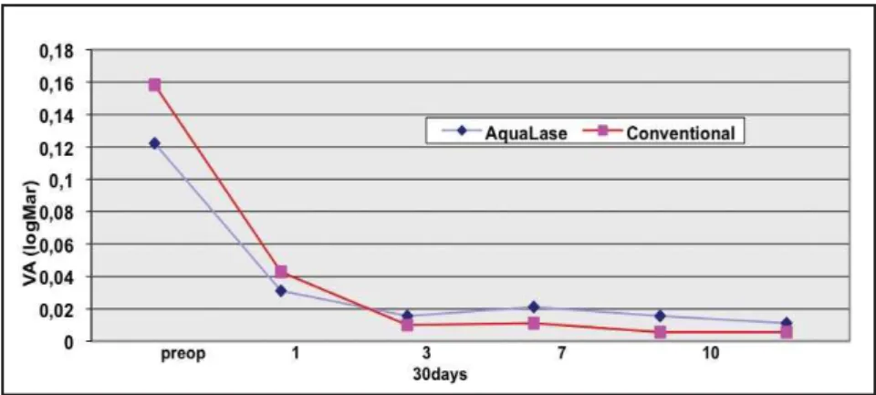
group and 0.043 logMAR in the conventional surgery group (approximately 20/20 on the Snellen table, for both groups). No statistical difference was detected in the assessment of visual acuity in the postoperative period; results are shown on Figure 1.
The surgery ultrasonic/liquefaction elapsed time in the conventional phacoemulsification were 49.12 ± 11.93 seconds and Aqualase time were 65.51 ± 12.19 seconds.
All number and magnitude were listed and recorded at the end of the surgery for conventional phacoemulsification (mean 11 ± 1.9; mean power time 29,33 ± 4.0 seconds) and for Aqualase (Mean 12 ± 2.12; pulses 3900 ± 1162; magnitude 68.23 ± 18.99).
Mean pre and postoperative central pachymetric values for both groups are shown on Table 1. The central corneal thickness average in both groups achieved 543.53 ± 32.16 microns before surgery and 550.62 ± 34.82 microns after surgery. There was no statistically significant difference between Aqualase®
and conventional ultrasonic phacoemulsification in the cen-tral pachymetry exam.
D
ISCUSSIONIn this study, we have compared the central corneal edema of patients submitted to cataract surgery performed with conventional phacoemulsification in one eye and with liquefaction, by Aqualase®
, in the fellow
eye. We found no statistically significant difference between the groups on the first day after surgery, considering the best corrected visual acuity average (0.031 logMAR in the Aqualase®
group and 0.043 logMAR in the conventional phacoemulsification group, both between 20/20 and 20/25 on the Snellen table). We observed that the postoperative visual acuity average reached approximately its maximal recovery on the 3rd
day postoperative, around 0.016 in the liquefaction group and 0.010 in the conventional technique group (mean 0.013 logMAR, equivalent to 20/20 on the Snellen table), as shown on Figure 1, suggesting that both techniques are effective in providing early visual restoration.
There was no statistically significant difference between the groups considering pre and postoperative corneal thickness average, measured by pachymetry with Orbscan II® (24)
. The pachymetric values variation, observed in both patients groups, remained within the normal limits for the test (24-26)
(Table 1).
In this study we have used corneal thickness to access corneal edema. Other authors has showed that the measurement of corneal thickness with Orbscan II®
has comparable accuracy to the ultrasonic method and a +/- 37.5 microns variability in the values found (27)
. Marisch et al. estimated that the variability of pachymetric values with Orbscan II®
was +/- 40 microns, based on measurements taken on the same day and on different days, in the same patients and the best reproducibility was found in the central corneal region
(25)
. In our research, we observed a maximum variation of 25.0 microns over the study period in the Aqualase®
group and of 13.8 microns in the conventional phacoemulsification group, i.e., within the normal range expected for the test. Considering that, in this study, pachymetry values varied within the normal range during the whole assessment period, we suggest that both
surgical techniques induced minimal corneal edema, which explains the quick postoperative recovery of visu-al acuity. This study has the limitation of smvisu-all number of eyes enrolled .
There was no statistically significant difference between the groups, but the conventional phaco-emulsification showed lower cornea edema, probably because the conventional phacoemulsifcation showed less total time surgery than lower intraoperative release of thermal energy and turbulent flow of fluid and lenticular particles within the anterior chamber.
The study indicates that both techniques (conventional phacoemulsification and Aqualase®
) are equally effective, in terms of time to visual recovery and induction of corneal edema, when used for cataract surgery in patients with up to grade 2 of nuclear opalescence (Lens Opacities Classification System, LOCS II), performed by an experienced surgeon, using manual fracture technique (Akahoshi pre chop)( 1)
. Recent article has suggested, using optical coherence tomography, that Aqualase®
liquefaction cataract extraction is as safe as standard US phacoemulsification cataract extraction and may carry less risk for the development of postoperative cystoid macular edema (28)
. Another article comparing Aqualase®
and Neosonix phacoemulsification on the corneal endothelium suggested that in patients younger than 80 years, the differences in the postoperative changes in ECC and in pachymetry were not statistically significant, although in patients of 80 years and older there were statistically significant differences, with the results being better in the Aqualase®
eyes (29)
. We believe further studies are necessary to compare other parameters, such as the length of the surgical procedure, apparently longer with the liquefaction mode, and the endothelial cell counting limits for different nuclear densities allowing the safe use of the Aqualase®
technique.
Aqualase® Conventional Mann-Whitney
Mean (microns) SD Mean (microns) SD P value
Preoperative 543.93 ±34.69 543.13 ±30.62 0.95
1st
day 568.93 ±45.67 546.07 ±35.28 0.14
3rd
day 564.07 ±41.13 549.93 ±28.54 0.28
7th
day 555.67 ±35,65 539.13 ±32,15 0.19
30th
day 545.08 ±25,67 536.08 ±34,89 0.47
Table 1
Comparison of the central pachymetric values between Aqualase®
R
ESUMOObjetivo: Avaliar o edema corneano central e a recupera-ção visual após cirurgia de catarata realizada através de duas tecnologias: ultrassônica convencional e liquefação (Aqualase®
). Métodos: Estudo retrospectivo com análise contralateral, onde 20 pacientes se submeteram à facectomia sob condições pré-operatórias comparáveis. A avaliação pré-operatória envolveu exame oftalmológico completo com graduação da catarata de acordo com o Lens Opacity Classification System II, e foram incluídos no estudo pacientes com catarata nucle-ar bilateral até grau II. A mesma técnica cirúrgica foi realizada em todos os casos pelo mesmo cirurgião, utili-zando as duas tecnologias: liquefação e facoemulsificação convencional. As medidas do edema corneano pós-ope-ratório foram realizadas com paquímetro óptico corneano (Orbscan II®
) nos dias 1, 3, 7 e 10 de pós-operatório. Re-sultados: Nenhum dos pacientes inicialmente inscritos no estudo foi excluído ou perdido durante o seguimento. No primeiro dia pós-operatório a acuidade visual média foi de 0.031 logMAR no grupo de Aqualase e 0.043 logMAR no grupo convencional. Nenhuma diferença estatistica-mente significativa foi detectada na análise de acuidade visual durante o período pós-operatório. A paquimetria corneana central variou de 543.93 + 34.69 no pré-opera-tório para 545.08 ± 25.67 no último dia de acompanha-mento no grupo de Aqualase e de 543.13 + 30.62 para 536.08 + 34.89 no grupo convencional, sem diferença es-tatisticamente significativa. Conclusão: O estudo sugere que as duas técnicas são igualmente eficaz para as cirur-gias nucleares graus I e II, além de apresentarem resulta-dos similares em relação ao edema corneano central e à recuperação visual.
Descritores: Facoemulsificação/methods; Extra-ção de catarata/métodos Técnicas, medidas e equipamen-tos de medição; Técnicas de diagnóstico oftalmológico; Biometria/instrumentação
R
EFERENCES1. Thompson AM, Sachdev N, Wong T, Riley AF, Grupcheva CN, McGhee CN. The Auckland Cataract Study: 2 year postoperative assessment of aspects of clinical, visual, corneal topographic and satisfaction outcomes. Br J Ophthalmol. 2004;88(8):1042-8.
2. Hirst LW, Snip RC, Stark WJ, Maumenee AE. Quantitative corneal endothelial evaluation in intraocular lens implantation and cataract surgery. Am J Ophthalmol. 1977;84(6):775-80.
3. Irvine AR, Kratz RP, O’Donnell JJ. Endothelial damage with phacoemulsification and intraocular lens implantation. Arch Ophthalmol. 1978;96(6):1023-6.
4. Duroviæ BM. [Endothelial trauma in the surgery of cataract]. Vojnosanit Pregl. 2004;61(5):491-7. Serbian.
5. Hoffman RS, Fine IH, Packer M. New phacoemulsification technology. Curr Opin Ophthalmol. 2005;16(1):38-43. 6. Kara-José Júnior N. Cirurgia de catarata: aspectos clínicos e
socio-econômicos [tese]. São Paulo: Faculdade de Medicina da Universade de São Paulo; 2002.
7. Grupcheva CN, Riley AF, Craig JP, Malik TY, McGhee CN. Analyzing small-incision cataract surgery by Orbscan II fourth-dimensional pachymetry mapping. J Cataract Refract Surg. 2002;28(12):2153-8.
8. Alió J, Rodríguez-Prats JL, Galal A, Ramzy M. Outcomes of microincision cataract surgery versus coaxial phacoemulsification. Ophthalmology. 2005;112(11):1997-2003. Comment in: Ophthalmology. 2006;113(9):1687; author reply 1687.
9. Alió JL, Schimchak P, Montés-Micó R, Galal A. Retinal image quality after microincision intraocular lens implantation. J Cataract Refract Surg. 2005;31(8):1557-60.
1 0 . Sippel KC, Pineda R Jr. Phacoemulsification and thermal wound injury. Semin Ophthalmol. 2002;17(3-4):102-9. Review.
11. Lundberg B, Jonsson M, Behndig A. Postoperative corneal swelling correlates strongly to corneal endothelial cell loss after phacoemulsification cataract surgery. Am J Ophthalmol. 2005;139(6):1035-41.
12. Jurowski P, Goœ R, Kuœmierczyk J, Owczarek G, Gralewicz G. Quantitative thermographic analysis of viscoelastic substances in an experimental study in rabbits. J Cataract Refract Surg. 2006;32(1):137-40.
13. Eda M, Matsushima H, Terauchi W, Mukai K, Izumida S, Obara Y, et al. [Protective effect on the corneal endothelium and remaining effect at the anterior chamber for three different kinds of viscoelastic devices]. Nippon Ganka Gakkai Zasshi. 2006;110(1):31-6. Japanese.
14. Jurowski P, Goœ R, Owczarek G, Gralewicz GZ. Corneal endothelial cells’ protection against thermal injury: influence of ophthalmic viscoelastic substances in experimental study on rabbits. Eur J Ophthalmol. 2005;15(6):674-9.
15. Kiss B, Findl O, Menapace R, Petternel V, Wirtitsch M, Lorang T, et al. Corneal endothelial cell protection with a dispersive viscoelastic material and an irrigating solution during phacoemulsification: low-cost versus expensive combination. J Cataract Refract Surg. 2003;29(4):733-40.
16. Mackool RJ, Brint SF. AquaLase: a new technology for cataract extraction. Curr Opin Ophthalmol. 2004;15(1):40-3. 17. Hughes EH, Mellington FE, Whitefield LA. Aqualase for
cataract extraction. Eye. 2007;21(2):194-4.
18. Doughty MJ, Müller A, Zaman ML. Assessment of the reliability of human corneal endothelial cell-density estimates using a noncontact specular microscope. Cornea. 2000;19(2):148-58.
19. Wirbelauer C, Wollensak G, Pham DT. Influence of cataract surgery on corneal endothelial cell density estimation. Cornea. 2005;24(2):135-40.
20. Arshinoff SA. Dispersive-cohesive viscoelastic soft shell technique. J Cataract Refract Surg. 1999;25(2):167-73. 21. Sanchis-Gimeno JA, Herrera M, Lleó-Pérez A, Alonso L,
22. Lui Netto A, Malta JBNS, Barros MAC, Giovedi Filho R, Alves MR. Confiabilidade das medidas de espessura central da córnea com Orbscan II e paquímetro ultra-sônico. Arq Bras Oftalmol. 2005;68(1):71-4.
23. Gherghel D, Hosking SL, Mantry S, Banerjee S, Naroo SA, Shah S. Corneal pachymetry in normal and keratoconic eyes: Orbscan II versus ultrasound. J Cataract Refract Surg. 2004;30(6):1272-7.
24. Fam HB, Lim KL, Reinstein DZ. Orbscan global pachymetry: analysis of repeated measures. Optom Vis Sci. 2005;82(12):1047-53.
25. Marsich MW, Bullimore MA. The repeatability of corneal thickness measures. Cornea. 2000;19(6):792-5.
26. Chakrabarti HS, Craig JP, Brahma A, Malik TY, McGhee CN. Comparison of corneal thickness measurements using ultrasound and Orbscan slit-scanning topography in normal and post-LASIK eyes. J Cataract Refract Surg. 2001;27(11):1823-8
27. Javaloy J, Vidal MT, Villada JR, Artola A, Alió JL. Comparison of four corneal pachymetry techniques in corneal refractive surgery. J Refract Surg. 2004;20(1):29-34.
ENDEREÇOPARACORRESPONDÊNCIA:
Wilson Takashi Hida
Rua Afonso de Freitas 488 - Apto. 61 Paraíso
CEP 04006-052 - São Paulo - SP E-mail [email protected]
28. Barsam A, Chandra A, Bunce C, Whitefield LA. Prospective randomized controlled trial to compare the effect on the macula of AquaLase liquefaction and ultrasound phacoemulsification cataract surgery. J Cataract Refract Surg. 2008;34(6):991-5.