4 5 4 5 4 5 4 5 4 5
Real e Benemérita Beneficência Portuguesa de São Paulo
Mailing address: Silas S. Galvão Fº - Rua Maestro Cardim, 1041 – 01323-001 São Paulo, SP, Brazil - E-mail: [email protected]
English version by Stela Maris C. e Gandour
Objective - The biventricular pacing (BVP) approach
has good results in the treatment of congestive heart fai-lure (CHF) in patients (pts) with disorders of intraventri-cular conduction.
Methods - We have applied BVP to 28 pts, with left
ven-tricular pacing using minitoracotomy in 3 pts and the trans-venous aproach via coronary sinus in 25 pts. The mean dura-tion of the QRS complexes was 187 ms, in the presence of the left branch block in 22 pts, and right branch block + divisio-nal hemiblock in 6 pts. All pts had been considerated can-didates to cardiac transplantation, and were under optimized drug therapy. Sixteen pts were in Functional Class (NYHA) IV, and 12 in class III. The ejection fraction varied from 22 to 46% (average = 34%). The pacing mode employed was biventricu-lar triple-chamber in 22 pts, and bi-ventricubiventricu-lar dual-cham-ber in 6 pts (one with ICD).
Results - The pts were followed up for a period that
ran-ged from 10 days to 14 months (mean 5 months). All pts presented clinical improvement after implant, chaging the NYHA Functional Class at the end of follow-up to Class I (9pts), Class II (10 pts) and Class III (6 pts). The initial mean ejection fraction have-raised to 37%. Two pts died suddenly. One patient died due to a pulmonary fungal infection.
Conclusion - Ventricular resynchronization
throu-gh BVP, improved significantly the Functional Class and, therefore, the quality of life. Assessments of myocar-dial function acutely performed do not reflect the clinical improvement observed.
Key words: biventricular pacing, ventricular resynchroni-zation, heart failure
Arq Bras Cardiol, volume 78 (nº 1), 45-50, 2002
Silas S. Galvão Fº, Cecília M. B. Barcellos, J. Tarcísio M. Vasconcelos, Jaime G. M. Arnez, Kátia N. Couceiro, Leu do Cam pos, Evan dr o Sbar ai n i , Mau r íci o G. Lyr a, Fer n an do S. O. Sou za
São Paulo, SP - Brazil
Ventricular Resynchronization Through Biventricular
Cardiac Pacing for the Treatment of Refractory Heart Failure
in Dilated Cardiomyopathy
Heart failure is currently considered a public health problem. In 1997, it accounted for 3.58% of all admissions in the Brazilian public health system, corresponding to 36.95% of the hospitalizations due to cardiocirculatory problems, with mortality of 6.39%, and a cost to the Brazilian government of 3 billion reais 1. In the USA, from 1970 to
1994, the rate of hospitalizations due to heart failure tripled in patients aged 45 to 65 years, and increased 4 times in those over the age of 65 years 2. Despite the great progress
in the medicamentous treatment of heart failure, many patients have dilated cardiomyopathy refractory to this type of therapy, with a poor prognosis and a very poor quality of life. Of the nonpharmacological procedures indicated for this type of patient, cardiac transplantation, despite its limitations, remains the gold standard, because other options, such as cardiomyoplasty and Batista’s surgery, have results that are somewhat controversial. At the beginning of the ‘90s, Hochleitner et al 3 proposed for
the first time artificial cardiac pacing (dual-chamber pacemaker with a short atrioventricular interval) as a treatment for refractory heart failure, with encouraging acute results; these results, however, proved catastrophic within a short period of follow-up 4,5. At the beginning of the
‘90s, Xiao et al 6,7 studied the effects of intraventricular
conduction disorders of the myocardial function in patients with dilated cardiomyopathies and showed that the larger the QRS, the longer the time required for contraction and relaxation of the fibers, impairing myocardial function. Bakker et al 8 were the first to report ventricular
resynchroni-zation through permanent biventricular pacing using DDD pacemakers with a bifurcated lead in the ventricular exit; the right ventricle was stimulated by anodic current through a conventional endocardial electrode, and the left ventricle by cathodic current through an epimyocardial electrode. In the first half of the ‘90s, Cazeau et al 9 in the classical study
4 6 4 6 4 6 4 6 4 6
its great value as adjuvant in the treatment of congestive heart failure refractory to medicamentous treatment in patients with dilated cardiomyopathy and intraventricular conduction disorders 10-12. In this article, we report our
experience with this technique in patients with dilated cardiomyopathy and decompensated congestive heart failure who were referred to our hospital for assessment of the possibility of cardiac transplantation.
Methods
From April 1999 to August 2000, we implanted pacema-kers with biventricular pacing in 28 patients (23 males and 5 females) with dilated cardiomyopathy, important intraven-tricular conduction disorders (QRS >140ms), and uncon-trollable congestive heart failure despite the optimized medicamentous treatment. Their ages ranged from 36 to 84 years (mean = 58.5). They were referred to our hospital by cardiologists who, not being able to control their congestive heart failure, asked for assessment aiming at cardiac transplantation. All patients had intraventricular conduc-tion disorders as follows: 6 had complete right bundle-branch block with the axis shifted to the left, 16 had left bundle-branch block, and 6 had interventricular conduction disorders (an electrocardiographic pattern of left bundle-branch block) consequent to exclusive left ventricular pacing (single-site pacing of the right ventricle). QRS width ranged from 140 to 220 (mean = 187) milliseconds. In regard to NYHA functional class, the patients were classified as follows: 12 were functional class III, 16 were functional class IV, and 6 were not discharged from the intensive care unit because of vasoactive-drug dependence. The sole indica-tion for the procedure was the need for ventricular resyn-chronization, and no primary indication for reestablishing cardiac rhythm or atrioventricular synchronism, or both, existed. Right ventricular pacing was performed with a conventional endocardial electrode in all patients, and left ventricular pacing was performed with an epimyocardial electrode in the first 3 patients, and through the coronary sinus in 26 patients (1 patient with an epimyocardial electrode had elevation of the pacing threshold and required implantation of a new electrode through the coronary sinus) (fig. 1). The electrodes used for left ventricular pacing through the coronary sinus were the Biotronic Corox CX LV-75 in 10 patients and the Medtronic 2187 - 75cm in 16 patients. For biventricular pacing, we used the following: 1 single-chamber pacemaker with a bifurca-ted lead in the exit; 22 dual-chamber pacemakers, 18 of which had a duplicator in the ventricular exit (for those without a duplicator in the ventricular exit, the atrial chamber was connected to the first ventricle to be activated during the patient’s own rhythm, right ventricle in the case of left bundle-branch block, and the ventricular chamber was connected to the other ventricle, the atrioventricular interval being programmed at 0 milliseconds); and 4 special triple-chamber pacemakers (single atrial exit and double ventricular exit). One patient with permanent atrial fibrilla-tion, severe ventricular arrhythmia, and adequate
ventricu-lar response underwent placement of an implantable cardioverter-defibrillator with biventricular antibradycardia function (fig. 2). All patients were informed that they were undergoing a new procedure that was still being investiga-ted. The etiologies of dilated cardiomyopathies were as fol-lows: ischemic in 13 patients, chagasic in 5, alcoholic in 2, hypertensive in 1, valvular in 2, and idiopathic in 5. The patients underwent clinical assessment and complementary tests in the preoperative period, on the 10th postoperative
day, and every 3 months after the procedure. All patients were able to perform the 6-minute walking test after the
Fig. 2 - Radiography on posteroanterior projection of a patient with implantable cardioverter-defibrillator with biventricular antibradycardia function. Note the pacemaker lead of the implantable cardioverter-defibrillator positioned in the inferoapical wall of the right ventricle through endocardial access, and pacemaker lead positioned in the lateral region of the left ventricle through the coronary sinus.
LV-CS
Fig. 1 – Radiography on right anterior oblique view of our first patient undergoing biventricular pacing. Note the epimyocardial pacemaker lead (LV-EPI) abandoned due to a threshold increase. Ventricular resynchronization was reestablished with implantation of a new electrode through the coronary sinus in the left ventricular la-teral wall (LV-CS). The right ventricular pacemaker lead was implanted through endocardial access in the inferolateral wall.
LV-CS
4 7 4 7 4 7 4 7 4 7
procedure, but only 13 could undergo this test prior to the procedure. The other 15 patients did not undergo this test preoperatively for the following reasons: 8 were not able be-cause of poor condition, and 6 due to a flaw in the protocol. E-jection fraction was assessed preoperatively on Doppler e-chocardiography in all patients, and ranged from 22% to 46% (mean = 34±0.57%). It is worth noting that all patients with ejection fraction above 40% had undergone treatment with vasoactive drugs in the intensive care unit a few days before the test; this value was, therefore, overestimated. My-ocardial scintigraphy was performed prior to the procedure in 18 patients and ranged from 8% to 39% (mean = 20±7.4%).
Results
Table I shows QRS width, the ejection fraction measured on Doppler echocardiography and on gated cardiac scan-ning, and the pre- and postoperative NYHA functional class. All patients improved their functional classes. In the preoperative period, most of them (16/28) were in functional class IV; after biventricular pacing, the great majority (19/ 28) were functional class I or II. Of the patients who were functional class IV before the procedure, 3 underwent cardiac transplantation in the 4th, 5th, and 6th postoperative
month. Three patients died, 2 suddenly on the 3rd and 10th
days after the procedure, and 1 due to pulmonary fungal
infection on the 5th day after the procedure. One patient had
dislocation of the electrode from the coronary sinus, which was solved with implantation of a second-generation elec-trode in the coronary sinus with a passive-fixation mecha-nism. One patient had infection at the site of pacemaker im-plantation requiring removal of the triple-chamber pacing system, treatment with antibiotics, and later reimplantation of a new system on the other side. The ejection fraction determined on Doppler echocardiography passed from the preoperative mean of 33% to 37% 10 days after the procedu-re. In 17 patients who were followed up for 3 months, the ejection fraction passed from 34% in the preoperative period to a mean of 43% 90 days after the procedure. In regard to the ejection fraction determined on myocardial scintigraphy, a 1st evaluation performed 10 days after the
procedure involving 18 patients showed that the initial preoperative mean passed from 20.8% to 21.2%, and a 2nd
evaluation carried out 90 days after the procedure in 10 patients from the 1st sample showed that the preoperative
mean passed from 22.9% to 23.4%.
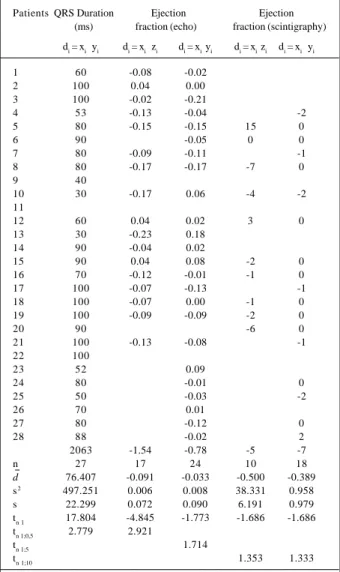
All differences observed between data prior to and after the procedure underwent a statistical test of signifi-cance, comparing the value of Student’s t calculated with n-1 degrees of freedom with the corresponding critical value obtained from statistical tables (tab. II). This comparison allowed the following conclusion: 1) in regard to QRS
Table I
Patients QRS duration (ms) Ejection fraction (Echo) Ejection fraction (gated blood pool) NYHA functional class
Pre Post Pre 10 days 3 months Pre 10 days 3 months Pre Post
1 180 120 0.33 0.35 0.41 IV III
2 200 100 0.45 0.45 0.41 IV II
3 220 120 0.28 0.49 0.40 IV III
4 163 110 0.31 0.35 0.44 21 23 IV III
5 180 100 0.32 0.47 0.47 39 39 24 III I
6 200 110 0.33 0.38 15 15 15 IV III
7 200 120 0.28 0.39 0.37 19 20 III III
8 180 100 0.32 0.49 0.49 35 35 42 IV II
9 180 140 III SD 10 d
10 160 130 0.27 0.21 0.44 16 18 20 III I
11 200 IV SD 3 d
12 200 140 0.41 0.39 0.37 16 16 13 III I
13 180 150 0.46 0.28 0.69 IV II
14 180 90 0.33 0.31 0.37 8 IV II
15 190 100 0.44 0.36 0.40 16 16 18 III I
16 160 90 0.31 0.32 0.43 18 18 19 III I
17 200 100 0.22 0.35 0.29 15 16 III II
18 200 100 0.35 0.35 0.42 21 21 22 III II
19 200 100 0.35 0.44 0.44 24 24 26 III I
20 200 110 0.45 29 29 35 III I
21 210 110 0.30 0.38 0.43 17 18 IV I
22 190 90 0.35 IV Death 5 d
23 212 160 0.40 0.31 16 IV II
24 180 100 0.30 0.31 25 25 IV I
25 140 90 0.29 0.32 12 14 IV III
26 160 90 0.35 0.34 23 IV II
27 180 100 0.33 0.45 21 21 IV III
28 204 116 0.29 0.31 15 13 III II
Χ 187 110 0.34 0.37 0.43 20.1 21.0 23.4
σ 18.35 19.14 0.057 0.069 0.079 7.45 6.68 8.53
4 8 4 8 4 8 4 8 4 8
width, a significant reduction in this value is statistically evident, even considering the 0.5% level of significance; 2) in regard to the ejection fraction measured on Doppler echocardiography, the improvement obtained over 3 months is statistically evident at the same level of signifi-cance; however, over the first 10 days, this evidence only manifested at the 5% significance level; 3) in regard to the ejection fraction measured on gated cardiac scanning, no statistical evidence of improvement was observed by the end of the 3rd month after the procedure, not even at the
10% significance level, which may be due to the reduced size of the sample (10 patients); over the first 10 days, however, a trend towards statistical improvement was observed at the latter significance level.
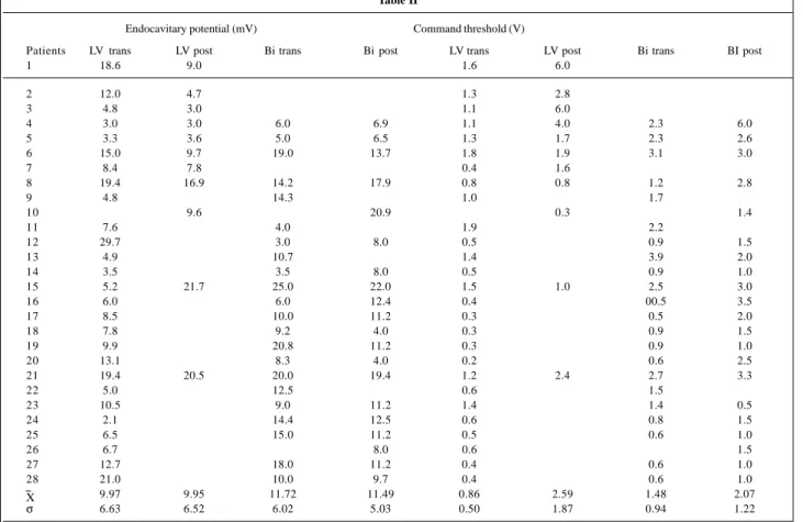
In regard to the implantations of pacemaker leads in the left ventricle through the coronary sinus, we obtained the following positions: anterior wall in 9 patients, lateral wall in 15, and posterior wall in 2. Left ventricular endoca-vitary potential and intraoperative and postoperative biventricular potential, and left ventricular command threshold and intraoperative and postoperative biventricu-lar command threshold are shown in table II. In 1 patient, phrenic stimulation with consequent diaphragmatic stimu-lation through the electrode in the coronary sinus made left ventricular pacing through this via impossible. Two increases in left ventricular pacing threshold occurred,
both in epimyocardial pacemaker leads, 1 immediately before cardiac transplantation, and the other repaired with implantation of a 2nd pacemaker electrode in the left ventricle
through the coronary sinus (fig. 1).
Discussion
Artificial cardiac pacing seems to be approaching a new era in which the objective of the procedure is not only to reestablish the usual cardiac rhythm, but also to contribute to hemodynamics by resynchronizing the cardiac chambers. Ventricular resynchronization through biventricular pacing has demonstrated good results in the treatment of refractory congestive heart failure of dilated cardiomyopathy in patients with intraventricular conduction disorders.
Because our hospital is a referral center for cardiac trans-plantations, most of our patients are candidates for cardiac transplantation with no possibility of waiting due to complete myocardial failure (patients in the intensive care unit, depending on parenteral vasoactive drugs), or are extremely limited patients with very poor quality of life, who await the procedure.
In our experience, reproducing other similar studies in the literature 13,14, we observed a significant acute clinical
improvement in all patients, whose functional classes chan-ged favorably after the procedure. This evident clinical im-provement, however, was not accompanied by a
proportio-Table II
Endocavitary potential (mV) Command threshold (V)
Patients LV trans LV post Bi trans Bi post LV trans LV post Bi trans BI post
1 18.6 9.0 1.6 6.0
2 12.0 4.7 1.3 2.8
3 4.8 3.0 1.1 6.0
4 3.0 3.0 6.0 6.9 1.1 4.0 2.3 6.0
5 3.3 3.6 5.0 6.5 1.3 1.7 2.3 2.6
6 15.0 9.7 19.0 13.7 1.8 1.9 3.1 3.0
7 8.4 7.8 0.4 1.6
8 19.4 16.9 14.2 17.9 0.8 0.8 1.2 2.8
9 4.8 14.3 1.0 1.7
10 9.6 20.9 0.3 1.4
11 7.6 4.0 1.9 2.2
12 29.7 3.0 8.0 0.5 0.9 1.5
13 4.9 10.7 1.4 3.9 2.0
14 3.5 3.5 8.0 0.5 0.9 1.0
15 5.2 21.7 25.0 22.0 1.5 1.0 2.5 3.0
16 6.0 6.0 12.4 0.4 00.5 3.5
17 8.5 10.0 11.2 0.3 0.5 2.0
18 7.8 9.2 4.0 0.3 0.9 1.5
19 9.9 20.8 11.2 0.3 0.9 1.0
20 13.1 8.3 4.0 0.2 0.6 2.5
21 19.4 20.5 20.0 19.4 1.2 2.4 2.7 3.3
22 5.0 12.5 0.6 1.5
23 10.5 9.0 11.2 1.4 1.4 0.5
24 2.1 14.4 12.5 0.6 0.8 1.5
25 6.5 15.0 11.2 0.5 0.6 1.0
26 6.7 8.0 0.6 1.5
27 12.7 18.0 11.2 0.4 0.6 1.0
28 21.0 10.0 9.7 0.4 0.6 1.0
9.97 9.95 11.72 11.49 0.86 2.59 1.48 2.07
σ 6.63 6.52 6.02 5.03 0.50 1.87 0.94 1.22
- mean; s - standard deviation; LV – left ventricle; Bi - biventricular.
4 9 4 9 4 9 4 9 4 9 Table III
Patients QRS Duration Ejection Ejection (ms) fraction (echo) fraction (scintigraphy)
d
i = xi yi di = xi zi di = xi yi di = xi zi di = xi yi
1 60 -0.08 -0.02
2 100 0.04 0.00
3 100 -0.02 -0.21
4 53 -0.13 -0.04 -2
5 80 -0.15 -0.15 15 0
6 90 -0.05 0 0
7 80 -0.09 -0.11 -1
8 80 -0.17 -0.17 -7 0
9 40
10 30 -0.17 0.06 -4 -2
11
12 60 0.04 0.02 3 0
13 30 -0.23 0.18
14 90 -0.04 0.02
15 90 0.04 0.08 -2 0
16 70 -0.12 -0.01 -1 0
17 100 -0.07 -0.13 -1
18 100 -0.07 0.00 -1 0
19 100 -0.09 -0.09 -2 0
20 90 -6 0
21 100 -0.13 -0.08 -1
22 100
23 52 0.09
24 80 -0.01 0
25 50 -0.03 -2
26 70 0.01
27 80 -0.12 0
28 88 -0.02 2
2063 -1.54 -0.78 -5 -7
n 27 17 24 10 18
d 76.407 -0.091 -0.033 -0.500 -0.389 s2 497.251 0.006 0.008 38.331 0.958
s 22.299 0.072 0.090 6.191 0.979
tn 1 17.804 -4.845 -1.773 -1.686 -1.686 tn 1;0,5 2.779 2.921
tn 1;5 1.714
tn 1;10 1.353 1.333
∑- sum of the differences; n- number of patients undergoing the test; d - mean of the differences; s2- variance, calculated with n-1 degrees of freedom;
s-standard deviation; t
n-1 – Student’s t, calculated with n-1 degrees of freedom;
t
n-1;0,5 - critical value of Student’s t, at 0.5% significance level; tn-1;5 - critical
value of Student’s t, at 5% significance level; t
n-1;10 - critical value of Student’s
t, at 10% significance level.
nal increment in the indices of ejection fraction measured on Doppler echocardiography and myocardial scintigraphy performed acutely (10th postoperative day). These indices
showed mild improvements, consisting of only statistical evidence on Doppler echocardiography performed on the 3rd month after the procedure.
Implantation of an epicardial pacemaker lead in the left ventricle, which previously required thoracotomy, has been very simplified with the adoption of the endocavitary access, being performed with local anesthesia. We believe that the development of new pacemaker leads with special guides for catheterization of the coronary sinus will make this method even simpler.
Recently, dual-site right ventricular pacing was proposed as an option to biventricular pacing in ventricular resynchroniza-tion 15. This procedure was attempted for the first time in 1997
when Depuis et al 16 were not able to show the benefits of right
ventricular dual-site pacing in relation to the isolated pacing of the right ventricular outflow tract. These results were also reproduced by LeHelloco et al 17. In our opinion, right ventricular
dual-site pacing may bring some benefits to ventricular resynchronization; these benefits, however, cannot be compared with those of complete ventricular resynchronization provided by biventricular pacing.
It is worth noting that even though the benefit provi-ded by ventricular resynchronization may be great, patients with cardiomyopathies persist with a severe myocardial di-sease, and they may experience decompensation with intra-ventricular conduction disorders. Therefore, this procedure may be a very good nonpharmacological option, mainly due to its minimally invasive feature.
Based on this initial experience, we conclude that ven-tricular resynchronization through bivenven-tricular pacing is an excellent nonpharmacological option for the treatment of congestive heart failure refractory to medicamentous treat-ment in patients with dilated cardiomyopathy with intraven-tricular conduction disorders.
Further in-depth studies may show the extension and duration of the benefits provided by this technique, and also identify the patients who will benefit the most from it.
References
1. Albanesi Filho FM. Insuficiência cardíaca no Brasil. Arq Bras Cardiol 1998:71: 561-2.
2. Thom TJ, Kannel WB, Silbershatz H, et al. Incidence, prevalence, and mortality of cardiovascular diseases in the United States. In: Alexander RW, Schlant RC, Fuster V. eds. Hurt’s The Heart. Vol. 1, 9th ed. New York: Editora Mc Graw-Hill, 1998: 3-17.
3. Hochleitner M, Hortnagl H, Ng C-K, et al. Usefulness of physiologic dual-chamber pacing in drug-resistant idiopathic dilated cardiomyopathy. Am J Cardiol 1990; 66: 198-202.
4. Breker SJ, Xiao HB, Sparrow J, et al. Effects of dual-chamber pacing with short atrioventricular delay in dilated cardyomiopathy. Lancet 1992; 340: 1308-11. 5. Hochleitner M, Hortnagl H, et al. Long-term efficacy of physiologic dual-chamber pacing in the treatment of end-stage idiopathie dilated cardiomyopa-thy. Am J Cardiol 1992; 70: 1320-5.
6. Xiao HB, Brecker SJD, Gibson DG. Effect of abnormal activation on the time course of the left ventricular pressure pulse in dilated cardiomyopathy. Br Heart J 1992; 68: 403-07.
7. Xiao HB, Roy C, Gibson DG. Nature of ventricular activation in patients with
dilated cardiomyopathy: Evidence for bilateral bandle branch block. Br Heart J 1994; 72: 167-74.
8. Bakker PF, Meijburg H, De Jonge N, et al. Beneficial effects of biventricular pacing in congestive heart failure. PACE 1994; 17: 820.
9. Cazeau S, Ritter P, Bakdach S, et al. Four chamber pacing in dilated cardiomyopa-thy. PACE 1994; 17: 1974-9.
10. Cazeau S, Ritter P, Lazzarus A, et al. Hemodynamic improvement provide by biventricular pacing in congestive heart failure: an acute study. PACE 1996; 19: 568. 11. Daubert C, Ritter P, Cazeal S, et al. Permanent biventricular pacing in dilated cardiomyopathy: Is a totally transvenous approach technically feaseble? PACE 1996; 19: 699.
12. Stellbrink C, Auricchio A, Djem B, et al. Potential benefit of biventricular pacing in patients with congestive heart failure and ventricular tachyarrhythmia. Am J Cardiol 1999; 83(5B): 143D-9D.
5 0 5 0 5 0 5 0 5 0
14. Leclercq C, Cazeau S, Breton H, et al. Acute hemodynamic effects of biventricular DDD pacing in patients with end-stage heart failure: J Am Coll Cardiol 1998; 32: 1825-31.
15. Pachon MJC, Albornoz RN, Pachon EI, et al. Estimulação ventricular direita bifocal no tratamento da miocardiopatia dilatada com insuficiência cardíaca. Arq Bras Cardiol 1999; 73: 485-98.
16. Depuis JM, Victor J, Pézard P, et al. Comparison of permanent right ventricular apex (RVA) pacing with right outflow tract (RVOT) and double right ventricular (DRV) pacing. PACE 1997; 20(II): 1130.