Ventricular Resynchronization – Comparing Biventricular and Bifocal
Right Ventricular Pacemakers
Eduardo Arrais Rocha, Tatiana Pereira Gondim, Sebastião Abreu, Roberto Farias, Vera Marques, Almino Rocha,
Demóstenes Ribeiro, Ricardo Pereira, Pedro Negreiros, Carlos Roberto M. Rodrigues, José Nogueira Paes Júnior
Hospital das Clínicas da Universidade Federal do Ceará, Hospital Prontocárdio, Hospital Monte Klinikum - Fortaleza, CE - Brazil
Summary
Objective: To analyze the conventional biventricular pacing (BV) and the bifocal (BF) right ventricular (RV) pacing, and to perform a comparative analysis of these two techniques in relation to clinical, functional and echocardiographic parameters in a population without the exclusion criteria of the major studies.
Methods: A prospective non-randomized analysis of 36 patients undergoing surgery for multisite pacemaker implantation due to QRS ≥ 130 ms, severe left ventricular dysfunction, and NYHA functional class III or ambulatory class IV congestive heart failure was performed.
Results: Favorable results of resynchronization were obtained with both techniques, with no signiicant diferences in the comparison of the two groups, except for a higher QRS narrowing in the BV group, and a trend of a lower number or hospital admissions in the BV group. When the groups were analyzed separately and compared before and after the procedures, we observed that improvement was much more signiicant in the biventricular group, as were the more statistically relevant rates.
Conclusion: Cardiac resynchronization therapy proved to be an eicient therapy in both groups analyzed, although with more signiicant outcomes in the biventricular group. (Arq Bras Cardiol 2007;88(6):596-603)
Key words: Ventricular resyncronization, pacemaker, artiicial; cardiac pacing, artiicial; heart failure, congestive.
Mailing address: Eduardo Arrais Rocha •
Av. Padre Antônio Tomás, 3535/1301 - 60190-020 - Fortaleza, CE - Brazil E-mail: [email protected]
Manuscript received July 18, 2006; revised received November 22, 2006; accepted May 2, 2007.
Introduction
The treatment of congestive heart failure (CHF) has improved with the great therapeutic advances of the past two decades, with clear reductions in morbidity and mortality of these patients. However, many patients remain with signiicant symptoms and a high number of hospital admissions. They have a poor prognosis and the treatment is costly1,2.
Conduction disturbances through the right or left bundle branches can be observed in 30 to 50% of the cases. Intra or interventricular conduction delay (dyssynchrony) can be observed in up to 80% of the patients with left bundle branch block (LBBB), and, less frequently, in right bundle branch block (RBBB) with associated hemiblocks (HB)3. Up to 20 to 30%
of the patients with CHF and narrow QRS (without bundle branch blocks) are also described to have dyssynchrony, and the echocardiographic study4 or phase analysis in nuclear
medicine are important to determine the presence and degree of dyssynchrony.
Several recent studies have demonstrated that the ventricular resynchronization therapy with multisite pacemaker implantation (right atrium, right ventricle,
and left ventricle) reduces symptoms and the number of hospital admissions, improves the functional class (FC) and quality of life, with signiicant changes in echocardiographic parameters and improvement of the systolic and diastolic functions, as well as a reduction in the degree of mitral regurgitation. These efects may be related to the correction of dyssynchrony, which induces the so-called reverse remodeling or diameter reductions and changes in the left ventricular morphology5,6.
More recently, two large randomized multicenter trials showed a signiicant impact of these therapies on the reduction of mortality, both with biventricular pacemakers alone7 and
combined with implantable deibrillators (ICD)8.
In smaller non-randomized series9,10, multisite pacing
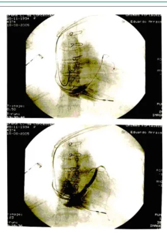
with two right ventricular leads (RV apex and RV outlow tract) and one right atrial lead, known as bifocal pacing (BP), has also proven to be a feasible alternative in the attempt to resynchronization, with faster and less costly procedures (Figure 1). However, studies with this technique are scarce, mainly when compared with the conventional resynchronization pattern with the lead in the coronary sinus pacing the left ventricle.
Fig. 1 - At the bottom: leads in RV and RA, and coronary sinus venogram. On top: lead in coronary sinus positioned in left lateral vein
with conventional indication for resynchronization, however without excluding populations of patients with right bundle branch block, atrial fibrillation, previous conventional pacemakers with RV pacing, and diferent cardiomyopathies, including Chagas’ disease with or without irst-degree AVB from the analysis.
Methods
A prospective non-randomized analysis of 36 patients undergoing surgery for multisite pacemaker implantation (ventricular resynchronization therapy) due to conduction disturbance (QRS ≥ 130 ms), severe left ventricular dysfunction, and NYHA functional class (FC) III or class IV congestive heart failure was performed during the past six months, on average. Patients should be duly controlled for at least 15 days to undergo the procedures. The operations were performed by the same medical team in diferent hospitals. The procedures were followed up by an electrophysiologist who helped catheterize the coronary sinus when necessary, and by two physicians specialized in cardiac pacing, as well as by one anesthetist for intravenous sedation. There were 15 patients in the bifocal group (BF) and 21 patients in the biventricular group (BV).
The biventricular pacemakers used were Insync (Medtronic™), Contak (Guidant™) and Frontier (St Jude™). Conventional dual-chamber pacemakers with adaptors
were used in some of the patients selected for bifocal pacing (26.6%); dual-chamber pacemakers with the atrial inlet serving as the entrance for the RV outlow tract lead were used in 13.3% of the BF group patients with atrial ibrillation, and the atrioventricular interval was shortened to the least possible value. The echocardiographic studies were performed by the same team of specialists, and the ejection fraction (EF) was preferentially analyzed using the Simpson’s rule and/or radionuclide ventriculography (Figures 2 and 3).
Bifocal pacing was chosen because of the following factors: older age, physician’s choice, or failed implantation using the conventional technique. The estimated procedural time ranged between two hours and four and a half hours for biventricular pacemakers, and was two hours for bifocal pacemakers, with a clear decrease in time with increasing number of procedures performed, and with the improvement of catheters and introducers for coronary sinus catheterization (Figure 1).
No in-hospital deaths occurred nor up to four months following the procedure (Tab. 1). One patient presented with acute pulmonary edema in the operating room, and the procedure was converted to RV bifocal pacing. Displacement of the coronary sinus lead occurred in one patient, who was not included in the present analysis. Five patients who already had conventional right pacemakers were implanted with biventricular pacemakers via a right internal jugular vein approach to facilitate the coronary sinus catheterization. In biventricular pacemakers, 20 to 50 ml of nonionic contrast medium diluted in 50 ml of saline solution were used for coronary sinus angiography. Five patients with biventricular pacemakers (BV) had a threshold higher than 3.0 volts, two of whom had a threshold higher than 5.0 volts. These patients did not require reoperation. Infection was not observed in none of the two groups.
Both groups were homogeneous in relation to the different variables analyzed, such as gender, etiology of the cardiomyopathy, presence of atrial fibrillation, right bundle branch block, and previous use of a pacemaker. A higher statistical trend (p = 0.102) was observed for older patients of the bifocal (BF) group, and statistically signiicant diferences were observed for a longer follow-up period in the bifocal group (p = 0.019). In relation to etiology, dilated cardiomyopathy predominated in both groups (61.9% BV vs 60% BF), followed by ischemic cardiomyopathy (33.3% BV vs 33.3% BF) and chagasic cardiomyopathy (4.8% BV vs 6.7% BF). The groups had similar incidence of patients with irst-degree AVB concomitantly with BBB (61.9% BV vs 61.5% BF). The patients were undergoing optimized treatment for CHF, and most of them were using two or more tablets of loop diuretics. Previous use of beta-blockers and angiotensin-converting-enzyme inhibitors had been attempted and optimized in all patients.
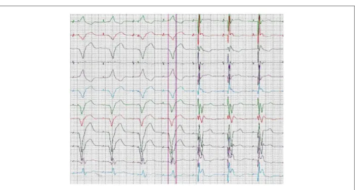
Fig. 2 - The three irst complexes represent RV bifocal (BF) pacing, and the last four represent isolated pacing in the RV outlow tract.
Fig. 3 - The irst four complexes represent pacing in the RV apex; the last three complexes represent biventricular (BV) pacing.
positioned in the RV apex, RV outlow tract (3p), and in the LV lateral, posterior or lateral posterior vein. In the BF group, the leads were positioned in the RV apex and in the RV outlow tract (RV free wall or high septal region) (Figure 4).
Patients with biventricular pacemaker implants underwent a parallel assessment for dyssynchrony, which was present in all of the 12 patients analyzed, with improvement or reversion in
11 patients. Previous identiication of ventricular dyssynchrony is important to determine patients’ improvement11.
The Levene’s test was used to compare the variability of continuous variables, and the Shapiro-Wilk test was used to verify the normality of distribution of these variables.
The paired Student’s t test was used to compare the means of variables with a normal distribution in paired populations, and the sign test was used to analyze the continuous variables with a non-normal distribution (non-Gaussian pattern). Parameters such as the functional class (FC) and degree of diastolic dysfunction (DD) were analyzed in the form of scores for the respective values, with Student’s t test analyses for non-paired populations, and the Mann-Whitney test for the comparison of mean scores in non-paired populations.
Table 1 - Incidence of acute/subacute complications related to the procedures
Complication Bivent = 21 Bifocal = 15
Infection 0 0
Pericardial efusion 1 0
Mortality 0 0
VLD 1 0
ARF 1 0
LVT 2 0
LH 0 0
APE 1 0
Mortality - peri-hospital mortality up to 4 months after implantation; VLD - ventricular lead displacement; ARF - contrast-induced acute renal failure; LH - large hematomas; LVT - LV or outlow tract thresholds > 5.0 volts; APE - pulmonary edema in operating room.
Some parameters were analyzed only descriptively due to the impossibility of an inferential analysis.
P values were considered signiicant when lower than 0.05 (5%), and with a statistical trend between 0.05 and 0.15 (5 a 15%).
Results
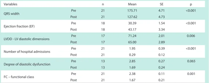
Biventricular pacemakers - The analysis of biventricular pacemakers showed significant improvements in all the parameters considered, with much more statistical signiicance than for bifocal pacemakers ( Table 2). The analysis of the diastolic function showed only a statistical trend of improvement. We observed a signiicant reduction in the number of hospital admissions, which is a very signiicant variable when the impact of treatment costs is considered.
Complete normalization of the systolic ventricular function and of the left ventricular dimensions were observed in two patients in the late follow-up after one and a half year. These patients were assessed with echocardiogram and radionuclide ventriculography (Figure 5). Both patients had severe chronic left ventricular dysfunction documented in several tests performed in the past years, with FC pre-III (1p), and FC IV (1p), with late progression to FC I in both.
Treatment failure rate was 9.5% when clinical criteria were used, and 19% when echocardiographic criteria such as lack of improvement (<15% in EF) and lack of ventricular remodeling were used. However, of these 19%, only one patient did not improve clinically. The remaining patients considered non-responders as assessed by systolic function and of LV dimensions may have improved thanks to the reduction in the degree of diastolic dysfunction.
Table 2 - Analysis of the variables of the biventricular (BV) pacemaker group
Variables n Mean SE p
QRS width Pre 21 175.71 4.71 <0.001
Post 21 127.62 4.73
Ejection fraction (EF) Pre 18 30.39 1.54 <0.001
Post 18 43.17 3.34
LVDD - LV diastolic dimensions Pre 17 71.24 2.01 0.006
Post 17 65.00 2.89
Number of hospital admissions Pre 21 1.95 0.39 <0.001
Post 21 0.29 0.12
Degree of diastolic dysfunction Pre 13 2.85 0.27 0.065
Post 13 1.69 0.24
FC – functional class Pre 21 2.38 0.11 0.001
Post 21 1.67 0.21
Using the paired Student’s t test for the pre and post comparison in relation to the means of the variables with a normal distribution, and the sign test for variables with a non-normal distribution.
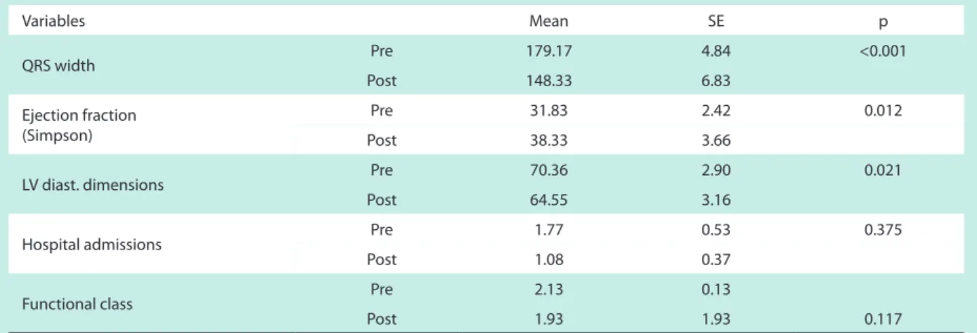
Bifocal pacemakers - The analysis of bifocal pacemakers showed statistical signiicance in QRS narrowing, improvement of ejection fraction, and reduction of left ventricular dimensions; only a trend of improvement was observed for the functional class. There was no signiicant reduction in the number of hospital admissions (Table 3). A detailed analysis of the diastolic function was not performed in the majority of the patients in this group.
When clinical criteria or lack of improvement in echocardiographic parameters were considered, 19.5% and 33% of the patients in this group were considered non-responders, respectively.
Using the paired Student’s t test for the pre and post comparison in relation to the means of the variables with a normal distribution, and the sign test for variables with a non-normal distribution.
Bifocal versus biventricular - The comparative statistical analysis between the two groups did not show statistically signiicant clinical or echocardiographic diferences, except for more QRS narrowing in the biventricular group, longer follow-up in the bifocal group, and a statistical trend of a lower number of hospital admissions in the biventricular group (Table 4).
Also, a greater number of clinical (19.5 % BF vs 9.5 % BV) and echocardiographic (33% BF vs 19 % BV) non-responders was observed in the bifocal (BF) group in relation to the biventricular (BV) group.
Discussion
The results presented in this study are, in general, quite consistent with those of the literature1,2. However,
several peculiarities are worth mentioning. Publications on comparative analyses between biventricular and bifocal pacing are scarce or absent in the review we performed, and the
same occurs with the assessment of patients without the usual exclusion criteria of most of the major studies, which eventually do not relect the clinical practice of cardiologists.
The irst major clinical studies with multisite pacing were conducted in Europe and in the United States between 1995 and 1998; epicardial leads were initially used, with several limitations of these procedures performed via thoracotomy due to the high surgical morbidity1,2,3. These studies were
followed by those of Cazeau et al in France12, now using the
endocardial approach, although still with many limitations because of the inappropriate material used at that time.
The three clinical trials (MIRACLE, MIRACLE ICD, and CONTAK CD) that led to the approval of biventricular pacemakers and of biventricular pacemakers coupled with defibrillators in the United States were randomized, controlled, double-blind studies in which the pacemaker was non-functioning for months to assess the placebo efect of this therapy. The objectives were the assessment of clinical and echocardiographic improvement of these patients5,13,14.
The main objective of the new generation of studies started in 2000 was to assess the efect of this therapy on mortality, like in the 2003 meta-analysis15 and in the COMPANION
study8 that showed reductions in mortality and in hospital
admissions for CHF. In this latter study mentioned, mortality reduction occurred in the group of deibrillators associated with resynchronizers. Other large meta-analyses corroborated these indings of reduction in morbidity and mortality, and in mortality alone16,17.
Some studies have demonstrated that a longer QRS duration is a good predictor of response to resynchronization, like in the subgroups of the CONTAK CD and MIRACLE studies18.
However, several other studies have not corroborated this inding19, and dyssynchrony started to be better assessed
patients had signiicant QRS narrowing. Only one patient presented with post-surgical QRS widening and progressed without improvement. Two other patients did not present QRS narrowing, but both had clinical and echocardiographic improvements, albeit mild.
Ventricular remodeling may occur even early in resynchronized patients (3 months after implantation)20, and is
fundamental as a predictor of sustained clinical improvement21.
Several clinical variables attempted to identify subgroups with possible favorable responses to resynchronization, however the indings were not uniform22,23. Patients with a restrictive pattern
may not achieve favorable results with resynchronization24,
unlike observed in our case series, in which many of these patients also obtained beneits.
Favorable results in resynchronization were observed with both techniques used, and biventricular pacing was veriied to promote much more signiicant statistical results than bifocal pacing. The latter demonstrated only a trend of improvement of functional class, and did not provide a reduction in the number of hospital admissions. The assessment of both ejection fraction and left ventricular dimensions showed signiicant statistical results. We also observed a greater number of clinical (19.5% BF vs 9.5% BV) and echocardiographic (33% BF vs 19% BV) non-responders in the bifocal (BF) group in relation to the biventricular (BV) group.
In the direct comparative analysis between the two groups, the BV group had more QRS narrowing (p = 0.016), and a lower number of hospital admissions than the BF group (p = 0.127). Based on these results, bifocal pacing is a good option for patients in whom the usual resynchronization via coronary sinus (BV) is not achieved. Some studies corroborate the results presented9,10. Right ventricular (RV) lead implantation in the
RV outlow tract was performed in three patients of the BV group, aiming at optimizing the ventricular resynchronization, a technique that is supported by some authors25.
The etiology of the cardiomyopathies may have contributed to the better results in these groups analyzed, considering
that some studies have demonstrated that patients with cardiomyopathy of ischemic etiology have a poorer response to resynchronization therapy. This would be due to the large areas of necrosis developed after previous myocardial infarctions26,27.
The comparative analysis of the groups in relation to mortality could not demonstrate criteria of statistical signiicance (p = 0.129) due to the study design and number of patients. However, the descriptive analysis showed that six patients (40%) in the bifocal (BF) group died in the mean follow-up of 18.5 months, whereas three patients (15%) in the BV group died in the mean follow-up of 10.1 months. Death causes in the BF group were: three from sudden death, two from CHF, and one from stroke. In the BV group, one death resulted from CHF, one from sudden death, and one from respiratory infection and renal failure not related to the procedure. Historically, in the selected and well-followed-up patients of the large trials, the overall mortality is higher than 30% in two years7.
The significant decrease in the number of hospital admissions of the BV group patients may reduce the efects of the initial costs with pacemaker implantation to a great extent, considering that in the cost-efectiveness analysis, the heavy cost burden results from hospitalizations. This analysis derives from cost-efectiveness studies involving resynchronizers28,29.
The option of upgrading patients who already have pacemakers to biventricular models was chosen in 19% of the BV group patients, and in 13.3% of the BF group patients, with improvement in these subgroups also; however, this statistical analysis was not performed because of the small number of patients. These indings are consistent with those of some series found in the world literature30.
Complete normalization of the echocardiographic parameters was obtained in two patients of the biventricular group at two years of follow-up. With less than one year of follow-up, a signiicant improvement of the ejection fraction and reduction of the LV dimensions were already observed in
Table 3 - Analysis of the variables of the bifocal (BF) pacemaker group
Variables Mean SE p
QRS width Pre 179.17 4.84 <0.001
Post 148.33 6.83
Ejection fraction (Simpson)
Pre 31.83 2.42 0.012
Post 38.33 3.66
LV diast. dimensions Pre 70.36 2.90 0.021
Post 64.55 3.16
Hospital admissions Pre 1.77 0.53 0.375
Post 1.08 0.37
Functional class Pre 2.13 0.13
Post 1.93 1.93 0.117
Table 4 - Analysis of pre and post-procedural variables in the biventricular and bifocal groups
Variables Pacemaker n Mean SE p
Age (years) Biventricular 21 67.24 1.92 0.102
Bifocal 15 72.20 2.23
QRS pre Biventricular 21 175.71 4.71 0.883
Bifocal 15 174.67 5.15
QRS post Biventricular 21 127.62 4.73 0.016
Bifocal 12 148.33 6.83
EF pre Biventricular 21 29.95 1.43 0.647
Bifocal 15 31.07 2.03
EF post Biventricular 18 43.17 3.34 0.348
Bifocal 12 38.33 3.66
LVDD pre Biventricular 19 71.74 1.90 0.873
Bifocal 13 71.23 2.59
LVDD post Biventricular 17 65.00 2.89 0.919
Bifocal 11 64.55 3.16
Hosp. admissions pre Biventricular 21 1.95 0.39 0.675
Bifocal 13 1.77 0.53
Hosp. admiss. post Biventricular 21 0.29 0.12 0.127
Bifocal 14 1.00 0.35
LV threshold (volts) Biventricular 21 2.10 0.34 0.441
Bifocal 13 1.56 0.20
Follow-up period (months) Biventricular 20 10.10 1.96 0.019
Bifocal 15 18.53 2.74
Grade diast. dysf pre Biventricular 15 2.87 0.26 0.467
Bifocal 5 2.40 0.68
Grade diast. dysf post Biventricular 17 1.65 0.19 0.976
Bifocal 9 1.89 0.42
Functional class pre Biventricular 21 2.38 0.11 0.172
Bifocal 15 2.13 0.13
FC post Biventricular 21 1.67 0.21 0.161
Bifocal 15 1.93 0.21
these patients. No other possible causes were found that could be responsible for such an improvement of these patients, but the resynchronization performed. In the BF group, this was not observed in any of the patients.
Conclusions
Cardiac resynchronization therapy proved to be an eicient therapy combined with the treatment with usual drugs in patients with severe ventricular dysfunction, conduction disturbances, and FC III or ambulatory class IV chronic heart failure. This therapy leads to QRS narrowing, improvement of the functional class and of the systolic and diastolic left ventricular function, reverse remodeling, with
signiicant reductions in the LV dimensions and reduction in the number of hospital admissions. These results are consistent with those demonstrated by the large studies on resynchronization. The treatment failure rate was lower than that described in the literature, perhaps because of the patient selection and type of analysis performed. The results were obtained within a population without the usual exclusion criteria of the large studies.
admissions in the biventricular group. Mortality was higher in the bifocal group (40%) in a longer follow-up period of this group, versus 15% in the biventricular group, however with no statistical diference.
When the groups were analyzed separately and compared before and after the procedures, we observed that the
improvement in the mean parameters of the variables analyzed was quite more significant in the biventricular group, as were the more statistically relevant rates. No reduction in the number of hospital admissions was observed in the bifocal group, but only a trend of improvement of the functional class.
References
1. Auricchio A, Abraham WT. Resynchronization therapy: current state of the art. Circulation. 2004; 109: 300-7.
2. Kadish A, Mehru M. Heart failure devices. Circulation. 2005; 111: 3327-35.
3. Saxon LA, Ellenbogen KA. Resynchronization Therapy for the treatment of heart failure. Circulation. 2003; 108: 1044-52.
4. Cho GY, Song JK, Park WJ, Han SW, Choi SH, Doo YC, et al. Mechanical dyssynchrony assessed by tissue doppler imaging is a powerful predictor of mortality in congestive heart failure with normal QRS duration. J Am Coll Cardiol. 2005; 46: 2237-43.
5. Abraham WT, Fisher WG, Smith AL, Delurgio DB, Leon AR, Loh E, et al. Cardiac resynchronization in chronic heart failure (MIRACLE). N Engl J Med. 2002; 346: 1845-53.
6. Linde C, Leclerq C, Rex S, Garrigue S, Lavergne T, Cazeau S, et al. Long–term beneits of biventricular pacing in congestive heart failure: results from the Multislice Stimulation in Cardiomyopathy (MUSTIC) study. J Am Coll Cardiol. 2002; 40: 111-8.
7. Cleland JGF, Daubert JC, Erdmann E, Freemantle N, Gras D, Kappenberger L, et al. The efect of cardiac resynchronization on morbidity and mortality - CARE – HF. N Engl J Med. 2005; 352 (15): 1539-49.
8. Bristow MR, Saxon LA, Boehmer J, Krueger S, Kass DA, De Marco T, et al. Cardiac resynchronizations therapy with or without an implantable defibrillator in advanced chronic heart failure. N Engl J Med. 2004; 350: 2140-50.
9. O’Donnel D, Nadurata V, Hamer A, Kertes P, Mohammed W. Bifocal right ventricular cardiac resynchronization therapies in patients with unsuccessful percutaneous lateral left ventricular venous acess. Pacing Clin Electrophysiol. 2005; 28: S27-S30.
10. Pachon JC, Pachon EI, Albornoz RN, Pachon JC, Kormann DS, Gimenes VM, et al. Ventricular endocardial right bifocal stimulation in the treatment of severe dilated cardiomyopathy heart failure with wide QRS. Pacing Clin Electrophysiol. 2001; 24: 1369-76.
11. Bax JJ, Bleeker GB, Marwick TH, Molhoek SG, Boersma E, Steendijk P, et al. Left ventricular dyssynchrony predicts response and prognosis after cardiac resynchronization therapy. J Am Coll Cardiol. 2004; 44: 1834-40.
12. Cazeau S, Leclerc C, Lavergne T, Walker S, Varma C, Linde C. Effects of multisite biventricular pacing in patients with heart failure and intraventricular conduction delay. N Engl J Med. 2001; 344: 873-80.
13. Young JV, Abraham WT, Smith AL, Leon AR, Lieberman R, Wilkoff B, et al. Combined cardiac resynchronization and implantable cardiovertor defibrillation in advanced chronic heart failure: The MIRACLE ICD trial. JAMA. 2003; 289: 2685-94.
14. US Food and Drug Administration. Summary of safety and effectiveness CONTAK-CD system including CONTAK CD CRT-D: PMA P010012. St Paul, Minn: Guidant Corp; 2002. p. 1-47.
15. Bradley DJ, Bradley EA, Baughman KL, Berger RD, Calkins H, Gooeman SN, et al. Cardiac resynchronization and death from progressive heart failure: a meta-analysis of randomized controlled trials. JAMA. 2003; 289: 730-40.
16. McAlister FA, Ezekowittz JA, Wiebe N, Rowe B, Spooner C, Crumley E, et al. Systematic review: cardiac resynchronization in patients with symptomatic
heart failure. Ann Intern Med. 2004; 141: 381-90.
17. Freemantle N, Tharmanathan P, Calvert MJ, Abraham WT, Ghosh J, Cleland JG. Cardiac resynchronization for patients with heart failure due to left ventricular systolic dysfunction – a systematic review and meta-analysis. Eur J Heart Fail. 2006; 8(4): 433-40.
18. Egoavil CA, Ho RT, Greenspon AJ, Pavri BB. Analysis of pooled data from the MIRACLE and Contak Trials. Heart Rhythm. 2005; 2: 611-5.
19. Kashani A, Barold S. Signiicance of QRS complex duration in patients with heart failure. J Am Coll Cardiol. 2005; 46: 2183-93.
20. Funk RC, Koelsch S, Waldhans S, Prinz H, Grimm W, Moosdorf R, et al. Marker improvement in left ventricular function and signiicant left ventricular remodeling witin 3 months of cardiac resynchronization therapy in patients with dilated cardiomyopathy. Pacing Clin Electrophysiol. 2005; 28: S5-S7.
21. Yu CM, Bleeker GB, Fung JWH, Schalij MJ, Zhang Q, van der Wall EE, et al. Left ventricular reverse remodeling but not clinical improvement predicts long-term survival after cardiac resynchronization therapy. Circulation. 2005; 112 (11); 1580-6.
22. Martinelli Filho M, Baggio JM, Nishioka SAD, Pedrosa A, Torres GG, Escarião A, et al. Ressincronização cardíaca em seguimento tardio: análise de preditores de resposta clínica. Reblampa. 2006; 19(1): 45-52.
23. Lecoq G, Leclerc C, Leray E, Crocq C, Alonso C, de Place C, et al. Clinical and electrocardiographic predictors of a positive response to cardiac resynchronization therapy in advanced heart failure. Eur Heart J. 2005; 26: 1094-100.
24. Salukhe T V, Francis DP, Clague JR, Sutton R, Poole-Wilson P, Henein MY. Chronic heart failures patients with restrictive LV filling pattern have signiicantly less beneit from cardiac resynchronization therapy than patients with late LV illing pattern. Int J Cardiol. 2005; 100: 5-12.
25. Riedlbauchova L, Cihak R, Bytesnik J, Vancura V, Fridl P, Hoskova L, et al. Optimization of right ventricular lead position in cardiac resynchronization therapy. Eur J Heart Fail. 2006; 8 (6): 609-14.
26. Mangiavacchi M, Gasparini M, Faletra F, Klersy C, Morenchi E, Galimbert P, et al. Clinical predictors of marked improvement in left ventricular performance after cardiac resynchonization therapy in patients with chronic heart failure. Am Heart J. 2006; 151 (2): 477e1- 477 e6.
27. Sutton MG, Plappert T, Hilpisch KE, Abraham WT, Hayes DL, Chinchoy E. Sustained reverse left ventricular structural remodeling with cardiac resynchronization at one year is a function of etiology: quantitative Doppler echocardiographic evidence from the Multicenter Insync Randomized Clinical Evaluation (MIRACLE). Circulation. 2006; 113: 266-72.
28. Calvert MJ, Freemantle N, Yao G, Cleland JG, Billingham L, Daubert JC, et al. Cost efectiveness of cardiac resynchronization therapy: results from the CARE-HF trial. Eur Heart J. 2005; 26: 2681-8.
29. Bantz K, Gras D. Cardiac resynchronization therapy: a model to assess the economical value of this new technology [abstract]. Eur Heart J. 2003; 24: 364.