Schistosoma mansoni Cercaria and
Schistosomulum
Antigens in Serodiagnosis
of Schistosomiasisl
HERM~NIA YOHKO KANAMURA,~ SUMIE HOSHINO-SHIMIZU,~
&
LUIZ
CAETANO DA SILVA~
Schistosoma mansoni cercaria and schistosomulum obtained in vitro were used in im-
munofluorescence (IF) tests and indirect hemagglutination (IHA) tests of 137 study sera,
44 from subjects infected with S. mansoni and 93 from healthy subjects residing outside areas endemic for the disease. The results of these tests were compared with those obtained by testing the same sera using conventional adult worm antigen, and also with the initial clinical and parasitologic diagnoses of the 137 subjects providing the study sera.
Regarding sera from acute versus chronic cases, IF testing of the acute sera consistently detected 1gA antibodies along with 1gM and IgG, the last two being found consistently in
chronic sera. Also, the geometric mean of the 1gM antibody titers found in the IF tests was
higher for acute than for chronic sera.
Excluding IF IgA, which was negative for chronic cases, the sensitivity of the other types
of tests (IF IgG, IF lgM, and IHA) using cercaria and schistosomulum antigens, both under
evaluation, ranged from 0.773 to 0.955, the specificity ranged from 0.957 to 1.000, the
efficiency ranged from 0.927 to 0.985, the predictive value of positives ranged from 0.909
to 1 .OOO, and the predictive value of negatives ranged from 0.903 to 0.979. No statistical differences were observed between these results and those obtained with conventional adult worm antigen. This suggests that cercaria and schistosomulum antigens, both of which can be produced more quickly and cheaply than adult worm antigen, could serve as reliable
alternatives to adult worm antigen in the serodiagnosis of schistosomiasis mansoni.
I
n areas endemic for schistosomiasiscaused by Schistosoma mansoni (known as schistosomiasis mansoni), diagnostic techniques involving direct observation
‘The work reported here has received financial sup- port from Brazil’s National Council of Scientific and Technologic Development (Conselho National de Desenvolvimento Cientifico e Tecnologico, CNl’q) .
2Department of Clinical and Toxicologic Analysis, Faculty of Pharmaceutical Sciences, University of Slo Paulo. Correspondence should be addressed to Dr. Herminia Yohko Kanamura, Departamento de Analises Clfnicas e Toxicologicas, Faculdade de Ciencias Farmaceuticas, Universidade de Sao Paulo, Av. Prof. Lineu Prestes, 580-Bloc0 17, Cidade Universitaria, CEP 05508-SPo Paulo-SP, Brazil. %stituto de Medicina Tropical de Sao Paulo, S&o
Paul0 (SP), Brazil.
of the parasite (eggs in stools) are gen- erally preferred. However, misleading diagnoses are frequent among patients having infections of low to moderate in- tensity, unisexual infections, and other sorts of infections in which factors such as the advanced age of the patient or lateness in the stage of the parasites’ life cycle considerably diminish the number of S. mansoni eggs in the patient’s feces (1, 4.
extent, in monitoring patient chemother- apy (3-5). At present an array of sero- logic tests is available (2). These generally employ adult worm or parasite egg an- tigens (either crude or pure); and while the potential has existed for use of other antigen sources, up to now the advan- tages involved have seemed uncertain.
Cercaria antigen was used in immu- nofluorescence (IF) testing during the late 1960s and early 1970s. However, until re- cently no further research was done to overcome this procedure’s low specificity and other features making it impractical for use in seroepidemiologic surveys (6). More recently, soluble cercarial extracts were used in indirect hemagglutination (IHA) and enzyme-linked immunosor- bent assay (ELISA) serology (7), but their diagnostic utility was not ascertained.
The diagnostic potential of antigen de- rived from schistosomulum, the larval stage of the parasite that develops after skin penetration (obtained either in vim or through in vitro cultivation), has not been assessed before, although anti- bodies against outer membrane schisto- somulum have been found in sera from patients with schistosomiasis mansoni (8).
Antigens from these early stages in the parasite’s development offer certain ad- vantages over antigens from the adult stages. Specifically, they can be obtained more quickly, easily, and cheaply than adult antigens because they require less time to develop and no expensive facili- ties for maintaining vertebrate hosts- points of particular interest to developing countries.
With this in mind, we sought to de- termine whether antigens from cercaria and schistosomulum cultivated in vitro could be used effectively to detect IgG, IgM, and IgA antibodies separately in IF tests and to detect total antibodies (class unspecified) in IHA tests. The sera ex- amined in these tests were obtained from patients with various different clinical
forms of schistosomiasis mansoni. The results obtained with the cercaria and schistosomulum antigens were com- pared to results obtained by testing the same sera with conventional IF and IHA procedures employing adult worm antigen.
MATERIALS
AND METHODS
Serum Samples
A total of 137 serum samples were studied. Forty-four of these came from patients with schistosomiasis mansoni who had yielded positive parasitologic results when tested by the Kato-Katz method (9). Of the 44 cases, 10 were clas- sified as acute and 34 as chronic, the lat- ter including 14 infections considered in- testinal, 10 hepatointestinal, and 10 hepatosplenic according to Neves’ crite- ria (20) for clinical forms of schistosom- iasis. The remaining 93 sera were ob- tained from clinically healthy individuals whose stool examinations yielded nega- tive results. These people included 60 residents of the municipality of SBo Paulo and 33 residents of the municipality of Iguape, both of these being areas that are nonendemic for schistosomiasis man- soni. All of the serum samples were di- vided into 1 ml aliquots and stored with glycerin at - 20°C (II).
Antigens
S. mansoni cercariae (LE strain, Belo Horizonte) were collected from speci- mens of the intermediate snail host Biom- phalaria glubrafa 40 days after infection. They were then concentrated in glass conical centrifuge tubes at 4°C as de- scribed previously (12). This cercaria sus- pension was divided into two parts, both of which were centrifuged. In one case the resulting sediment was embedded in a gel (Tissue-Teck OCT Ames Co., Miles
Laboratories, U.S.A.), frozen, and sec- tioned in a cryostat for use in the IF test. In the other the sediment was stored at
-20°C until used as antigen for the IHA test (13).
Schistosomula cultivated in vitro were obtained as previously described (14). In brief, the cercaria suspension was cen- trifuged and resuspended in Earle’s medium4 enriched with lactalbumin (ELAC). Cercaria bodies were prepared by stirring the suspension for 60 seconds in a Vortex Jr. homogenizer (Scientific In- dustries, Inc., Queens Village, New York, U.S.A.). After allowing this suspension to settle for 10 minutes, the tail-rich su- pernatant was discarded, and the sedi- mented bodies were resuspended in ELAC medium. This washing procedure was repeated two or three times, after which the cercaria bodies in ELAC medium were incubated for two hours at 37°C. The schistosomula obtained in this manner were then divided into two portions for preparation of IF and IHA antigens using the procedure described above for cercaria.
Adult worms were processed in a sim- ilar manner (22) to provide IF and IHA antigens.
Serologic Tests
The cryostat sections of cercaria, schis- tosomulum, and adult worm concen- trates were assayed in IF tests with anti- human IgG (y-chain), anti-human IgM (k-chain), and anti-human IgA (a-chain) fluorescent conjugates supplied commer- cially (Hyland Div. Travenol Lab., U.S.A.), after checking the latter’s monospecificity by immunoelectrophoresis.
4Earle’s balanced salt solution containing 0.68% NaCI, 0.04% KCl, 0.0125% NaH,PO,H,O, 0.02% CaCI,, 0.01% MgSO,, 0.22% NaHCO,, 0.005% phenol red, and 0.1% glucose.
Before attempting to detect IgM anti- bodies, the rheumatoid factor was re- moved by absorption of sera with human gamma globulin aggregates (25).
For the IHA test, formalin-stabilized human group 0 red blood cells were coated with alkaline-solubilized cercaria, schistosomulum, and adult worm prep- arations (13).
Statistical Analyses
Each serologic test using different S. mansoni antigens was evaluated with re- spect to its sensitivity, specificity, effi- ciency, and the predictive values of pos- itive and negative results (16). The kappa
(K) index of agreement between each test
and the true (clinical/parasitologic) diag- nosis was determined. In addition, the kappa index of agreement was deter- mined for each test using cercaria or schistosomulum antigen with respect to the corresponding reference test using adult worm antigen. The McNemar test (27) was applied to determine whether the positive and negative results of the tests under investigation differed signif- icantly from those of the reference test or the true diagnosis. Student’s t test was used to compare the geometric mean IgM and IgG antibody titers (GMT) detected in acute infections with the GMT found in chronic infections (18).
RESULTS AND DISCUSSION
tions employed (notably cryostat sections for the IF tests and alkaline-solubilized extracts for the IHA tests) have been thoroughly explored.
The IF tests of the 10 acute case sera that used cercaria and schistosomulum antigens succeeded in detecting IgG, IgM, and IgA antibodies in a fashion similar to reference tests using adult worm an- tigen. The former antigens caused flu- orescent staining of the parasite mem- brane and, to a lesser extent, the parenchyma. In tests with conventional adult worm antigen the fluorescence was limited to the gut and occasionally the membrane.
The IF tests of sera from chronic cases using the two study (cercaria and schis- tosomulum) antigens detected IgG and IgM antibodies that yielded fluorescence patterns similar to those observed with the acute sera. The IF tests of chronic sera with adult worm antigen also detected IgG and IgM antibodies, the IgG strongly staining the parasite parenchyma while the IgM only reacted with the gut.
In general, the IgA and IgM antibodies detected with the cercaria and schisto-
somulum antigens produced good flu- orescent staining, easily visualized in the microscope’s dark field. This was clearly superior to the fluorescent staining of the gut obtained with the adult worm anti- gen, which was sometimes weak and was not present in all sections.
The cutoff titer established for each se- rologic test was selected so as to yield the highest diagnostic efficiency in discrimi- nating between individuals who were and were not infected with S. mansoni. Thus, for IF tests using the cercaria and schis- tosomulum antigens, the cutoff titer was 80 for IgG but 10 for both IgM and IgA. For IHA tests using the same antigens, the cutoff titer was 20. For the conven- tional IF tests using adult worm antigen, the cutoff titer was 20 for IgG but 10 for IgM and IgA. And for the conventional MA tests using adult worm antigen, the cutoff titer was 20.
The values indicated by the data for sensitivity, specificity, and efficiency, as well as the predictive values of positives (PV + ) and negatives (PV - ), are shown in Table 1. The values found for all the serologic tests using the cercaria and
Table 1. Diagnostic performance of IgG, IgM, and IgA immunofluorescence (IF) tests and indirect hemagglutination (IHA) tests using different S. mansoni antigens in the study of 137 patients with schistosomiasis mansoni and uninfected individuals.
Test Antigen Sensitivity Specificity Efficiency pv+ PV- Cercaria 0.909 0.957 0.942 0.909 0.957 IgC IF Schistosomulum 0.955 1 .ooo 0.895 1.000 0.979 Adult worm 1 .ooo 1 .ooo 1.000 1.000 1 .ooo
i
Cercaria 0.955 1 .ooo 0.985 1 .ooo 0.979 IgM IF Schistosomulum 0.864 1.000 0.956 1 .ooo 0.939
Adult worm 0.977 1 .ooo 0.993 1 .ooo 0.989
{
Cercaria 0.227 1 .ooo 0.752 1 .ooo 0.732
IgA IF Schistosomulum 0.227 1 .ooo 0.752 1 .ooo 0.732
Adult worm 0.295 1 .ooo 0.774 1 .ooo 0.750
i
Cercaria 0.886 1 .ooo 0.964 1.000 0.949 IHA Schistosomulum 0.773 1 .ooo 0.927 1 .ooo 0.903
Adult worm 0.977 0.892 0.920 0.811 0.988
Note- Sensitwity = true positives detected/(true positives + false negatives); specificity = true negatives detected/(true negatives + false positives); efficiency = (true positives detected + true negatives detected)/total number tested; predictive value of positives (PV+) = true positives detected/(true positives + false positives); and predictive value of negatives (PV-)
= true negatives detected/(true negatives + false negatives).
schistosomulum antigens are close to those provided by reference tests using adult worm antigen. Within this context, it is worth noting that the sensitivity and specificity values obtained in the IgG IF test with the cercaria antigen employed here are higher than those values re- ported from tests carried out with the whole cercaria body (6).
As can be seen, the sensitivity of the IgA IF test with all three antigens was relatively low because most of the sera tested were obtained from patients with chronic cases. In general, our past re- search (12) as well as the work reported here has shown a positive correlation be- tween the presence of IgA antibodies and acute schistosomiasis infection. Thus, IgA antibodies can be considered better im- munologic indicators of acute schisto- somiasis than IgM antibodies because they are detected at higher titers in acute in- fections than in chronic infections. It is true that the present study did detect IgA antibodies in five serum samples from patients with chronic infections, but the positive titers involved were relatively low, ranging from 20 to 40. IgA antibody in four of these samples reacted only with adult worm antigen, while that in the remaining sample reacted with both cer- caria and schistosomulum antigens but not with adult worm antigen. In contrast, IgA antibody in samples from patients with acute cases invariably reacted with all three types of antigens, yielding titers ranging from 10 to 640 with adult worm antigen and from 40 to 640 with cercaria and schistosomulum antigens (see Table 4). Because study of other unrelated dis- ease has associated the presence of IgA antibodies with mucosal involvement (29), it seems likely that the small number of chronic patients testing positive by IgA IF might have had lesions of the intes- tinal mucosa.
Although other investigators (20, 21) reported some time ago that IgA anti-
bodies were present in sera from patients with undefined clinical forms of schis- tosomiasis, the finding that IgA anti- bodies are present in sera from patients with acute infections was only recently confirmed (22).
It has been determined that the ob- served IgA antibodies are reactive against worm gut, cercaria, and schistosomulum membranes, and that they probably share the same reported (23) carbohydrate ep- itopes of IgM and IgG antibodies, both of which are observed in sera from acute infections. Since these carbohydrates from schistosomulum antigens cross-react with keyhole limpet hemocyanin (24, 25), a search for cross-reactivity with other bacterial polysaccharide antigens seems relevant.
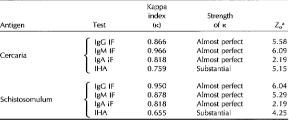
Table 2 shows the kappa (K) agreement
index, as well as the degree of K agree-
ment, between the diagnosis indicated by each variety of test and the true di- agnosis. All the K indices calculated for
tests using cercaria and schistosomulum antigens were similar to those calculated for the reference tests using adult worm antigen, In addition, all the tests except the IgA IF tests had statistically signifi- cant K indices of agreement that were
rated as “almost perfect.”
Table 3 shows the K agreement index
and degree of K agreement between the
test using cercaria and schistosomulum antigens and the corresponding refer- ence tests using adult worm antigen. All the IF tests using cercaria and schisto- somulum antigens exhibited high K in-
dices of agreement rated as “almost per- fect,” while the IHA tests yielded slightly lower K indices of agreement rated as
“substantial.” All the K indices obtained
in this comparison were statistically sig- nificant.
Table 2. The degree of kappa (K) index agreement between (a) the test results using cercaria, schistosomulum, and adult worm antigens and (b) the true (clinical and parasitologic) diagnoses of the subjects providing the 137 test sera.
Antigen Test
Kappa
index (4
Strength of K IgG IF
Cercaria IgM IF IgA IF IHA
Schistosomulum
IgG IF 0.950 IgM IF 0.879 IgA IF 0.271 IHA 0.806 f- IgC IF 1 .ooo 0.983 0.363 0.825 Adult worm IgM IF
IgA IF IHA
0.866 Almost perfect 5.58 0.950 Almost perfect 6.04 0.271 Slight 1.30 0.897 Almost perfect 5.58 Almost perfect 6.04 Almost perfect 5.40 Slight 1.30 Almost perfect 5.58 Almost perfect 6.44 Almost perfect 6.27 Slight 1.78 Almost perfect 5.78 “Z, = observed 2 (critical Z = 1.96 for 0.05 level). See references 26-28.
the numbers of sera in each group testing positive in each type of test performed. Within each variety of test, each of the three antigens yielded positive and neg- ative results that did not differ signifi- cantly from those obtained with the two other antigens. (When the McNemar test was applied, the x2 obtained were all lower
than the critical x2 = 1.36, degrees of freedom (d.f.) = 1, at the 0.05 level.)
The overall analysis indicated that the geometric mean titers (GMT) of the IgM antibodies detected in sera from acute cases were significantly higher than those detected in sera from chronic cases, ir- respective of the type of antigen used.
Table 3. The degree of kappa (K) index agreement between (a) the test results
using cercaria and schistosomulum antigens and (b) the test results using adult worm antigen.
Antigen Test
Kappa index
bd
Strength
of K L”
IgG IF 0.866 Almost perfect 5.58 Cercaria IgM IF 0.966 Almost perfect 6.09 IgA IF 0.818 Almost perfect 2.19 IHA 0.759 Substantial 5.15 IgC IF 0.950 Almost perfect 6.04 Schistosomulum IgM IF 0.878 Almost perfect 5.29 IgA IF 0.818 Almost perfect 2.19 IHA 0.655 Substantial 4.25 Note: The observed 2 for IgA antibodies were lower than the observed Z for other antlbody isotypes because the proportion of IgA positive results was low in relation to the total number of sera studled. Since the ability of cercaria and schistosomulum antigens to detect IgA antibodies was similar to that of worm antigen, the K indices were higher.
“Z, = observed Z (critical 2 = 1.96 for 0.05 level). See references 26-28.
% 2 8
k Table 4. The numbers of test sera found positive and the geometric mean titers obtained in the IF and IHA tests, by classification of the sera G
.? according to the clinical form of the disease experienced by the subjects providing the sera. E
E Acute (IV = 10) Intestinal (N = 14) Hepatointestinal (N=lO) ’ Hepatosplenic (N = 10) Uninfected (Iv = 93) Test Antigen No. pos. GMT No. pos. GMT No. pos. GMT No. pos. GMT No. pos. GMT
i
Cercaria 10 3.3 11 2.1 10 2.5 9 2.4 4 co.1 IgG IF Schistosomulum 10 3.1 12 2.3 10 2.7 10 2.7 0 0
Adult worm IO 3.1 14 2.5 10 2.9 10 3.2 0 0
i
Cercaria 10 2.6 13 1.5 10 1.7 9 1.8 0 0 IgM IF Schistosomulum 10 2.5 12 1.5 7 1.6 9 1.8 0 0 Adult worm 10 2.9 13 1.8 10 2.1 10 2.0 0 0
1
Regarding IgG antibodies, the tests US-
ing cercaria antigen gave a higher GMT for acute sera than for the various classes of chronic sera (Student’s t test values were all higher than 2.101, d.f. = 18, at the 0.05 level.)
With respect to the IHA tests, those using the cercaria antigen yielded results generally similar to those published else- where (7, 29, 30), despite other investi- gators’ use of a sonicated cercaria extract as antigen. In general, cercaria antigen showed greater reactivity with acute serum antibodies than did the adult worm an- tigen; conversely, the adult worm anti- gen was more reactive than the cercaria antigen with chronic serum antibodies.
False positive results were obtained with four sera in the IgG IF test using cercaria antigen and with 10 sera in the IHA test using adult worm antigen (see Table 4). Although the specificity of both the IF and IHA tests needs to be studied fur- ther, the rates of false positive results in our previous5 as well as in our present work are lower than those reported else- where using different immunoenzyme assays (31).
False positive results are thought to de- rive from cross-reactivity prompted by other unrelated helminthiases (mainly in multiple parasitic infections) (32), aller- gies to other nonpathogenic cercariae (1, 32), and host-like tissue components present on adult worm tegmentum (33). This could help to explain why, in the tests assessed here, schistosomulum an- tigen cultivated in vitro, rather than the cercaria or adult worm antigens, seemed to yield more specific results. It should also be mentioned that both cercaria and schistosomulum antigens are now being
5Leal-Bacelar M; Equistossomose-mansbnica: pad- ronizaslo e avalia@o da t&mica de ELIEDA para fins diagnbsticos, e acompanhamento de pacientes tratados; master’s thesis; 50 Paula, Brazil: LJni- versity of SC0 Paulo; 1989.
evaluated by means of immunoenzyme assays such as ELISA, ELIEDA,6 and Dot- ELISA .
CONCLUSION
The test results described here point up the possibility of utilizing cercaria or schistosomulum antigen as an alternative to conventional adult worm antigen in serologic tests for the diagnosis of schis- tosomiasis mansoni. Despite the stage- specific antigens present in each of the developmental parasite stages investi- gated in this study, common antigenic components seem to participate predom- inantly in the IF and IHA tests, giving all three types of antigens similar diag- nostic efficiency in all the tests per- formed.
Acknowledgments. We wish to thank Ms. Lucia Maria F.R. Eid, Maria Amelia da Cruz Romano, Clarice I’. Abrantes, Dime Mary C.L. Misel, and Dr. Carlos A. Tavares for technical assistance. Also, we are grateful for the help of Mrs. Maria In& Cardilho Serafim, Sueli Providelo, Miriam Marta Barbosa, and Almir Rob- son Ferreira in the preparation of this manuscript.
REFERENCES
Hoshino-Shimizu S, Camargo ME, Kan- amura HY, et al. Aspectos sorologicos e soroepidemiologicos da esquistossomose mansbnica. Anais Acad Mineira Med.
1986;14(suppl):67-89.
Mott KE, Dixon H. Collaborative study on antigens for immunodiagnosis of schistosomiasis. Bull WHO. 1982;60:729- 53.
Dias LCS, Camargo ME, Hoshino-Shim- izu S, et al. Inqukitos populacionais da
6ELIEDA = enzyme-linked immunoelectrodiffu- sion assay (a type of counterimmunoelectropho- resis associated with an immunoenzymatic assay).
esquistossomose mansoni por tecnicas so- rologicas de imunofluores&ncia e de hemaglutina@o. Rev lnst Med Trop Sao 14. Paulo. 1971;13:37-44.
4. Yogore Jr MG, Lewert RM, Blas BL. Sero- epidemiology of schistosomiasis japonica by ELISA in the Philippines. Am J Trop
Med Hyg. 1983;32:1322-34. 15.
5. Silva LC, Hoshino-Shimizu S, Kanamura HY, et al. Serum antibody changes in re- peated chemotherapeutic series in “par- asitologically cured” patients with schis- tosomiasis mansoni. Rev lnst Med Trop S&o 16. Paulo. 1975;17:344-49.
6. Sadun EH, Gore RW. Relative sensitivity and specificity of soluble antigens (met- 17. abolic and somatic) and whole cercariae in fluorescent antibody tests for schisto- somiasis mansoni in humans and rabbits.
Exp Parasitol. 1967;20:131-37. 18.
7. Feldmeyer H, Buttner DW. Immunodi- agnosis of schistosomiasis haematobium and schistosomiasis mansoni in man: ap- plication of crude extracts from adult worm
i9
and cercariae in the IHA and ELISA. Zen- tralbl Bakteriol Mikrobiol Hyg [A]. 1983; 255413-21.
8. Dunne DW, Grabouska AM, Fulford AJC, et al. Human antibodv resuonses to Schis- 2.
g lutination test. Rev Inst Med Trop Slio Paula. 1’ 981;23:92-95.
R R tt I: 9. 10. 11. 12. 13.
tosoma mansoni: the influence of epitopes shared between different life cycle stages on the response to the schistosomulum. Eur J Immunol. 1988;18:123-31.
Katz N, Chaves A, Pellegrino J. A simple 21. device for quantitative stool thick smear technique in schistosomiasis mansoni. Rev lnst Med Trop Stio Paula. 1972;14:397-400. 22. Neves J. Quadro clfnico. In: Cunha AS, ed. Esquistossomose mansoni. SBo Paulo: Sarvier e Editora Universidade de SBo
Paulo; 1970:131-91. 23.
Hoshino-Shiiizu S, Nagasse-Sugahara TK, Castilho EA, et al. A control chart method for evaluating hemagglutination reagent used in Chagas’ disease diagnosis. Bull Pan Am Health Organ. 1986;20:170-78. 24. Kanamura HY, Hoshino-Shimizu S, Ca- margo ME, et al. Class specific antibodies and fluorescent staining patterns in acute and chronic forms of schistosomiasis 25. mansoni. Am J Trop Med Hyg. 1979;28:242- 48.
Kanamura HY, Hoshino-Shimizu S, Silva LC. Solubilization of antigens of S. man- soni adult worms for the passive hemag- 26.
Lamalho-Pinto FJ, Gazzinelli G, Howells :E, et al. Schistosoma mansoni: defined sys- em for stepwise transformation of cer- ariae to schistosomula “in vitro.” Exp
‘arasitol. 1974;36:360-72.
zamargo ME, Leser PG, Rocca A. Rheu- natoid factors as a cause for false positive :gM anti-Toxoplasma fluorescent test: a :echnique for specific results. Rev Inst Med Trap Stio Paula. 1972;14:310-13.
zalen RS, Gambino SR. Beyond normality: !he predictive value and efficiency of medical diagnosis. New York: Wiley; 1975. Siegel S. Estatistica nao-parametrica. SBo Paulo: Editora McGraw-Hill do Brasil; 1979. 59 PP.
White C. Statistical methods in serum surveys. In: Paul JR, White C, eds. Ser- ological epidemiology. New York: Academic Press; 1973:19-31.
Primavera KSC, Hoshino-Shimizu S, Umezawa ES, et al. Immunoglobulin A antibodies to T. cruzi antigens in digestive forms of Chagas’ disease. J Clin Microbial. 1988;26:2101-04.
Deelder AM, Snoijink JJ, Ploem JS. Im- munoprecipitation and class-specific im- munofluorescence titration of human serum antibodies to Schistosoma mansoni antigens. Z Parasitenkd. 1975;46:195-201. Jassin A, Hassan K, Catty D. Antibody isotypes in human schistosomiasis man- soni. Parasite lmmunol. 1987;9:627-50. Evengard B, Hammarstrijm L, Smith CIE. Early antibody responses in human schis- tosomiasis. Clin Exp Immunol. 1990;80:69- 76.
Deelder AM, Kornelis D. Immunodi- agnosis of recently acquired Schistosoma mansoni infection: a comparison of var- ious immunological techniques. Trop Geogr Med. 1981;33:36-41.
and misuse of the kappa statistic. Am J Epidemiol. 1987;126:161-69.
27. Fleiss JL. Stafistical methods in rates and pro- portions. New York: Wiley; 1981.
28. Fleiss AR. Clinical epidemiology. In: The architecture of clinical research. Philadelphia: Saunders; 1985:185-86.
29. Lunde MN, Ottesen EA, Cheever AW. Serological differences between acute and chronic schistosomiasis mansoni detected by enzyme-linked immunosorbent assay (ELISA). Am J Trop Med Hyg. 1979;28:87- 89.
30. Lunde MN, Ottesen EA. Enzyme-linked
immunosorbent assay (ELISA) for detect- ing IgM and IgG antibodies in human schistosomiasis. Am J Trap Med Hyg. 1980; 29:82-85.
31. Correa-Oliveira R, Dusse LMS, Viana IRC, et al. Human antibody responses against schistosomal antigens: Am J Trop Med Hyg. 1988;38:348-55.
32. Kagan IG. Serologic diagnosis of schis- tosomiasis. Bull N Y Acad Med. 1968;44:262- 77.
33. Smithers SR. Recent advances in the im- munology for schistosomiasis. Br Med Bull. 1972;28:49-54.
Environment and Health
The United Nations Conference on Environment and Development (3-14 June 1992) in Rio de Janeiro underlined health as a key factor in global sustainable development. Several chapters of Agenda 21, the framework for action produced by the “Earth Summit,” address issues affecting health. Chapter 6, which is devoted exclusively to health, states in its introduction, “Health and development are intimately in- terconnected. Both insufficient development, leading to poverty, and inappropriate development, resulting in overconsumption, coupled with an expanding world population, can result in severe environmen- tal health problems in both developing and developed nations. . . . Countries ought to develop plans for priority actions, drawing on the program areas in this chapter. . . . An appropriate international orga- nization, such as WHO, should coordinate these activities.”
This challenge provides support for a new WHO environmental health strategy, which the 45th World Health Assembly in May 1992 requested the Director-General to formulate. This strategy will be based on the report “Our Planet, Our Health” of the WHO Commis- sion on Health and Environment.
Source: World Health Organization, Press Release WHOM2, 19 June 1992.