197 197 197 197 197 Mem Inst Oswaldo Cruz, Rio de Janeiro, Vol. 100(2): 197-203, April 2005
Evaluation of enzyme-linked immunosorbent assay using crude
Leishmania
and recombinant antigens as a diagnostic marker for
canine visceral leishmaniasis
Eliza Yoshie do Rosário, O dair Genaro†, João C França-Silva, Roberto T da Costa, W ilson M ayrink, Alexandre Barbosa Reis* , M ariângela Carneiro/+
Departamento de Parasitologia, Instituto de Ciências Biológicas, Universidade Federal de Minas Gerais, Av. Antonio Carlos 6627, 31270-901 Belo Horizonte, MG, Brasil *Laboratório de Parasitologia e Histopatologia, Núcleo de Pesquisas de Ciências
Biológicas, Universidade Federal de Ouro Preto, Ouro Preto, MG, Brasil
The performances of ELISA assays with different antigen preparations, such as Leishmania amazonensis or L.
chagasi lysates and the recombinant antigens rK-39 and rK-26, were compared using sera or eluates from dried
blood collected on filter paper to detect anti-Leishmania antibodies in dogs from a visceral leishmaniasis-endemic area in Brazil. Of 115 IFAT-reactive dogs at 1:40 titre, 106 (92.2%) were positive in parasitological exams (skin and/or spleen). These animals were compared to healthy animals (n = 25), negative for IFAT at a titre of 1:40 and parasitological exams. The sensitivities of crude and recombinant antigens were similar and remarkably high for both sera and eluates (97-100%). Specificity was higher than 96% for sera and eluates for different antigens, except for L. chagasi antigen using eluates (88%). Concordance values among the tests were higher either for sera or eluates (J = 0.95-1.00). High concordances were observed between sera and eluates tested with different antigens (kappa = 0.93-0.97). Crude and recombinant antigens identified different clinical phases of canine leishmaniasis.
These results show that eluates could be used in canine surveys to identify L. chagasi infection. Recombinant antigens added little when compared to crude antigen in identifying positive dogs. Cross-reactivity with other diseases whose distribution often overlaps VL-endemic areas is a limitation of crude antigen use however.
Key words: canine visceral leishmaniasis - Leishmania (Leishmania) chagasi - diagnosis - recombinant antigens
Visceral leishmaniasis (VL) is a zoonosis caused by Leishmania (Leishmania)chagasi and transmitted by the phlebotomine sand fly Lutzomyia(Lutzomyia) longipalpis in Brazil.VL occurs in both rural and urban areas and has become a serious public health problem in several large Brazilian cities in recent decades (Arias et al. 1996). Do-mestic dogs play an important role in VL maintenance in
man-made environments byserving as reservoirs of the
parasite (Deane & Deane 1962). In Brazil, canine visceral leishmanisis (CVL) prevalence ranges from 1.9 to 35% in endemic areas (Evans et al. 1990, Nunes et al. 1991, França-Silva et al. 2002). The parasite can be isolated by means of tissue biopsies in 40-50% of dogs with positive immunof-luorescence titres(Lanotte et al. 1979, Pozio et al. 1981) as well as asymptomatic animals (Marzochi et al. 1985). Con-sequently, asymptomatic and symptomatic infected dogs are both infective to the sand fly vectors (Molina et al. 1994).
Recommended VL control strategies in Brazil involve systematic treatment of human cases, elimination of se-ropositive dogs and residual spraying of houses and ani-mal shelters with insecticides(Vieira & Coelho 1998). Due to the difficulty of direct detection of the parasite and the
+Correspondingauthor. E-mail: mcarneir@icb.ufmg.br †Deceased in 8 March 2002
Received 30 August 2004 Accepted 7 March 2005
high proportion of asymptomatic dogs,serological meth-ods are essential for CVL diagnosis. Although the immu-nofluorescent antibody test (IFAT) is the most widespread diagnostic method, cross-reactivity with other diseases and low sensitivity in detecting asymptomatic dogs are the main limitations of this technique. In addition, IFAT is not readily adaptable to large-scale seroepidemiological studies (Mancianti et al. 1995). The enzyme-linked immunosorbent assay (ELISA) has proved to be at least as sensitive and specific as IFAT and is suitable for such surveys (Evans et al. 1990, Paranhos-Silva et al. 1996, Braga et al. 1998). Cross-reactivity with other canine parasites and bacterial pathogens has also been reported for ELISA (Roffi et al. 1980, Mancianti et al. 1996). Crude antigen preparations made from whole promastigotes or their soluble extracts limit standardization of the tests and re-producibility of the results (Hommel et al. 1978, Moha-mmed et al.1986, Burns et al. 1993). Recombinant antigens have been developed and tested using ELISA to identify active subclinical and asymptomatic leishmaniasis (Badaró et al. 1996, Braz et al. 2002, Carvalho et al. 2003). These include the recombinantantigens rK-39 and rK-26 from L. chagasi, whichhaveshown high sensitivity and speci-ficity inCVL serodiagnosis (Burns et al. 1993, Bhatia et al. 1999, Scalone et al. 2002).
198 198 198 198
198 D iagnostic m arker for canine VL • Elisa Yoshie do Rosário et al.
This is mainly because of the lower sensitivity of IFAT using eluates compared with sera in CVL diagnosis (Evans et al. 1990, Braga et al. 1998). However, some studies have shown similar results for IFAT usingeluates and sera for CVL (Coutinho et al. 1985) and with ELISA for human VL (Gomes et al. 2001).
The aim of the present study was to compare ELISA using lysates of L. amazonensis and L. chagasi pro-mastigotes and the recombinant antigens 39 and rK-26, performed with either sera and eluates to detect anti-Leishmania antibodies in dogs from a VL-endemic area of Minas Gerais, Brazil.
METHODS
Dogs and blood sample collection - In the present
study ELISA performed onsera was compared with that
using eluates from blood dried on filter paper from 140 dogs. These were made up of 115 dogs reactive for IFAT at a titer of 1:40, identified in a field survey conducted in Janaúba, MG and 25 healthy animals obtained from the kennels of the UFMG Parasitology Department, IFAT-nega-tive at 1:40. In addition a further 101 serum samples were also tested, including 27 from negative healthy dogs kept in the Leishmaniasis Laboratory ofthe UFMG Parasitol-ogy Department and 74 from dogs infected with other pathogens (40 with Trypanosoma cruzi, 24 with Babesia canis and 10 with Dirofilaria immitis).
Blood samples were collected by venipuncture from the jugular radial vein. A drop of blood was spread imme-diately onto Klabin no. 125 absorbent filter paper (15 × 6 cm). The filter papers were dried at room temperature, placed in plastic bags with silica gel to control humidity, labeled and stored at –20°C. The remaining blood was centrifuged and stored in aliquots at –20°C.
Clinical and parasitological examinations - The dogs were clinically classified based on presence/absence of signs of infection as being asymptomatic,
oligoasymp-tomatic and sympoligoasymp-tomatic,as described by Mancianti et
al. (1988). Euthanasia of the 115 dogs from Janaúba reac-tive for L. chagasi was performed according to the stan-dard operating practices of the National Health Founda-tion, Ministry of Health in the Diseases Control Centre of Montes Claros. Necropsies were performed and tissue fragments from the skinat the border of the ears and from the spleen were collected for parasitological examinations. Skin fragments from the ears were also collected from the 25 healthy dogs. The fragments were smeared onto slides, stained with Giemsa and examined under optical micros-copy to identify Leishmania amastigotes.
Serological assessment - ELISA test was performed for the IgG assessment against whole parasite extracts and for r-K39 and r-K26. Promastigotes of L. amazonensis
(MHOM/BR/1960/BH6) and L. chagasi (MHOM/BR/1070/
BH46) were harvested from LIT (Liver Infusion Tryptose) cultures in stationary phase. The parasites were washed three times by centrifugation at 2000 rpm in phosphate buffer solution (PBS) at pH 7.2, for 10 min. This was fol-lowed by three cycles of 1 min at 40Win an ice bath in ultra-sound (Sonifer Cell Disruptor® – Branson Sonic Power Co, US). Sonicated culture material was then
cen-trifuged at 18,500 rpm for 90 minat 4°C. The supernatant was transferred to dialysis tubes and dialyzed through PBS for 36 h, involving four PBS changes every 6 h. Fi-nally, the remaining material was filtered in disposable sterile 22 µm filters under aseptic conditions and a single aliquot removed for calculation of protein dosage, after being adjusted to a concentration of 1 mg/ml and stored at –70ºC until use.
The antigens rK-39 and rK-26 were prepared accord-ing to the procedures described by Burns et al. (1993) and Bhatia et al. (1999) and provided by Heska Corporation, US.
A canine anti-IgG peroxidase conjugate (Sigma Co., US) was used for the ELISA tests. Reactions were read at 492 nm. The cut-off value was established as the mean absorbancy value +2 SD from 52 known negative sera as well as 25 eluates from blood dried on filter paper of dogs whose sera was negative at a titre of 1:80. In order to perform the reactions, sera and filter paper were thawed and 5 µm diameter spots eluted in casein-PBS for testing by ELISA.
Test reproducibility - Twenty to 30% of the samples of sera and filter paper were randomly selected in order to evaluate testreproducibility. Each duplicate received a different number from the original sample and the assays were performed blind.
Statistical Analysis - Sensitivity and specificity for ELISA performed with different antigens were compared with parasitological examinations (gold standard) using a
2 × 2 table. The agreements among these comparisons
were expressed by Youlden index (Szklo & Nieto 2000). Comparisons among the reactions performed with sera and eluates for each antigen and the reliability for the duplicates were evaluated by Kappa statistics (Szklo & Nieto 2000). Absorbance values between sera and elu-ates for each antigen were compared by Pearson correla-tion. A one-way ANOVA test was used to compare the mean absorbancy values of sera and eluates for different clinical forms.
RESULTS
Sensitivity and specificity - Of 115 IFAT-reactive dogs from Janaúba, 106 (92.2%) were positive in parasitologi-cal exams (skin and/or spleen) andconsidered to be truly positive. The 25 healthy dogs that gave negative results in parasitological exams were considered to be truly nega-tive. A total of 131 dogs wasincluded in sensitivity and specificity analyses.
199 199 199 199 199 Mem Inst Oswaldo Cruz, Rio de Janeiro, Vol. 100(2), April 2005
Cross-reactivity analyses - Cross-reactivity was ana-lyzed for 74 sera of different parasitic diseases. The ELISA
performed with L. amazonensis antigen showed
cross-reactivity with T. cruzi (25/40) and D. immitis (6/10). ELISA performed with L. chagasi werereactive with sera of T. cruzi (34/40), D. immitis (4/10) and B. canis (1/24). The recombinant antigens rK-39 and rK-26 did not show any cross-reactivity with other parasiticdiseases.
Comparison among serological results and clinical form - Clinical features allowed the 115 animals to be sepa-rated into three groups. The first one was composed of 48
(41.7%) asymptomatic dogs. The second consistedof 46
(40%) oligosymptomatic dogs, each with a maximum of three clinical signs (opaque bristles, localized alopecia, and moderate weight loss). The third group was composed of 21 (18.3%) symptomatic dogs with characteristic clini-cal signs of VL, such as opaque bristles, severe weight loss, onychogriphosis, cutaneous lesions, apathy, and keratoconjunctivitis.
Comparisons among the mean absorbancy values of sera and eluates for different clinical forms were performed by ANOVA. The differences between the mean absor-bancy values of the three clinical phases for the four an-tigens assayed were not statistically significant (p > 0.05).
The ELISA values for the different antigen prepara-tions for sera and eluate applied to detect the clinical
phases are shown in Table II. Crude antigens of L.
amazonensis andL. chagasi detected the majority of the infected dogs, irrespective of the clinical phase, for both sera and eluate samples. The recombinant antigens were more sensitive in detecting symptomatic dogs, indepen-dent whether sera or eluate samples were used. However, no statistically significant difference was observed be-tween the two types of antigen in this respect (p > 0.05).
Comparisons between sera and eluates - These
com-parisons were performed using140 dogs, of which 115
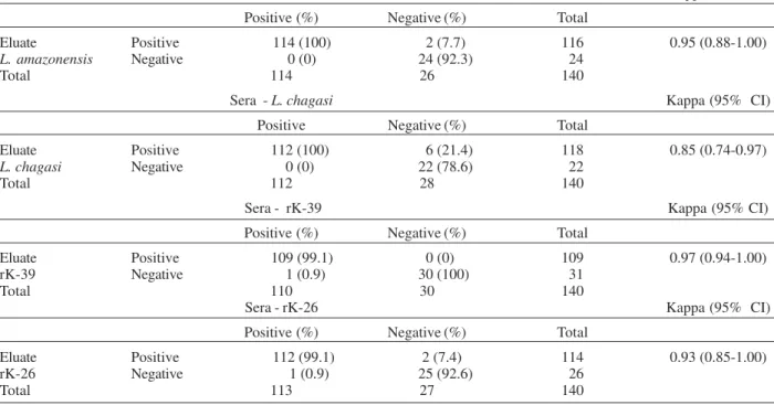
were positive and 25 healthy. Among the 115 IFAT-positive dogs the ELISA test performed with different antigens in sera identified 114 dogs (91.2%) with L. amazonensis, 112(89.6%) with L. chagasi, 110 (88.0%) for rK-39 and 113 (90.4%) for rK-26. Concordances between sera and eluates for different antigens are shown in Table III: L. amazonensis (Kappa = 0.95), L. chagasi (Kappa = 0.85), rK-39 (Kappa = 0.97), andrK-26 (Kappa = 0.93).
Correlations for absorbancy values between sera and eluate using each antigen were estimated by Pearson cor-relation. The absorbance results were transformed to log absorbance + 1. The Pearson correlations for the different
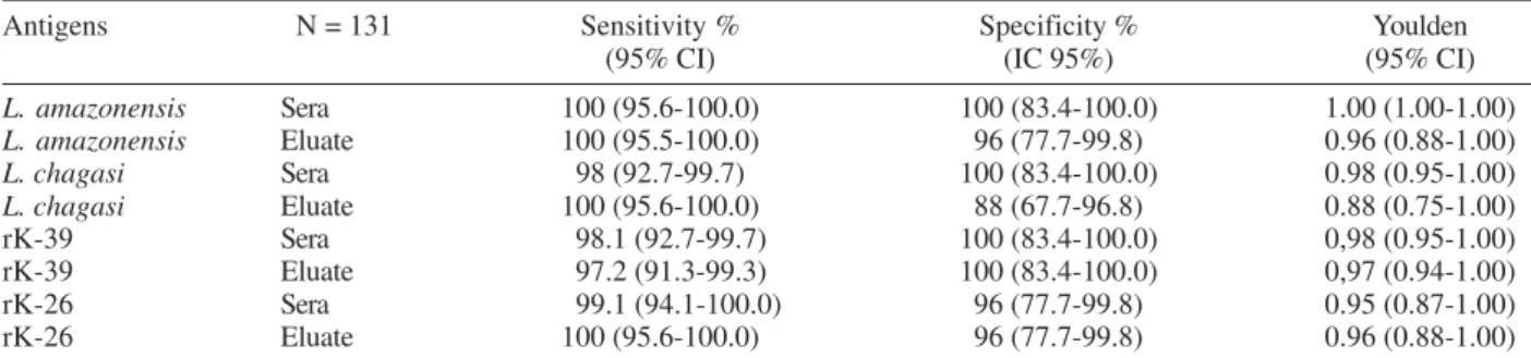
TABLE I
Sensitivity, specificity and Youlden coefficient (J) of ELISA performed with crude antigens and the recombinant antigens rK-39 and rK-26 in sera and eluates in diagnosis of canine visceral leishmaniasis
Antigens N = 131 Sensitivity % Specificity % Youlden
(95% CI) (IC 95%) (95% CI)
L. amazonensis Sera 100 (95.6-100.0) 100 (83.4-100.0) 1.00 (1.00-1.00)
L. amazonensis Eluate 100 (95.5-100.0) 96 (77.7-99.8) 0.96 (0.88-1.00)
L. chagasi Sera 98 (92.7-99.7) 100 (83.4-100.0) 0.98 (0.95-1.00)
L. chagasi Eluate 100 (95.6-100.0) 88 (67.7-96.8) 0.88 (0.75-1.00)
rK-39 Sera 98.1 (92.7-99.7) 100 (83.4-100.0) 0,98 (0.95-1.00)
rK-39 Eluate 97.2 (91.3-99.3) 100 (83.4-100.0) 0,97 (0.94-1.00)
rK-26 Sera 99.1 (94.1-100.0) 96 (77.7-99.8) 0.95 (0.87-1.00)
rK-26 Eluate 100 (95.6-100.0) 96 (77.7-99.8) 0.96 (0.88-1.00)
The results of sensitivity, specificity and Youlden coefficients were estimated with a 95% confidence interval; L: Leishmania
TABLE II
Comparison between clinical phases and ELISA performed with crude antigens of Leishmania amazonensis and L. chagasi and the recombinant antigens rK-39 and rK-26 in sera and eluates
ELISA test
L. amazonensis L. chagasi rK-39 rK-26
Clinical phases Sero (%) Eluate (%) Sero (%) Eluate (%) Sero (%) Eluate (%) Sero (%) Eluate (%)
Asymptomatic 100 100 98 100 93.7 93.7 98 100
(n = 48)
Oligoasymtomatic 97.8 100 97.8 100 95.6 95.6 97.8 95.6 (n = 46)
Symptomatic 100 100 95.2 100 100 95.2 100 100
(n = 21)
200 200 200 200
200 D iagnostic m arker for canine VL • Elisa Yoshie do Rosário et al.
antigens were: L. amazonensis (r = 0.76) and L. chagasi (r = 0.25). The recombinant antigens produced strong cor-relations between sera and eluate for rK-39 (r = 0.88) and rK-26 (r = 0.63).
Test reproducibility - To evaluate the reliability of the tests, 55 (33%) duplicate samples in sera and 28 (20%) duplicate samples in eluates for each antigen were ana-lyzed (Table IV). The agreement varied from 0.74-1.0 for the different antigens. The lowest agreement was
ob-served with ELISA performed with L. chagasi in sera
(Kappa = 0.74) and eluates (Kappa = 0.63). Highest agree-ment was detected for r-K39 antigens in both sera (Kappa = 0.81) and eluates (Kappa = 1.0).
DISCUSSION
Among the 115 dogs IFAT-positive at titers of 1:40, 106 (92.2%) were positive in parasitological exams and were considered to be truly positive, while 25 IFAT-nega-tive, clinically healthy animals were considered to be truly negative. Sensitivities were similar between crude and recombinant antigens and remarkably high for both sera and eluates (97-100%). Specificity of sera and eluates ex-ceeded 96% for all antigens exceptL. chagasi in eluate, for which the value was 88%. Concordances between the tests were higher either for sera or eluates (J = 0.95- 1.00) except for L. chagasi antigen ineluates (J = 0.88).
Similar sensitivity and specificity values in ELISA tests for CVL diagnosis were found by Mancianti et al. (1995), who reported higher sensitivity (99.5%) and specificity (97.1%) with L. infantum antigen. However, Ashford et al.
(1993) found sensitivity to be lower than 88% when they used L. chagasi antigen in FAST-ELISA performed with sera.
Sensitivityand specificity of ELISA is greatly influ-enced by the antigen used to perform the tests (Singh & Sivakumar 2003). Although the L. chagasi antigen is spe-cies-specific, in the present study its performance was slightly inferior to L. amazonensis in ELISA carried out with eluates. Crude antigensare traditionally derived from promastigotes cultivated in vitro and consist of a reper-tory of somatic antigens and several surface components that vary according to parasite species. The different spe-cies and strains that have been used in crude antigens may therefore limit standardization of tests and reproduc-ibility of results (Hommel et al. 1978, Mohammed et al. 1986, Burns et al. 1993).
TABLE III
Comparison between sera and eluates used in ELISA with crude and the recombinant antigens rK-39 and rK-26 to diagnose canine visceral leishmaniasis
Sera - L. amazonensis Kappa (95% CI)
Positive (%) Negative (%) Total
Eluate Positive 114 (100) 2 (7.7) 116 0.95 (0.88-1.00)
L. amazonensis Negative 0 (0) 24 (92.3) 24
Total 114 26 140
Sera - L. chagasi Kappa (95% CI)
Positive Negative (%) Total
Eluate Positive 112 (100) 6 (21.4) 118 0.85 (0.74-0.97)
L. chagasi Negative 0 (0) 22 (78.6) 22
Total 112 28 140
Sera - rK-39 Kappa (95% CI)
Positive (%) Negative (%) Total
Eluate Positive 109 (99.1) 0 (0) 109 0.97 (0.94-1.00)
rK-39 Negative 1 (0.9) 30 (100) 31
Total 110 30 140
Sera - rK-26 Kappa (95% CI)
Positive (%) Negative (%) Total
Eluate Positive 112 (99.1) 2 (7.4) 114 0.93 (0.85-1.00) rK-26 Negative 1 (0.9) 25 (92.6) 26
Total 113 27 140
CI: confidence intervals; L.: Leishmania
TABLE IV
Reproducibility of ELISA performed with crude and recombinants antigens rK-39 and rK-26 in sera and eluates to
diagnose canine visceral leishmaniasis
Antigens Serum duplicates Eluate duplicates Kappa (95% CI.) Kappa (95% CI)
L. amazonensis 0.76 (0,6-0.95) 0.84 (0.53-1.00)
L. chagasi 0.74 (0.54-0.95) 0.63 (0.15-1.00)
rK-39 0.81 (0.63-0,99) 1.00 (1.00-1.00) rK-26 0.74 (0.54-0.95) 0.84 (0.53-1.00)
201 201 201 201 201 Mem Inst Oswaldo Cruz, Rio de Janeiro, Vol. 100(2), April 2005
Several purified and recombinants have recentlybeen developed and tested, of which rK-39 antigen (Badaró et al. 1996) ishighly sensitive and predictive of the onset of disease manifestation in human VL patients. Badaró et al. (1996) and Scalone et al. (2002) found high sensitivity (98%) and specificity (99%) for CVL. However, there is some dispute about the predictive onset of the disease in CVL. Scalone et al. (2002), showed that rK-39 antigen did not predict the onset of disease manifestation while Rhalem et al. (1999) found that asymptomatic dogs were negative for rk-39 antigen. Scalone et al. (2002), consid-ered the efficacy of rK-39 in detecting asymptomatic cases to be markedly different between CVL and human VL.
In our study, crude and recombinant antigens detected the anti-leishmanial antibodies of mostasymptomatic and oligoasymptomatic dogs when ELISA was performed with sera. For asymptomatic dogs the highest success rate was observed for L. amazonensis antigen (100%), followed by L. chagasi and rK-26 antigens (both 98%). The lowest proportion was observed for rK-39 in eluate (93.7%). With respect to oligoasymptomatic dogs the proportions were 97.8% for L. amazonensis andL. chagasi and 95.6% for rK-26 and rK-39. The results obtained in ELISA with elu-ates for different antigens were similar to those observed in tests performed in sera. The proportion detected by rK-39 was similar to that found by Scalone et al. (2002) when they assayed asymptomatic dogs (92.4%).
Although both crude antigens (L. amazonensis and L. chagasi) showed high sensitivity, they presented
cross-reactivity with other microorganisms as T. cruzi, D.
reconditum, and B. canis. Thereare two primary draw-backs to tests performed with crude antigens, i.e., cross-reactivity and the inability to discriminate between past and current infections. The recombinant antigens rK-39 and rK-26 performed similarly whether in sera or eluate. They showed high sensitivity but no cross-reactivity with other microorganisms. High specificities of recombinant antigens have also been reported by other authors (Badaró et al. 1996, Scalone et al. 2002).
High concordances were observed between sera and eluates performed with different antigens (Kappa = 0.93-0.97). The lowest concordance was observed in L. chagasi antigen (Kappa = 0.88). According to Guimarães (1983),
errors in conducting tests with filter papers could occur for two possible reasonss : difficulties in precisely esti-mating eluate dilutions and denaturing of antibodies dur-ing filter paper storage at high environmental temperature and relative humidity. The good concordances observed in our study with filter paper are most likely: (1) homoge-neity of the blood spread on absorbent filter paper; (2) storage at –20°C and use of silica gel to control humidity; (3) elution of filter papers in casein-PBS on the same day that the tests were carried out.
The evaluation of testreproducibility revealed a good and regular agreement for ELISA in the duplication of elu-ates (Kappa = 0.63-1.00). Eluelu-ates of blood dried on filter paper are commonly used in laboratory tests and the va-lidity of this technique has been confirmed for the diag-nosis of leishmaniasis (Coutinho et al. 1985, Gomes et al. 2001) and for other infections such as toxoplasmosis and Chagas disease (Chiari et al. 1987, Machado-Coelho et al. 1995).
It is also important to note improvements in the leish-maniasis control program during recent decades in Brazil. The number of dog samples collected increased from 526 to 1,353,812 between 1980 and 1996 (Vieira & Coelho 1998). Collecting blood on filter paper makes collection, trans-port and storage of samples easier for local health ser-vices (Guimarães 1983).
The use of only one antigen to perform both condi-tion screening and confirmatory tests could increase the proportion of false-positive results, particularly in cases of low prevalence (Gart & Buck 1966, Costa & Vieira 2001). One important point that merits consideration is the need to standardize antigens with respect to source and purity in control programs, in order to improve the reproducibil-ity of results in different regions of Brazil.
In summary, the results of this investigation showed that ELISA using crude and recombinant antigens gave results to similar degrees of sensitivity and specificity in identifying different clinical phases of CVL. High concor-dance among sera and eluates confirm that filter paper could be used to collect blood in canine surveys to iden-tify L. chagasi infection. Use of recombinant antigens did not markedly improve results compared with use of crude antigen in dogs identified as being positive. However, the main limitation of crude antigens is their cross-reactivity with other diseases that often occur in VL-endemic areas. The association of crude and recombinant antigens is a strategy that could be used in control programs. This could involve an initial screening using ELISA with crude anti-gen in eluate from blood collected on filter paper, followed by a confirmatory test using a recombinant antigen also in eluate. However recombinant antigens would need to be widely available. Recombinant antigens could also improve differential diagnosis in clinical veterinary medi-cine.
ACKNOWLEDGEMENTS
To Fundação Nacional de Saúde, Minas Gerais, and Dr Marília Rocha, Centro de Controle de Zoonoses de Montes Claros, Minas Gerais for cooperation and logistical support. To Heska Corporation, US for providing the recombinants an-tigens rK-39 and rK-26 and Dr Terezinha Bahia and Dr Gilberto Fontes for providing the serum of dogs infected with other pathogens. The Conselho Nacional de Desenvolvimento Cientifico e Tecnológico sponsored EY Rosario scholarships (Master Degree - Programa de Pós-Graduação em Parasitologia, Universidade Federal de Minas Gerais).
REFERENCES
Arias JR, Monteiro PS, Zicker F 1996. The reemergence of visceral leishmaniasis in Brazil. Emerg Infect Dis2: 145-146.
Ashford D, Badaró R, Eulálio C, Freire M, Miranda C, Zalis MG, David JR 1993. Studies on the control of visceral leishmanisis: validation of the falcon assay screening test-enzyme-linked immunosorbent assay (FAST-ELISA) for field diagnosis of canine visceral leishmaniasis. Am Soc Trop Med Hyg48: 1-8.
Leishma-202 202 202 202
202 D iagnostic m arker for canine VL • Elisa Yoshie do Rosário et al.
nia chagasi that predicts active visceral leishmaniasis. J Infect Dis173: 758-761.
Bhatia A, Daiffalla NS, Jen S, Badaró R, Reed SG, Skeiky YAW 1999. Cloning, characterization and serological evaluation of K39 and K26: two related hydrophilic antigens of Leish-mania chagasi. Mol Biochem Parasitol 102: 249-261.
Braga MDM, Coelho ICB, Pompeu ML, Evans TG, MacAullife IT, Teixeira MJ, Lima JWO 1998. Controle do calazar canino: comparação dos resultados de um programa de eliminação rápida de cães sororreagentes por ensaio imunoenzimático com outro de eliminação tardia de cães sororreagentes por teste de imunofluorescência indireta de eluato de papel filtro. Rev Soc Bras Med Trop 31: 419-424.
Braz RF, Nascimento ET, Martins DR, Wilson ME, Pearson RD, Reed SG, Jeronimo SM 2002. The sensivity and speci-ficity of Leishmania chagasi recombinant K39 antigen in the diagnosis of American visceral leishmaniasis and in dif-ferentiating active from subclinical infection. Am J Trop Med Hyg67: 344-348.
Burns JM, Shreffer WG, Benson DR, Ghalib HW, Badaró R, Reed SG 1993. Molecular characterization of a kinesin-related antigen of Leishmania chagasi that detects specific antibody in African and American visceral leishmaniasis.
Proc Natl Acad Sci USA90: 775-779.
Carvalho SF, Lemos EM, Corey R, Dietze R 2003. Perfor-mance of recombinant K39 antigen in the diagnosis of Bra-zilian visceral leishmaniasis. Am J Trop Med Hyg68: 321-324.
Chiari CA, Antunes CMF, Lima JD 1987. Utilização do eluato de sangue dessecado em papel filtro no diagnóstico sorológico da toxoplasmose caprina. Rev Bras Med Vet Zootec39: 471-483.
Costa CHN, Vieira JBF 2001. Changes in the control program of visceral leishmaniasis in Brazil. Rev Soc Bras Med Trop 34: 223-228.
Coutinho SG, Nunes MP, Marzochi MCA, Tramontano N 1985. A survey for American cutaneous and visceral leish-maniasis among 1.342 dogs from areas in Rio de Janeiro (Brazil) where the human diseases occur. Mem Inst Os-waldo Cruz80: 17-22.
Deane L, Deane M 1962. Visceral leishmaniasis in Brazil. Geo-graphic distribution and transmission. Rev Inst Med Trop São Paulo 4: 149-212.
Evans TG, Vasconcelos IAB, Lima JW, Teixeira JM, McAullife IT, Lopes UG, Pearson RD, Vasconcelos AW 1990. Canine visceral leishmaniasis in northeast Brazil: assessment of serodiagnosis methods. Am J Trop Med Hyg42: 118-123.
França-Silva JC, Costa RT, Siqueira AM, Machado-Coelho GLL, Costa CA, Mayrink W, Vieira EP, Costa J, Genaro O, Nascimento E 2002. Epidemiology of canine visceral leish-maniosis in the endemic area of Montes Claros municipal-ity, Minas Gerais state, Brazil. Vet Parasitol2474: 1-13.
Gart JJ, Buck AA 1966. Comparison of a screening test and a reference test in epidemidemiologic studies. II. A probabi-listic model for the comparison of diagnostic tests. Am J Epidemiol 83: 593-602.
Gomes HR, Rodrigues MS, Silva MP, Nascimento EG, Moreira ED, Pontes-de-Carvalho LC, Santos WLC 2001.
Com-paração entre ELISA de soro e de eluato de sangue para o imunodiagnóstico da leishmaniose visceral canina (LVC).
Rev Soc Bras Med Trop34(Suppl. I): 197.
Guimarães MCS 1983. Inquéritos soroepidemiológicos. Coleta, transporte e armazenamento de amostras. Rev Inst Med Trop São Paulo25: 93-96.
Hommel M, Peters W, Ranque J, Quilici M, Lanotte G 1978. The micro-ELISA technique in the serodiagnosis of visceral leishmaniasis. Ann Trop Med Parasitol72: 213-218.
Lanotte G, Rioux JA, Vollhardt Y 1979. Ecologie des leishmani-oses dans le sud de la France. Les formes evolutives de la leishmaniose canine Ann Parasitol Hum Comp54: 277-295
Machado-Coelho GLL, Vitor RWA, Chiari CA, Antunes CMF 1995. Validity of serological for American trypanosomiasis with eluates from filter paper. Mem Inst Oswaldo Cruz90: 59-64.
Mancianti F, Gramiccia M, Gradoni L, Pieri S 1988. Studies on canine leishmaniasis control. 1. Evolution of infection of different clinical forms of canine leishmaniasis following antimonial treatment. Trans R Soc Trop Med Hyg82: 566-567.
Mancianti F, Falcone ML, Giannelli C, Poli A 1995. Compari-son between an enzyme-linked immunosorbent assay using a detergent-soluble Leishmania infantum antigen and indi-rect immunofluorescence for the diagnosis of canine leish-maniasis. Vet Parasitol 59: 13-21.
Mancianti F, Pedonese F, Poli A 1996. Evaluation of dot en-zyme-linked immunosorbent assay (dot-ELISA) for the serodiagnosis of canine leishmaniosis as compared with in-direct immunofluorescence assay. Vet Parasitol65: 1-9.
Marzochi MCA, Coutinho SG, Souza WJS, Toledo LM, Grimald Jr. G, Momen H, Pacheco RS, Sabrosa PC, Souza MA, Rangel Jr. FB, Tramontano N 1985. Canine visceral leish-maniasis in Rio de Janeiro, Brazil. Clinical, parasitological, therapeutical and epidemiological findings (1977-1983).
Mem Inst Oswaldo Cruz80: 349-357.
Mohammed EAER, Wright EP, Rahman AMB, Kolk A, Laarman JJ, Pondman KW 1986. Serodiagnosis of sudanese visceral and mucosal leishmaniasis: comparison of ELISA, immun-ofluorescence and indirect haemagglutination. Trans R Soc Med Hyg80: 271-274.
Molina R, Amela C, Nieto J, San-Andres M, Gonzalez F, Castillo JA, Lucientes J, Alvar J 1994. Infectivity of dogs naturally infected with Leishmania infantum to colonized Phleboto-mus perniciosus. Trans R Soc Trop Med Hyg88: 491-493.
Nunes MP, Jackson JM, Carvalho RW, Furtado NJ, Coutinho SG 1991. Serological survey for canine cutaneous and vis-ceral leishmaniosis in area for transmission in Rio de Janeiro where prophylactic measures had been adapted. Mem Inst Oswaldo Cruz86: 411-417.
Paranhos-Silva M, Freitas LAR, Santos WC, Grimaldi Jr G, Pontes-de-Carvalho LC, Oliveira-dos-Santos AJ 1996. A cross-sectional serodiagnosis survey of canine leishmania-sis due to Leishmania chagasi. Am J Med Hyg 55: 39-44.
Pozio E, Gradoni S, Bettini S, Gramicia M 1981. Leishmaniasis in Tuscany (Italy): VI. Canine leishmaniasis in the focus of Monte Argentario (Grosseto). Acta Trop38: 383-393.
203 203 203 203 203 Mem Inst Oswaldo Cruz, Rio de Janeiro, Vol. 100(2), April 2005
Riyard M, Berrag B 1999. Immune response against Leish-mania antigens in dogs naturally and experimentally in-fected with Leishmania infantum. Vet Parasitol81: 173-184.
Roffi J, Dedet JP, Desjeux P, Garré MT 1980. Detection of circulating antibodies in cutaneous leishmaniasis by enzyme-linked immunosorbent assay (ELISA). Am J Trop Med Hyg29: 183-189.
Scalone A, De Luna R, Oliva G, Baldi L, Satta G, Vesco G, Mignone W, Turilli C, Mondesire RR, Simpson D, Donoghue AR, Frank GR, Gradoni L 2002. Evaluation of
Leishmania recombinant K39 antigen as a diagnostic marker for canine leishmaniasis and validation of a standardized enzyme-linked immunosorbent assay. Vet Parasitol 104: 275-285.
Singh S, Sivakumar R 2003. Recent advances in the diagnosis of leishmaniasis. J Postgrad Med 49: 55-60.
Szklo M, Nieto FJ 2000. Epidemiology: Beyond the Basis. Aspen Publishers, Inc., Gaithersburg, 495 pp.