Sputum culture yield: comparing an automated
diagnostic system to Löwenstein-Jensen medium in the
diagnosis of pulmonary tuberculosis*
ELISABETE APARECIDA DE ALMEIDA, MANOEL ARMANDO AZEVEDO DOS SANTOS, JORGE BARROS AFIUNE, DELURCE TADEU DE ARAÚJO SPADA, FERNANDO AUGUSTO FIUZA DE MELO**
*Study carried out at the Instituto Clemente Ferreira - Coordenadoria de Contole de Doenças - Secretaria de Estado da Saúde - São Paulo **Specialist, as designated by the Sociedade Brasileira de Pneumologia e Tisiologia
Correspondence to: Elisabete Ap. Almeida. Rua da Consolação, 717. CEP: 01301-000, São Paulo, SP. Phone: 55 11 3257-0624. E-mail: [email protected]
Submitted: 18 May 2004. Accepted, after review:20 January 2005
J Bras Pneumol 2005; 31(3): 231-6.
Key words: Mycobacterium Tuberculosis. Diagnostic. Automated systems. Background: Tuberculosis continues to be a global health problem.
Objective: To evaluate an automated system designed to diagnose tuberculosis, comparing it to sputum microscopy and culture in Löwenstein-Jensen medium.
Method: A comparative study using 844 sputum samples, collected between September and December of 1999 at a reference center for tuberculosis in São Paulo, Brazil, to draw distinctions between the results obtained through the use of the automated system and those obtained through sputum microscopy and culture in Löwenstein-Jensen medium.
Results: Of the 844 samples evaluated, 27.1% tested positive for acid-fast bacilli, and 72.9% tested negative. In Löwenstein-Jensen culture, 34.7% were positive and 63% were negative, compared with 37.1% positivity and 56.9% negativity using the automated system. Sensitivity was 98.1% for the automated system and 91.9% for Löwenstein-Jensen culture. Specificity and positive predictive value were 100% for both methods. Negative predictive value was 98.9% for the automated system and 95.5% for Löwenstein-Jensen culture. The degree of accuracy was 99.3% for the automated system and 97% for Löwenstein-Jensen culture, and the Kappa was 0.99 for the automated system and 0.94 for Löwenstein-Jensen culture. The difference between the mean time to detection of mycobacteria using the automated system (10.5 days) and that found using Löwenstein-Jensen culture (34.7 days) was statistically significant.
Conclusion: The difference between the culture yield obtained using the automated system and that achieved with Löwenstein-Jensen culture was statistically significant. Mean time to detection of mycobacteria was significantly shorter with the automated system. The higher yield provided by this new system justifies its use in a reference center for tuberculosis in São Paulo
INTRODUCTION
The idea that tuberculosis (TB) could be controlled with the advances attained in the middle of the last century prevailed until recently(1). The
re-emergence of TB has been observed in developed countries, and the situation has worsened in developing countries, leading the World Health Organization to declare TB a "global emergency" in its annual report of April 1993(2-4).
In Brazil, TB cannot be considered a re-emergent disease because it never disappeared. Neither is it an emergent disease since the high level of incidence has been maintained. It may be said that Brazil inhabits an intermediate terrain between the situation seen in developing countries and the severe situation observed in a number third world countries such as some in Africa. In the 1980s, it was expected that TB incidence in Brazil would decrease steadily and progressively (by approximately 4% to 6% per year). However, in the early 1990s, a lower rate of decrease was observed, with a discrete worsening of morbidity and mortality, especially in the states where rates of TB/human immunodeficiency virus coinfection were higher(4-7).
The Brazilian Tuberculosis Control Program is based on the sputum smear microscopy and culture, and states that the presumptive diagnosis should be based on clinical and radiographic findings of the disease. The results obtained, although sufficient from the epidemiological point of view, are nonspecific, not allowing, in isolation, a conclusive diagnosis of TB. More complex bacteriological tests are necessary for the definition of the etiology(7-9).
In laboratory testing, the most important contribution to the final diagnosis is made by identification of the bacillus, either through direct examination (sputum smear microscopy) or through its isolation in culture (culture medium). Mycobacteria can be cultivated in either solid or liquid media. In Brazil, the Lowenstein-Jensen (LJ) medium is considered the standard. The average time to formation of M. tuberculosis colonies on LJ medium is four weeks, this being its greatest disadvantage(7).
Currently, the issue of diagnosis has become more complicated due to the symbiosis between TB and human immunodeficiency virus, as well as to the emergence of multidrug-resistant forms. The classic resources for the diagnosis of TB no longer meet the demand. There is a need for more precise and faster laboratory resources.
Advances in technology have spawned automated methods such as the BACTEC 460 System, a radiometric
system which utilizes Middlebrook 7H9 liquid culture medium with Carbon-14 palmitic acid. This method has the considerable disadvantage of utilizing radioactive material. The automated system that measures oxygen consumption, the BACTEC® Mycobacteria Growth Indicator TubeTM 960 (BM960), utilizes the same culture medium but has a fluorescent sensor that reacts to the concentration of oxygen in culture medium(9,10).
The objective of this study was to determine the performance and reliability of an automated diagnostic system which measures oxygen consumption for the laboratory diagnosis of pulmonary TB, comparing it to smear microscopy and culture on LJ medium, with the goal of assessing its implementation in a referral center for the disease in the city of São Paulo (SP).
METHODS
Between September and December 1999, 851 sputum samples of patients were examined in the Mycobacteriology Laboratory of the Instituto Clemente Ferreira in the city of São Paulo. Smear microscopy for acid-fast bacilli was performed using the Ziehl-Neelsen method (on LJ medium) and the BM960 oxygen consumption monitor system (BACTEC MGIT 960; Becton Dickinson, Sparks, MD, USA). A total of 7 samples were excluded after being identified as nontuberculous mycobacteria.
S p u t u m s a m p l e s w e r e e x a m i n e d a f t e r decontamination using the Petroff method(11), with
smear microscopy for acid-fast bacilli and culture on LJ medium according to the guidelines established by the Brazilian Ministry of Health(12).
Culture in the oxygen consumption monitor used Middlebrook 7H9 broth medium enriched with oleic acid-albumin-dextrose-catalase as well as with polymyxin B-amphotericin B-nalidixic acid-trimethoprim-azlocillin. Culture tubes contain fluorescent ruthenium salt sensors, which emit light when concentrations of oxygen, in this case consumed by the bacilli, are low. A photoelectric cell assesses the luminescence of the tubes once every hour, registering the positive results upon light emission(9). The positive results were confirmed by a
cultures in the oxygen consumption monitor medium, which tested positive in subculture on LJ medium.
Mycobacterium species were identified through recent culture on solid medium. To that end, a 0.1 bacterial suspension was seeded on LJ culture medium containing p-nitrobenzoic acid, and in another tube with thiophene-2-carboxylic acid hydrazide. The vials were incubated at 37°C for 28 days. After this time, the inoculates which were sensitive to the presence of p-nitrobenzoic acid (presenting no growth) and resistant to thiophene 2 carboxylic acid hydrazide (presenting growth)(12) were interpreted as belonging to the M.
tuberculosis complex.
Contamination rates between the two culture systems were statistically assessed using the McNemar test and the time to M. tuberculosis growth was analyzed in each of them using the Student's t-test. The study was approved by the Ethics in Human Research Committee of the Instituto de Ciências Biomédicas da Universidade de São Paulo (University of São Paulo Institute of Biomedical Sciences). There was no conflict of interest since the present study was funded entirely by the Clemente Ferreira Institute.
RESULTS
During the pre-established study period, 851 sputum samples were collected, 7 of which were excluded for presenting nontuberculous mycobacteria. Therefore, 844 samples from 489 Instituto Clemente Ferreira patients were analyzed, most of which were submitted to more than one test.
Smear microscopy results for acid-fast bacilli among the 844 sputum samples were: 229 positive (27.1%) and 615 negative (72.9%). On LJ culture medium, 293 samples were positive (34.7%) and 532 negative (63.0%), whereas in the oxygen consumption monitor medium 313 (37.1%) were positive and 480 (56.9%) were negative (Table 1).
Regarding the contaminated cultures, 19 occurred on LJ medium (2.3%) and 51 in the oxygen consumption monitor medium (6.0%), the latter being higher, a statistically significant difference (p < 0.0001, McNemar test) (Table 2).
Of the 51 contaminated oxygen consumption monitor medium cultures, only 1 sample was also contaminated on LJ medium. Likewise, of the 19 cultures contaminated on LJ medium, only the above
TABLE 1
Distribution of results of the analysis of 844 sputum samples of 489 patients treated at the Instituto Clemente Ferreira, in São Paulo, from September to December of 1999, provided by sputum smear
microscopy (Ziehl-Neelsen), culture (Lowenstein Jensen) and automated diagnostic system
Sputum smear microscopy LJ medium BM960
N % N % N %
Positive 229 27,1 293 34,7 313 37,1
Negative 615 72,9 532 63,0 480 56,9
Contaminadet - - 19 2,3 51 6,0
Total 8 4 4 1 0 0 8 4 4 100 844 100
LJ: Lowenstein Jensen; BM960: automated diagnostic system
TABLE 2
Comparative results regarding contamination on Lowenstein Jensen medium and through automated diagnostic system of the analysis of 844 sputum samples from 489 patients treated at the Instituto
Clemente Ferreira, in São Paulo, from September to December of 1999
Contaminated BM960 Uncontaminated BM960 TOTAL
Contaminated LJ 1 18 9 (2,3%)
Uncontaminated LJ 50 775 825 (97,7%)
TOTAL 51 (6,0%) 793 (94%) 844 (100%)
p < 0.0001 (McNemar test)
mentioned sample was contaminated in the oxygen consumption monitor medium.
The contaminated cultures were excluded from the global results, resulting in a total of 775 samples, of which 289 presented LJ medium positivity and 307 oxygen consumption monitor medium positivity, with discordance in 28 specimens (5 cultures considered negative in the oxygen consumption monitor medium and 23 on LJ medium). Therefore, the oxygen consumption monitor system recovered 18 samples that had tested negative on LJ medium (Table 3).
In the case of more than one culture per patient, the result was considered positive when at least one culture was positive and negative only when all were negative. With regard to the 161 patients diagnosed from culture (158 positive in the oxygen consumption monitor medium and 3 positive on LJ medium), the samples were typified and the charts were assessed and found to be in accordance with the diagnosis of pulmonary TB. The 3 samples in disagreement presented smear microscopy that was positive in 2 cases and negative in 1 case (Table 4).
Statistical calculations demonstrated sensitivity of 98.1% for the oxygen consumption monitor medium and 91.9% for LJ medium. Specificity and positive predictive value were 100% for both methods. Negative predictive value was 98.9% for the oxygen consumption
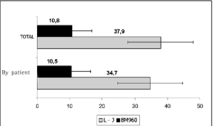
monitor medium and 95.5% for LJ medium. Accuracy was 99.3% for the oxygen consumption monitor medium and 97% for LJ medium. Kappa statistics (corrected degree of concordance) was 0.99 for the oxygen consumption monitor medium and 0.94 for LJ medium. Time to M. tuberculosis detection was analyzed in all samples and by patient, with statistically significant differences between the two culture media tested (p < 0.001). The average growth time in the oxygen consumption monitor medium was 10.8 days (90% ranging from 4 to 16 days) and 10.5 days (90% ranging from 5 to 15 days) for all 844 samples and for the 438 per-patient samples, respectively, compared with 37.9 days (90% ranging from 29 to 45 days) and 34.7 days (90% ranging from 28 to 42 days) on LJ medium. Of the excluded samples, 51 presented contaminated oxygen consumption monitor medium (Figure 1).
In general, a reduction of more than one-third was observed in the growth time of the mycobacteria in the oxygen consumption monitor medium compared to the culture on LJ solid medium. A variation of 2 to 42 days (system limit) was found for the oxygen consumption monitor medium versus 23 to 60 days (established limit) for the LJ medium.
DISCUSSION
TABLE 3
Comparative results of Lowenstein Jensen medium and an automated diagnostic system in the analysis of 775* sputum samples from 489 patients treated at the Instituto Clemente Ferreira, in
São Paulo, from September to December of 1999
Positive BM960 Negative BM960 TOTAL
Positive LJ medium 284 (92,5%) 05 (1,1%) 289 (37,3%)
Negative LJ medium 23 (7,5%) 463 (98,9%) 486 (62,7%)
TOTAL 307 (100%) 468 (100%) 775 (100%)
*Contaminated cultures were excluded p = 0.001 (McNemar test)
LJ: Lowenstein Jensen; BM960: automated diagnostic system
TABLE 4
Comparative results of sputum culture on Lowenstein Jensen medium and through an automated diagnostic system of the analysis of 438* patients treated at the Instituto Clemente Ferreira, in
São Paulo, from September to December of 1999
Positive BM960 Negative BM960 TOTAL
Positive LJ medium 145 (91,8%) 3 (1,1%) 148 (33,8%)
Negative LJ medium 13 (8,2%) 277 (98,9%) 290 (66,2%)
TOTAL 158 (100%) 280 (100%) 438 (100%)
*Contaminated cultures were excluded and, in the case of multiple cultures per patient, the result was considered positive when at least one of the results was positive
In the middle of the twentieth century, TB was brought under control in developed countries, therefore becoming primarily restricted to developing countries. Until recently, the conception prevailed that TB could be controlled with the existing resources: simple and economical diagnostic methods and potent chemotherapy(1).
In the last two decades, however, the TB situation has undergone significant changes. Some of the contributing factors were the appearance of the acquired immunodeficiency syndrome pandemic (with profound implications for TB epidemiology) and increased poverty (and the consequential low educational levels, poor-quality housing, large families, high density communities, malnutrition, alcoholism and associated infectious diseases). In addition, lack of investment in health care and deterioration of the medical system infrastructure (a situation more commonly found in third world countries) have made it difficult to gain access to treatment. Furthermore, elderly patients infected with TB during the era of high TB incidence have increased the rates of TB prevalence in developed countries by virtue of increased longevity, constituting sources of infection not found previously. Moreover, migration from poor countries to rich countries has resulted in globalization of the disease. Along with other infirmities, these migrants bring with them endemic diseases, including TB(2-7).
In a way, the recent advances in diagnostic methods have come about as a result of the reemergence of TB in advanced countries, which have the technical resources for such development, impelled by the necessity of
obtaining faster results from tests that identify the mycobacterium species involved and its resistance profile, especially in view of the need for procedures to deal with the emergence of acquired immunodeficiency syndrome. The implementation of these methods in routine practice, however, should always undergo validation of its applicability in a certain region, defining its efficiency in relation to patterns of use and cost-benefit ratios, especially in developing countries(13).
The efficiency of the oxygen consumption monitor medium was assessed in a high-traffic referral center serving patients with complicated diagnoses and therapeutic problems of varying natures, comparing it to the traditional methods in use, such as smear microscopy for acid-fast bacilli and culture on LJ medium.
One of the motivations for this study was the number of tests carried out over a short period of time, whether for diagnosis or for treatment control. The oxygen consumption monitor system represents an advance in the means of rapid detection of M. tuberculosis. It replaces the radiometric system known as BACTEC 460, designed in 1977, which detects mycobacteria through culture. Results are obtained within an estimated 9 to 14 days for samples of the M. tuberculosis complex and in less than 7 days for those of atypical mycobacteria. The BACTEC 460 measures the concentration of radioactive carbon dioxide produced by mycobacteria in Middlebrook 7H9 medium enriched with carbon-14 palmitic acid(14).
In addition to the use of radioactive material, the method involves needle inoculation.
The new oxygen consumption monitor system has the additional advantages of having a nonradioactive methodology, assessing mycobacterial growth through oxygen production (measured by the fluorescence emitted by ruthenium salt), being automated and measuring concomitantly more than twice the number of samples measured by its predecessor.
In Brazil, there have been articles and studies regarding BACTEC 460 and, in isolation, the Mycobacteria Growth Indicator Tube (oxygen consumption monitor) system(15-17). However, to date,
there have been no studies addressing the validation or applicability of the oxygen consumption monitor system. Nevertheless, in the international literature, there are several reports on the use of this system, some comparing it with the LJ medium(18,19).
In the analysis of the 844 sputum samples evaluated in the present study, the differences between the positive results obtained by the two methods were statistically significant, with positivity percentages of Figure 1. Time, in days, to growth of mycobacteria colonies in the BM960
(oxygen consumption system) medium and on Lowenstein-Jensen (LJ) medium in all samples and by patient
in the oxygen consumption monitor medium, a significance also found in cultures individualized by patient (n = 438), of 36% and 34%, respectively. These results are consistent with those of international studies, which have documented the same similarity(18,19) (Tables 1 and 2).
Of the 5 discordant samples referenced in Table 2 (total sampling), in which the oxygen consumption monitor medium culture was negative and the LJ medium culture was positive, 4 presented only a few colonies grown on LJ medium, 2 presented negative smear microscopy, and 2 tested positive with few bacilli. Only 1 of the 5 samples presented smear microscopy (++) with growth on LJ medium (+) (from 20 to 100 colonies).
Among the 3 discordant cases shown in Table 4 (one sample per patient), smear microscopy was positive in 2 and negative in 1. One of the positive smear microscopy samples presented few colonies on LJ medium, and the other (with ++ smear microscopy) presented a (+) result on LJ medium.
Since it utilizes a medium richer in nutrients, the oxygen consumption monitor system tends to present higher contamination rates than does the LJ solid medium, as evidenced by the fact that, in the present study, we found a significantly higher contamination rate in the oxygen consumption monitor system medium (5l/844) than on LJ medium (19/844). There are studies in the literature, such as the one by Chien et al.(20), in which no such difference was found. This
discordance may be related to the characteristics of the clientele treated in the center, where chronic patients, protracted diagnoses and extensive lesions may facilitate contamination.
Another important finding was that the time to detection of M. tuberculosis was much shorter in the oxygen consumption monitor medium. The average time was 10.8 days for all samples and 10.5 days for per-patient samples, less than one-third of the average 37.9 days and 34.7 days, respectively, required on LJ medium (Figure 1).
In this study, the efficiency of the culture in the oxygen consumption monitor medium presented significant similarity with the LJ medium. In some cases, the oxygen consumption monitor system even presented higher sensitivity and specificity. The average time to detection of mycobacteria was significantly shorter in the oxygen consumption monitor medium than on the LJ medium, which has been adopted as the standard.
We can conclude that the efficiency of the oxygen consumption monitor system justifies its implementation in the operational protocols of a laboratory at an outpatient referral center for TB in the city of São Paulo.
1. Fox W, Mitchison DA. Quimioterapia de la tuberculosis. [Opas, publicacion cientifica, 310]. Washington: OPAS/OMS, 1975.
2. World Heath Organization. TB: a global emergency. [WHO.TB 94.177] Geneve, 1994.
3. Chaisson RE, Benson CA. Tuberculosis and HIV infection.
In: Rossman MD, MacGregor RR. Tuberculosis: clinical
mangement and new challenges. New Yourk: McGraw-Hill; 1996:Chap.14:223-238.
4. Rosemberg J. Tuberculose. Panorama global. Óbices para seu controle. Fortaleza: Secretaria de Estado da Saúde do Ceará; 1999.
5. Ruffino Netto A. Brasil e a Tuberculose Doença Emergente ou Reemergente? Bol Pneumol Sanit 1997;5:3.
6. Campos HS, Fiuza de Melo FA. Efetividade do esquema 3 (3SZEEt/9EEt) no retratamento da tuberculose na rotina das Unidades de Saúde. Bol Pneumol Sanit 2000;8:7-14. 7. Ministério da Saúde FUNASA Centro de Referência Prof. Hélio
Fraga – Sociedade Brasileira de Pneumologia e Tisiologia. Controle da tuberculose: uma proposta de integração ensino-serviço. 5a Rio de Janeiro; 2002: Chap. 5:61-96. 8. Fiuza de Melo FA, Afiune JB, Kritski AL, Seiscento M, Hijjar
MA. Tuberculose. In: Veronesi R, Focaccia R, editors. Tratado
de infectologia. São Paulo: Atheneu; 1996. p. 914-59. 9. Hanna BA. Diagnosis of tuberculosis by microbiologic
techiniques. In: Rom WN, Garay SM, editors. Tuberculosis.
New York: Little, Brown and Co; 1995; Cap.11: 149-60. 10. Becton-Dickson. Manual do usuário, BACTEC® MGIT™;1998. 11. Petroff AS. A new rapid method for the isolation and cultivation of tubercle bacille directly from sputum and fees. J Exp Med 1915.;21:38.
12. Ministério da Saúde/FUNASA/Centro de Referência Prof. Hélio Fraga. Manual de bacteriologia da tuberculose 2a. ed. Rio de Janeiro; 1994.
13. Kritski AL, Conde MB, Souza GRM. Tuberculose: do ambulatório a enfermaria. São Paulo: Atheneu; 1999. 14. Becton-Dickson. Manual do usuário, BACTEC
460-System®;1985.
15. Palaci M, Ueki SYM, Sato DN, Telles MAS, Curcio M, Silva EAM. Evaluation of Mycobacteria Growth Indicator Tube for recovery and drug susceptibility testing of Mycobacterium tuberculosis isolates from respiratory specimens. J Clin Microbiol 1996;34:762-4.
16. Machado AMO. Avaliação de meio de cultura líquido BBL
Mycobacteria Growth Indicator Tube (MGIT) em rotina de detecção de micobactérias em amostras de escarro de pacientes com suspeita de tuberculose pulmonar. [tese]. São Paulo: Escola Paulista de Medicina, Universidade do Estado de São Paulo; 1998.
17. Martins FM, Caldas PC, Barreto AMW. Avaliação do antibiograma pelo método radiométrico para M. tuberculosis.
Livro de Resumos do XX Congresso Brasileiro de Microbiologia. VIII Simpósio Brasileiro de Micobactérias 1999; :432. 18. Oplustil CP, Sinto SI, Marins M, Mendes CMF. Avaliação de
um novo sistema para detecção de micobactérias:
Mycobacterium Growth Indicator tube (MGIT). J Bras Patol
1997;3:70-5.