PRODUCTION AND PURIFICATION OF AN ENDO–1,4-
βββββ
-XYLANASE FROM
HUMICOLA GRISEA VAR. THERMOIDEA BY ELECTROELUTION
Rubens Monti
1*; Leonardo Cardello
1; Marcos F. Custódio
1; Antonio J. Goulart
1; Adriana H. Sayama
1; Jonas Contiero
21
Departamento de Alimentos e Nutrição, Faculdade de Ciências Farmacêuticas de Araraquara, Universidade Estadual Paulista,
Araraquara, SP, Brasil;
2Departamento de Bioquímica e Microbiologia, Instituto de Biociências de Rio Claro, Universidade
Estadual Paulista, Rio Claro, SP, Brasil.
Submitted: September 03, 2002; Returned to authors for corrections: January 07, 2003; Approved: May 06, 2003
ABSTRACT
Humicola grisea
var. thermoidea produces two forms of extracellular xylanase. The component form 1 was
purified using the electroelution method, due to the very small production of this extracellular enzyme. The
apparent molecular mass was 61.8 kDa by SDS-PAGE.
Key words: electroelution, enzyme purification, extracellular xylanase, Humicola grisea var. thermoidea
INTRODUCTION
A wild type strain of a thermophilic fungus isolated from
Brazilian soil was identified as
Humicola grisea
var. thermoidea
by Monti
et al.
(8). This stain produces several extracellular
enzymes (3,4,10,15,16). Besides cellulose, xylans are the most
abundant renewable polysaccharides (2,5,12). Endo-1,4-
β
-D-xylanases (EC 3.2.1.8) are produced by numerous
microorganisms and several xylanases from fungus have been
purified and biochemically characterized. Purkarthofer
et al.
(13)
suggested that the use of xylanases in pulp and paper industry
will probably be one of the major achievements of the
biotechnology. Furthermore, Poutanen (11) observed that
endogenous and added enzymes have an important effect on
the quality of cereal foods. Great progress has been reached
due to the result of the studies on xylanolytic enzymes in wheat
and germinated wheat.
There are two ways in which macromolecules separated by
gel electrophoresis can be purified for further study. The first
involves performing electrophoresis in an identical way as that
used for purely analytical evaluation and then, once the
components of interest have been located, the gel is cut up and
the material extracted. The second consists of allowing the
components to migrate the whole length of the gel and then
* Corresponding author. Mailing address: Rodovia Araraquara–Jaú, Km 1, Caixa Postal 502. 14801-902, Araraquara, SP, Brasil. Fax: (+5516) 222-0073, E-mail: montiru@fcfar.unesp.br
collecting them at the end, usually by some suitable elution
technique. Some workers refer to only the latter as preparative
electrophoresis. Extraction of separated material can be achieved
using either stained or unstained gels in three principal ways:
simple extraction with an appropriate buffer; solubilization of
the gel matrix; electrophoretic elution (1). The aim of the present
study was to purify xylanase form 1 from this microorganism by
electroelution. This method showed to be efficient for the
purification of microbial enzymes up to homogeneity, when they
are produced in small amounts.
MATERIALS AND METHODS
Microorganism
The fungus was maintained on slants containing 4% of
oatmeal baby food Quaker and solid medium containing 2% of
agar.
Culture media
Enzyme production and culture medium proteins concentration
Conidia were harvested from a 10-days-old slant and
inoculated in 25 mL of the culture medium. The pH of the medium
was adjusted to 6.0 using 1.0 M HCl. The inoculated medium was
incubated in 130 rpm/min at 40ºC. At specific time intervals the
cultures were filtrated through nº 1 Whatman paper and treated
with Kaolin Sigma (40 mg/mL). The culture medium proteins were
precipitated with 70% ammonium sulfate and dialyzed overnight
to be applied to ion exchange chromatography.
Specific enzymatic activity determination
Xylanase activities were determined according to Miller’s
method. Briefly, the enzyme was incubated with 1% birchwood
xylan at 40ºC; samples were removed at specific intervals for
quantification of reducing sugar (between 0 to 45 min). One
unit (U) of xylanase activity was defined as the amount of
enzyme required to release reducing sugar at an initial rate of
1.0 µmol.min
-1at 40ºC using xylose as standard. The
concentration of soluble proteins were determined according
to Hartree (6). The amount of protein was calculated through
BSA as standard.
Polyacrylamide gel electrophoresis (PAGE)
The non-denaturing electrophoresis was carried out as
described by Davis (pH 8.9, 7.5% acrylamide) and denaturing
conditions according to Laemmli (pH 8.3, 12% acrylamide). To
purify the xylanase form 1 from the electrophoresis gels (not
stained), the bands were sliced, macerated, centrifuged, dialyzed
and assayed to determine which of them presented xylanolytic
activity. The gels were stained using 0.5% Coomassie Brilliant
Blue R250 (Sigma). Apparent molecular weight was determined
using kit Amersham Pharmacia Biotech (range between 14.6
and 90.0 kDa).
Ion-exchange chromatography
Two columns of ion-exchange were used: DEAE-cellulose
column (30 x 1.1 cm) and DEAE-Trisacryl column (20 x 0.8 cm).
Both were equilibrated and eluted with 50 mM potassium
phosphate buffer pH 8.8 to verify the two forms of xylanase in
medium with corncobs. Samples of filtrate of the culture were
applied into the first column and fractions of 1.5 mL were
collected with a flow rate of 30 mL/h and the samples of proteic
band with enzymatic activity obtained from polyacrylamide gels
were applied into the second column and fractions of 1.0 mL
were collected with a flow rate of 15 mL/h.
Electroelution protein purification
The xylanase form 1 was purified as detailed by Ohshita
et
al.
(9), using non-denaturing gels instead of denaturing gels.
Similar methodology involving electroelution for the purification
of a high weight xylanase excreted by a strain of
Bacillus sp.
was described by Sá-Pereira
et al.
(14).
RESULTS AND DISCUSSION
Xylanolytic enzyme production
Humicola grisea
var. thermoidea produces and secretes at
least two extracellular xylanases. In previous studies, the main
xylanolytic component (form 2; 90% of recovered activity) was
purified to homogeneity by Monti
et al.
(8). The xylanolytic
enzymes production by
Humicola
was studied using the medium
described in Table 1. Purkarthofer
et al.
(13) showed under
submerged conditions in shake flasks that carbon and nitrogen
sources influenced the enzyme yield. These authors observed
that the best xylanase production by
T. lanuginosus
was found
using corncobs (coarse), xylan birchwood and corncobs powder.
Kulkarni
et al.
(7) showed that the main factor for the production
of xylanolytic enzymes is the choice of an appropriate inducing
substrate and an optimal composition of the medium. In this
research, the effect of the medium composition in the
extracellular enzyme production at different times was studied
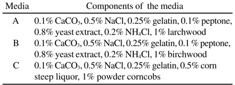
for comparative purposes. The data show that there is a
significant difference in the enzyme production when different
culture medium and sources of xylans were used. According to
Monti
et al.
(8), the maximum production of xylanase activity
using larchwood as carbon source was obtained at 24 hours of
culture (medium A, Table 1); when birchwood was used in the
medium (medium B, Table 1), the xylanase activity 0.542 ± 0.005
U/mg protein was obtained at 28 hours (Table 2). The substitution
of ammonium chloride, yeast extract and peptone by corn steep
liquor in the medium (medium C) produced maximum xylanase
activity at 22 hours (Fig. 1). These results showed that the
composition medium and xylan types had a great influence on
the enzyme production (Table 2) and the best medium was the
medium C. Furthermore, corncobs is a carbon source of low
cost. Fig. 1 shows that the extracellular xylanase activity
increases during the growth of the culture, achieving a maximum
level at 22 hours and decreasing slowly around 40 hours. The
protein determination during the growth of the culture was used
because of the difficulties that involve the mycelium separation
using this carbon source. That decrease can be explained by
the pellets development at 40 hours of growth in corncobs
cultures and the enzyme immobilization in those pellets. During
Media
Components of the media
A
0.1% CaCO
3, 0.5% NaCl, 0.25% gelatin, 0.1% peptone,
0.8% yeast extract, 0.2% NH
4Cl, 1% larchwood
B
0.1% CaCO
3, 0.5% NaCl, 0.25% gelatin, 0.1 % peptone,
0.8% yeast extract, 0.2% NH
4Cl, 1% birchwood
C
0.1% CaCO
3, 0.5% NaCl, 0.25% gelatin, 0.5% corn
steep liquor, 1% powder corncobs
the period of 22 hours, there is a drastic decrease of the protein
quantity of the initial medium and there is an increase on the
protein quantity which is then produced by the microorganism.
Ion exchange chromatography
To detect the two xylanase forms in the filtrate of the culture
containing corncobs (medium C), the proteins were precipitated
with 70% ammonium sulfate and dialyzed overnight and applied
into a DEAE-cellulose column. Fig. 2 shows that two activity
peaks were obtained and labeled as form 1 and form 2, both
eluted in the elution buffer. Xylanase form 2 had previously
been purified and characterized by Monti
et al.
(8).
Purification of xylanase form 1 through electroelution
The dialyzed sample from medium C (250 mg prot.) was
submitted to a preparative non-denaturing 7.5% PAGE pH 8.9
using glass tubes 13 x 1.4 cm. Monti
et al.
(8) determined that
xylanase form 2 did not migrate into the 7.5% PAGE; on the
other hand, xylanase form 1 migrated in this gel. After 6 hours at
2 mA/tube, one of the gels was stained with Coomassie Brilliant
Blue R250. After revelation, it was placed side by side with
another not stained gel in order to cut the protein bands for the
identification of the band with xylanase form 1 activity. The
enzyme extracted from the proteic band with activity was applied
into DEAE-Trisacryl. Figure 3 shows that xylanase form 1 did
not bind to the resin and was eluted in the void volume of the
equilibrating buffer. To have a greater quantity of form 1, four
similar columns were made and fractions with xylanase form 1
activity were collected, dialyzed and concentrated by vacuum
desiccator. The material obtained (2.25 U/mg prot.
-1) was put in
distilled water and submitted to a non-denaturing 7.5% PAGE
pH 8.9. After the migration, the band that showed xylanolytic
activity obtained of four gels was sliced, macerated, and added
to another 5% PAGE functioning as a heaping up gel to the
macerated gels. The protein was extracted electrophoretically.
On the side where the migration occurred (bottom of the glass
tube) a membrane for dialysis was connected with 5 mL buffer
elution (electroelution). This procedure was performed according
Figure 1.
Specific activity of xylanase form 1 in function of time
of culture in liquid medium C containing 1% of powder corncobs.
Figure 2.
DEAE-cellulose chromatography of ammonium sulfate
precipitated protein from the culture with carbon source 1%
corncobs at a flow rate of 30 mL/h. Fractions of 1.5 mL were
collected and assayed.
Figure 3.
DEAE-Trisacryl chromatography of sliced, macerated
and dialyzed polyacrylamide gels eluted with potassium
phosphate buffer at a flow rate of 15 mL/h. Fractions of 1.0 mL
were collected and assayed.
Media
Protein (mg/mL) Xylanase (U/mL) Xylanase (U/mg)
A
2.217 ± 0.030
1.605 ± 0.022
0.722 ± 0.010
B
2.58 ± 0.026
1.385 ± 0.014
0.542 ± 0.005
C
2.11 ± 0.046
1.809 ± 0.040
0.864 ± 0.019
Table 2.
Effect of the composition of the medium in the
production of xylanase form 1.
to Ohshita
et al.
(9). After 6 hours of electrophoretic migration
at 10 mA, the protein eluted from the heaping up gel was dialyzed
against distilled water. The sample obtained was concentrated
by vacuum desiccator, dissolved in 200 ml of water and the
enzymatic activity showed the presence of xylanase form 1.
After obtaining the enzyme, it was applied to a 7.5% PAGE pH
8.9, where only one proteic band was detected.
The apparent
molecular mass of purified xylanase form 1 was 61.8 kDa by
SDS-PAGE (Fig. 4), suggesting that the preparation was
homogeneous and showing that this enzyme was different from
form 2 (25.5 kDa). These results complement what was described
by Monti
et al.
(8), where
H. grisea
produced two forms of
xylanase.
CONCLUSIONS
In this study the best medium for the production of
xylanolytic enzymes was composed of 0.1% CaCO
3, 0.5% NaCl,
0.25% gelatin, 0.5% corn steep liquor and 1% powder corncobs
(medium C). This media is of low cost and allows the production
of both xylanases (form 1 and form 2). The purification of form
1 was exclusively possible through electroelution technique,
which allowed its molecular mass determination. It would not
have been possible to obtain through other techniques because
of the small production of xylanase form 1. The existence of
multiples forms of xylanases has been well described in various
researches, but this was the only research that purified the
xylanase form 1 produced by
Humicola grisea
. These results
will eventually permit more detailed biochemical characterization
and genomic studies.
ACKNOWLEDGEMENTS
Adriana Hitomi Sayama was supported by Conselho de
Desenvolvimento Científico e Tecnológico (CNPq). Rubens
Monti was supported by PADC (FCF-Araraquara – UNESP).
We thank Osmar Redondo for technical assistance.
RESUMO
Produção e purificação de uma Endo-1,4-
βββββ
-Xilanase de
Humicola grisea var. thermoidea por eletroeluição
Humicola grisea
var. thermoidea produz duas formas de
xilanase extracelular. A componente forma 1 foi purificada
usando o método de eletroeluição devido à baixa produção
desta enzima extracelular. A aparente massa molar foi
determinada 61,8 kDa por SDS-PAGE.
Palavras-chave:
eletroeluição,
Humicola grisea
var. thermoidea,
purificação de enzimas, xilanase extracelular
REFERENCES
1. Andrews, A.T. Preparative gel electrophoresis. In: Andrews, A.T.
Electrophoresis: Theory, techniques, and biochemical and clinical applications. Oxford, New York, 1992, p. 178-204.
2. Biely, P. Microbial xylanolytic system. Trends Biotechnol., 3: 286-290, 1985.
3. Cardello, L.; Terenzi, H.F.; Jorge, J.A. A cytosolic trehalase from the thermophilic fungus Humicola grisea var. thermoidea.
Microbiology, 140: 1671-1677, 1994.
4. Chaves, V.M.G.; Silva, D.O.; Brune, W.; Moreira, M.A. Cellulolytic activities of Humicola sp. Rev. Microbiol., 20: 460-465, 1989. 5. Haltrich, D.; Preiss M.; Steiner, W. Optimization of a culture medium
for increased xylanase production by a wild strain of Schizophylum commune. Enzyme Microb. Technol., 15: 854-860, 1993. 6. Hartree, E. F. Determination of protein: a modification of the Lowry
method that gives a linear photometric response. Anal. Biochem., v. 48, p. 422-427, 1972.
7. Kulkarni, N.; Shendye, A.; Rao, M. Molecular and biotechnological aspects of xylanases. FEMS Microbiol Rev., 23: 411-456, 1999. 8. Monti, R.; Terenzi, H.F.; Jorge, J.A. Purification and properties
of an extracellular xylanase from the thermophilic fungus
Humicola grisea var. thermoidea. Can. J. Microbiol., 37: 675-81, 1991.
9. Ohshita, T.; Sakuda, H.; Nakasone, S.; Iwamasa, T. Purification, characterization and subcellular localization of pig liver α-L-iduronidase. Eur. J. Biochem., 179: 201-207, 1989.
10. Peralta, R.M.; Terenzi, H.F.; Jorge, J.A. β-D-glycosidase activities of Humicola grisea var. thermoidea: biochemical and kinetic characterization of a multifunctional enzyme. Biochim. Biophys. Acta, 1033: 243-249, 1990.
11. Poutanen, K. Enzymes: An important tool in the improvement of the quality of cereal foods. Trends Food Sci. Technol., 8: 300-306, 1997. 12. Prade, R.A. Xylanases: from Biology to Biotecnology Biotechnol.
Genet. Eng. Rev., 13: 100-131, 1995.
13. Purkarthofer, H.; Sinner, M.; Steiner, W. Cellulase-free xylanase from Thermomyces lanuginosus: optimization of production in submerged and solid–stabte culture. Enzyme Microb. Technol., 15: 677-68, 1993.
14. Sá-Pereira, P.; Duarte, J.; Costa-Ferreira, M. Electroelution as a simple and fast protein purification method: isolation of an
extracellular xylanase from Bacillus sp. CCMI 966. Enzyme Microb. Technol., 27: 95-99, 2000.
15. Tosil, L.R.O.; Terenzi, H.F.; Jorge, J.A. Purification and characterization of an extracellular gluco-amylase from the thermophilic fungus Humicola grisea var. thermoidea. Can. J. Microbiol., 39: 846-852, 1993.