Vol.55, n. 1: pp. 1-6, January-February 2012
ISSN 1516-8913 Printed in Brazil BRAZILIAN ARCHIVES OF
BIOLOGY AND TECHNOLOGY
A N I N T E R N A T I O N A L J O U R N A L
A Rapid and Reliable Method for the Clonal Isolation of
Acanthamoeba
from Environmental Samples
Janice Zanella
1,2, Sergio Olavo Pinto da Costa
3, Jucimar Zacaria
1and Sergio
Echeverrigaray
1*1Instituto de Biotecnologia; Universidade de Caxias do Sul; Caxias do Sul - RS - Brasil. 2Departamento de
Citopatologia e Microbiologia; Faculdade de Biomedicina; Universidade de Cruz Alta; RS - Brasil. 3Universidade Católica de Santos; São Paulo – SP - Brasil
ABSTRACT
Acanthamoeba are abundant in a wide range of environments, and some species are responsible for cutaneous infections, keratitis, and granulomatous amoebic encephalitis (GAE). The conventional detection and isolation of amoeba from clinical and environmental samples involves sampling and culture on non-nutrient Ágar medium. Although efficient, this system requires several transfers in order to eliminate contaminants, and is not appropriate for the isolation of individual amoeba from samples with a biodiverse community. In this study we propose an alternative method for the isolation of monocystic clones of Acanthamoeba. The propose method involves sampling,
enrichment, encystment induction, and direct cysts micromanipulation and culture on Ágar plates.
Key words:Acanthamoeba, monocystic culture, micromanipulation
*Author for correspondence: [email protected]
INTRODUCTION
Free-living amoebae are abundant in soil, dust and water. These organisms have been isolated from fresh water, seawater, surface water, swimming pools, mineral water, dust, contact lens solutions,
among other environments (Khan 2006;
Visvesvara et al. 2007; Caumo et al. 2009; Costa et al. 2010). Several amphizoic amoebae such as
Acanthamoeba spp., Balamuthia spp. and
Naegleria spp. occasionally invade hosts and cause infections (Khan 2003; Schuster and
Visvesvara 2004; Khan 2006). Acanthamoeba can
invade the human´s skin, eye and central nervous system where they can be responsible for cutaneous infections, keratitis, and granulomatous amoebic encephalitis (GAE), respectively (Khan,
2003, 2006). In Brazil, a twenty year survey of
Acanthamoeba keratitis showed that amoebic corneal infection could be considered a
well-established and increasing ophthalmological
disease (Carvalho et al. 2009). Moreover, Acanthamoeba serves as a carrier for different microorganisms to humans, acting as a vehicle for the circulation of pathogens between the environment and humans (Krusnell and Linder, 2001; Huws et al. 2008).
Various Acanthamoeba species have been reported
to be able to cause keratitis and other human
infections, among which the most prevalent are A.
castellani, A. polyphaga, A. rhysodes and A. hatchetti (Walochnik et al. 2000a; Khan 2006). Although easily recognized at the genus level by
their polygonal cysts, the accurate Acanthamoeba
DNA sequencing) and several behavioral tests
(osmotolerance, temperature tolerance and
cytotoxicity assays) which can require axenic and clonal amoeba cultures (Walochnik et al. 2000b). Agar culture is the mainstay for laboratory
detection of Acanthamoeba from clinical and
environmental samples. Amoeba isolation and culture procedures involves sampling, plating on
non-nutrient agar medium covered by Escherichia
coli, incubation for 14 days at 30-37ºC with
periodical examination, amoeba harvesting at a non-contaminated site of the plate, and transfer to a fresh plate in order to eliminate ciliates, flagellates, bacteria and fungi. The overall
isolation of Acanthamoeba is time consuming (7 to
60 days) and several samples, particularly environmental samples, are often lost due to fungal or bacterial contamination.
In order to solve these problems, efforts were made in this work to develop a rapid and reliable
protocol for the clonal isolation of Acanthamoeba
from environmental samples.
MATERIAL AND METHODS
Acanthamoeba strains and sampling
An Acanthamoeba polyphaga strain isolated from a non-hospital environment in Santos, SP, Brazil (Teixeira et al. 2009) was used in initial
experiments to optimize the culture and
encystment conditions. Ten samples were
collected in community bathrooms of a public hospital in Caxias do Sul, RS, Brazil. Fresh water samples (200ml) were collected directly from the taps, and dust samples were collected by scrapping
the surface (100 cm2) of basin benches using
sterile swabs. All the samples were processed within 2h.
Optimization of procedures for the clonal isolation of Acanthamoeba
In order to evaluate the best condition for the
enrichment and encystment of Acanthamoeba
samples, three culture media were tested: (1) NN-EI, non-nutritive medium (Page saline solution-
2.5 mM NaCl, 1 mM KH2PO4, 0.5 mM Na2HPO4,
40 mM CaCl2, and 20mM MgSO4) seeded with 0.1
ml of a thermal inactivated 48-h culture of E. coli
ATCC 11775, (2) NN-E, non-nutritive medium
seeded with 0.1 ml of an active 48-h culture of E.
coli, and (3) PYG medium [proteose peptone
0.75% (w/v), yeast extract, 0.75% (w/v) and
glucose 1.5% (w/v)]. Acanthamoeba growth was
monitored by direct microscopic counting of
trophozoites and cysts using a Neubauer
hemocytometer.
Otherwise specified, Acanthamoeba were cultured
at 30-32ºC, and encystment was induced by incubation at low temperature (4ºC). Cyst isolation was performed using an Eclipse E 50i microscope with a TDM 50i yeast tetrads micromanipulator (Schuett-Biotec, Denmark) that allowed direct cyst isolation on the solid media. The three enrichment media described before were solidified with 1.0% agar (NNA-EI, NNA-E, and PYGA) and tested for micromanipulation. In order to compare the efficiency of the proposed method, the samples were also processed by the conventional plating method adopted by several authors (Caumo et al. 2009; Teixeira et al. 2009; Costa et al. 2010), and preconized by the UK National Health Service (www.hpa-standardmethods.org.uk).
Processing and culture of environmental samples
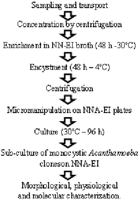
Fresh water samples (50 ml) were centrifuged in Falcon tubes (1000xg, 5 min) and the pellet was suspended in 1.0ml NN-EI broth. Dust samples were directly suspended in 1.0ml NN-EI broth. For the conventional method, these samples were immediately plated on pre-seeded NNA-EI, sealed with plastic film, and incubated at 30ºC. For the proposed method, the samples were enriched by incubation for 48 h at 30ºC, transferred to 4ºC for 24 h to induce cyst formation, and microscopically monitored. Encysted samples were centrifuged (1000 x g, 5 min) and suspended in 50-100 µl of Page´s saline. For micromanipulation, a loop (approx. 5 µl) of the suspension was spread in a straight line along the edge of a well dried
NNA-EI plate. Under the microscope, individual cyst
were picked up with a micromanipulator needle and transferred to new positions on the plate. After manipulation, the plates were sealed with plastic film and incubated at 30ºC. Plates were daily examined with an inverted microscope, and actively growing amoebas from individual clones
were transferred to fresh NNA-EI medium. Up to
ten individual clones of positive samples were isolated and evaluated.
Morphological and molecular characterization of Acanthamoeba isolates
al. 2003), directly observed under the microscope at a 400X magnification, and identified as belonging to one of the cyst morphological groups (Acanthamoeba sp. I to III) as established by
Pussard and Pons (1977). Molecular
characterization of Acanthamoeba cultures was
performed by RFLP-PCR of ssu rDNA according to Kong and Chung (1996).
RESULTS
Optimization of Acanthamoeba isolation
method
Initial experiment of enrichment and cyst induction performed with an environmental isolate of A. polyphaga indicated that NN-EI broth supported better growth of trophozoites than NN-E
and PYG media. Static culture of A. polyphaga
trophozoites for 24, 48 and 72 h in NN-EI broth
allowed 2x, 5x and 6x increase of A. polyphaga,
respectively. The transfer of active enrichment cultures to low temperature (4ºC) for a period of 48 h induced up to 80% encystment. This percentage was not significantly increased by longer incubation (48h).
For micromanipulation, NNA-EI allowed a rapid
and efficient isolation of Acanthamoeba cysts, as
well as their hatching and trophozoites
multiplication. Conversely, the presence of active E. coli on NNA-E posse several problems for the
separation of cysts by drastic modification on surface tension that made it difficult for the attachment of the cyst to the needle. PYG did not
support a good growth of Acanthamoeba
increasing from 96 h to more than 168 h the growing period necessary for the first sub-culture of the clones.
Application of the proposed method to environmental samples
After established the above basic conditions, ten samples (five fresh water and five dust samples) were processed by the conventional plating method and the proposed method summarized in Figure 1. Five of the ten environmental samples examined using the proposed method (Fig. 1) were
positive for Acanthamoeba (Table 1). Overall cyst
viability was 82% (37 growing clones from 45 isolated cysts). Two samples (W1 and D2)
exhibited just one Acanthamoeba species, while in
three samples (W3, D1 and D5), two species were
identified. The most prevalent species was A.
castellanii identified in four out of five positive
samples. This species was the only Acanthamoeba
isolated from the samples W1 and D1, or was
found associated with A. polyphaga (samples W3
and D1) and A. lenticulata (sample D5). A.
castellanii was characterized by small cysts with stellate endocysts (Group II) and a typical RFLP-PCR pattern (Fig. 2).
Table 1- Samples and clonal isolates of Acanthamoeba obtained by micromanipulation method.
Sample Origin Nº clones Clone
Name
Cyst Type2
Molecular Classification3
W1 Water 5 W1.1 II A. castellanii
W2 Water 0 - - -
W3 Water 4
2
W3.1 W3.2
II A. castellanii
A. polyphaga
W4 Water 0 - -
-W5 Water 0 - - -
D1 Dust 2
6
D1.1 D1.2
II II
A. castellanii A. polyphaga
D2 Dust 10 D2.1 II A. castellanii
D3 Dust 0 - - -
D4 Dust 0 - - -
D5 Dust 7
1
D5.1 D5.2
II III
A. castellanii A. lenticulata 1
Samples with the same number were collected from the same bathroom (ex. W1 and D1).
2
Based on cyst morphology (Pussard and Pons, 1977).
3
Based on the restriction patterns (MboI, HaeIII and TaqI) of the ssu rRNA amplicon (Kong and Chung, 1996).
Figure 2 - ssu rDNA RFLP-PCR profiles of Acanthamoeba clones- A. castellanii (1- W1.1, 2- W3.1, 4- D1.1, 5- D2.1, 6- D5.1) and A. polyphaga (3- W3.2). P- Molecular marker 100bp ladder.
A. polyphaga was identified in three samples: W3
and D1 associated with A. castellanii, and D2, in
which it was the only Acanthamoeba species
identified (Table 1). A. lenticulata was present in
the sample D5 associated with A. castellanii.
Using the conventional plating method, sample
W1 contaminated with fungi preventing
Acanthamoeba isolation, only A. castellani was isolated from sample D5, and at least three subcultures were necessary to decontaminate the positive samples.
DISCUSSION
Despite the lack of information about the
efficiency of Acanthamoeba isolation methods,
most of the works report difficulties with fungal, flagellate protozoa and bacterial contamination that demand several sub-cultures in order to obtain pure cultures (Walochnik et al. 2000b; Caumo et al. 2009; Teixeira et al. 2009; Costa et al. 2010). In the proposed method, these difficulties were overcome by the direct micromanipulation of induced cysts on NNA-EI plates. Although
Acanthamoeba micromanipulation has been
reported in other works (Fritsche et al. 1993; Kong and Chung 1996; Petry et al. 2006), this is the first report that used cyst induction and a yeast tetrad micromanipulator for the isolation and culture of individual cyst clones in a single step.
efficient and rapid than the plating method. The
proposed method allowed isolating Acanthamoeba
clones from all the positive environmental samples in 10 to 12 days, whereas the conventional plating method required 27 to 32 days to obtain axenic cultures. Even after several transfers, it was not possible to clean up one of the samples processed by the conventional method.
The main objective of this work was not to
determine the incidence of Acanthamoeba, but to
evaluate the efficiency of the proposed method. Nevertheless as observed in Table 1, five out of ten samples (50%) collected at community bathrooms of a public hospital (two fresh water and three dust samples) exhibited the presence of Acanthamoeba. Compared with other Brazilian
studies, the prevalence of Acanthamoeba was
lower than that obtained by the analysis of dust samples from a university hospital in Curitiba, PR (Costa et al. 2010), but higher than that reported in the hospitals of Porto Alegre, RS (Carlesso et al. 2007, 2010) and Santos, SP (Teixeira et al. 2009).
Considering the ubiquitous nature of
Acanthamoeba, and the large number of factors that influenced their presence in a hospital environment, it was not surprising to obtain high but variable incidence of these protozoa.
Three out of five positive samples (60%) exhibited
the presence of two Acanthamoeba species. The
simultaneous presence of Acanthamoeba species
has been reported in clinical as well as in
environmental samples (Khan 2006).
Cystmorphology and RFLP-PCR analysis made it
possible to detect three species of Acanthamoeba:
A. castellanii, A. polyphaga and A. lenticulata. A. castellanii was the most prevalent and present in all the positive samples.
The present results showed that 50% of the samples (fresh water and dust) collected from a hospital environment in the Southern Brazil harbored potentially pathogenic species of Acanthamoeba (A. castellanii and A. polyphaga), representing a risk for patients and visitors. These data corroborated those of previous works
developed in different Brazilian hospitals
(Carlesso et al. 2007; Texeira et al. 2009; Carlesso et al. 2010; Costa et al. 2010), and emphasized the need for increasing the public awareness and hospital disinfection measures in order to reduce
the incidence of Acanthamoeba infections
(Carvalho et al. 2009).
REFERENCES
Carvalho FRS, Foronda AS, Mannis MJ, Höfling-Lima AL, Belfort RJr, Freitas D, Twenty years of
Acanthamoeba keratitis. Cornea. 2009; 28: 516-519. Carlesso AM, Simonetti AB, Artuso GL, Rott MB.
Isolamento e identificação de amebas de vida livre potencialmente patogênicas em amostras de ambientes de hospital público da cidade de Porto Alegre, RS. Rev Soc Bras Med Trop. 2007; 40: 316-320.
Carlesso AM, Artuso GL, Caumo K, Rott MB. Potentially pathogenic Acanthamoeba isolated from a hospital in Brazil. Curr. Microbiol. 2010; 60: 185-190.
Caumo K, Frasson AP, Pens CJ, Panatieri LF, Frazzon AP, Rott MB. Potentially pathogenic Acanthamoeba in swimming pools: a survey in southern Brazilian city of Porto Alegre. Ann Trop Med Parasitol. 2009; 103: 477-485.
Costa AO, Castro EA, Ferreira GA, Furst C, Crozeta MA, Thomaz-Soccol V. Characterization of
Acanthamoeba isolates from dust of a public hospital in Curitiba, Paraná, Brazil. J Eukar Microbiol. 2010; 57: 70-75
Fritsche TR, Gautom RK, Seyedirachti S, Bergeron DL, Lindquist TD. Occurrence of bacterial endosymbionts in Acanthamoeba spp. Isolated from corneal and environmental specimens and contact lenses. J Clin
Microbiol., 1993; 31: 1122-1126.
Grossniklaus HE, Waring GO, Akor C, Castellano-Sanchez A, Bennett K. Evaluation of hematoxylin and eosin and special stains for the detection of
Acanthamoeba keratitis in penetrating keratoplasties.
Am J Ophthalmol. 2003;136: 520-526.
Huws SA, Morley RJ, Jones MV, Brown MRW, Smith AW. Interactions of some common pathogenic bacteria with Acanthamoeba polyphaga. FEMS Microbiol Lett. 2008; 282: 258-265.
Khan NA. Pathogenesis of Acanthamoeba infections.
Microb Pathog. 2003; 34: 277-328.
Khan NA. Acanthamoeba: biology and increasing importance in human health. FEMS Microbiol Rev. 2006; 30: 564-595.
Kong HE, Chung DI. PCR and RFLP variation of conserved region of small subunit ribosomal DNA among Acanthamoeba isolates assigned to either A.
castellanii or A. polyphaga. Korean J Parasitology 1996; 34: 127-134.
Krusnell J, Linder E. Bacterial infections of free-living amoebae. Res Microbiol. 2001; 152: 613-619. Petry F, Torzewski M, Bohl J,
Pussard M, Pons R. Morphologie de la paroi kystique et taxonomie du genre Acanthamoeba (Protozoa, Amoebida). Protistologica. 1977; 8: 557-598. Schuster FL, Visvesvara GS. Free-living amoebae as
opportunistic and non-opportunistic pathogens of humans and animals. Int J Parasitol. 2004; 34: 1001-1027.
Teixeira LH, Rocha S, Pinto RMF, Caseiro MM, Costa SOP. Prevalence of potentially pathogenic free-living amoebae from Acanthamoeba and Naegleria genera in non-hospital, public, internal environments from the city of Santos, Brazil. Braz J Infect Dis. 2009; 13: 395-397.
Visvesvara GS, Moura H, Schuster FL. Pathogenic and opportunistic free living amoebae: Acanthamoeba spp., Balamuthia mandrillaris, Naegleria fowleri, and
Sappinia diploidea. FEMS Immunol Med Microbiol. 2007; 50: 1-26.
Walochnik J, Haller-Shober EM, Kölli H, Picher O, Obwaller A, Aspöck H. Discrimination between clinically relevant and nonrelevant Acanthamoeba strains isolated from contact lens-wearing keratitis patients in Austria. J Clin Microbiol. 2000a; 38: 3932-3936.
Walochnik J, Obwaller A, Aspöck H. Correlations between morphological, molecular biological, and physiological characteristics in clinical and nonclinical isolates of Acanthamoeba spp. Appl
Environ Microbiol. 2000b; 66: 4408-4413.