485
AbstractObjective: To ascertain the distribution of alpha 1 antitrypsin genotypes and correlate it with the severity of pulmonary disease in patients with cystic fibrosis.
Method: A clinical and laboratory cross sectional study of 70 patients at the Universidade Estadual de Campinas teaching hospital. Cystic fibrosis diagnoses was confirmed by both clinical and laboratory methods. The severity of cystic fibrosis was evaluated by Shwachman score. All the patients were tested for the presence of S and Z alleles for alpha 1 antitrypsin deficiency using polymerase chain reaction.
Results: Nine (12.8%) patients were heterozygous for S or Z alleles or the heterozygote compound (SZ). No significant differences were found in clinical severity of Cystic fibrosis between genotypes of alpha 1 antitrypsin. No significant differences were found when the patients were divided according to the presence or absence of the ∆F508 mutation.
Conclusion: In this study, the first undertaken in Brazil into the association of alpha 1 antitrypsin deficiency and cystic fibrosis, we did not find an association between the deficiency and cystic fibrosis severity.
J Pediatr (Rio J). 2005;81(6):485-90: Cystic fibrosis, alpha 1 antitrypsin.
Association between alpha 1 antitrypsin deficiency
and cystic fibrosis severity
Elisangela Jacinto de Faria,1 Isabel Cristina Jacinto de Faria,1
Alfonso Eduardo Alvarez,2 José Dirceu Ribeiro,3
Antônio Fernando Ribeiro,4 Carmen Silvia Bertuzzo5
Copyright © 2005 by Sociedade Brasileira de Pediatria
O
RIGINALA
RTICLE1. MSc. (Biology), Departamento de Genética, Faculdade de Ciências Médicas, Universidade Estadual de Campinas (UNICAMP), Campinas, SP, Brazil.
2. MSc. Pediatric pulmonologist, Faculdade de Ciências Médicas, UNICAMP, Campinas, SP, Brazil.
3. PhD. Pediatric pulmonologist. Professor, Departamento de Pediatria, Faculdade de Ciências Médicas, UNICAMP, Campinas, SP, Brazil. 4. PhD. Pediatric Gastroenterologist. Professor, Faculdade de Ciências
Médicas, UNICAMP, Campinas, SP, Brazil.
5. PhD. Geneticist. Professor, Faculdade de Ciências Médicas, UNICAMP, Campinas, SP, Brazil.
Manuscript received Jul 21 2004, accepted for publication Jul 27 2005.
Suggested citation: de Faria EJ, de Faria IC, Alvarez AE, Ribeiro JD, Ribeiro AF, Bertuzzo CS. Association between alpha 1 antitrypsin defici-ency and cystic fibrosis severity. J Pediatr (Rio J). 2005;81:485-90. Introduction
Cystic fibrosis (CF) is the most common and lethal autosomal recessive genetic diseases and affects one in 2,500 Caucasians. The risk of its heterozygote in the general population is one to 25. More than 1,000 mutations have been identified to the CFTR gene,1 which codes for a protein containing 1,489 amino acids.
Cystic fibrosis is characterized by abnormal chlorine flow at the apical membrane of epithelial cells, causing diverse clinical manifestations including pancreatic insufficiency, lung disease, meconium ileus, elevated sweat chlorine levels and obstruction of the vas deferens.
The extreme diversity of CF phenotypes is probably influenced by other genetic areas, distant from the CFTR locus. Many of the genes that are studied nowadays as modifiers of CF, particularly among those that influence the severity of the lung disease, are involved in controlling infection, immunity and inflammation. Some of these include the class II HLA antigens, mannose-binding lectinand alpha 1 antitrypsin.2
although not all are associated with low or undetectable serum A1AT levels. The normal variant is PiM. Currently the most common deficient alleles are PiZ (Z) and PiS (S). The Z allele causes a single point mutation which changes a guanine to an adenine guanine adenine on exon 5 of the gene, and results in the substitution of glutamic acid to lysine in A1AT.
Homozygotes for Z have 15% of normal A1AT levels and their risk of developing pulmonary emphysema are higher,6 and less often, liver diseases in newborns. The S allele is more common than the Z allele results in a single point mutation where adenine is swapped for timine at exon 3, resulting in the substitution of glutamic acid by valine. Levels of A1AT are reduced in SS homozygotes (60% of normal). With SZ, the quantity of A1AT (37% of normal) is low enough to put affected individuals at risk of chronic lung disease.6
With relation to the progression of pulmonary status in CF, it is known that there is noteworthy proteases-antiproteases imbalance in favor of neutrophil elastase within the lungs. These proteases have deleterious effects, destroying elastin in the lungs, stimulating mucus production and cleaving immunoglobulins and fibronectin.7 In patients with CF and alpha 1 antitrypsin deficiency (DA1AT), this imbalance could be exacerbated and pulmonary damage be even greater.
Studies into the association between DA1AT and CF are rare and have returned controversial results.
In contrast with what might be expected, one study of CF patients demonstrated that those with the association with DA1AT exhibited better pulmonary function parameters than individuals CF without DA1AT (VEF1 62.55% of normal and VEF 51.15% of normal, respectively).8,9 Another study with a significant patient sample, did not find any association between the gravity of CF and the presence of DA1AT.10 Bearing in mind the elevated quantity of elastase from dead neutrophils and present in the airway secretion of CF sufferers, it would be expected that an association between DA1AT and CF would confer a more severe phenotype on CF patients. Our study objective was to investigate whether the presence of S and Z alleles of A1AT was related to the severity of lung disease at a reference center for CF treatment in Brazil.
Patients and methods
This was a clinical and laboratory based, cross-sectional study of patients from the Cystic Fibrosis Clinic at the Hospital de Clínicas of the Universidade Estadual de Campinas (UNICAMP) from March 2000 to August 2002. All patients were recruited if they were on outpatients treatment during this period, had a Cystic Fibrosis diagnosis confirmed by compatible clinical history and at least two sweat test results with chlorine levels greater than or equal to 60 mEq/l, performed by iontophoresis sweat stimulation with pilocarpine according to the method described by Gibson & Cooke.11
Those responsible for the patients signed informed consent before the study began.
The study was approved by the Committee for Ethics in Research at the Medical Sciences Faculty/UNICAMP.
The chi-square test was used to compare categorical variables. Fishers exact test was applied if one of the 2 X 2 tables cell values less than or equal to 5. Significance was set at 5%.
The clinical criteria analyzed included pulmonary manifestations, digestive manifestations and Shwachman score.12 The laboratory evaluation included pulmonary function tests, sweat sodium and chlorine, chest x-ray and computerized tomography of the thorax. The Shwachman score assesses physical activity, the physical examination, nutrition and x-ray findings. For each item the maximum score is 25 points and the lower the score, the worse the clinical status of the patient. The score is graded from excellent (86-100), through good(71-85), medium (56-70), moderate (41-55) and severe (40 or less), according to total number of points. The Shwachman score is in Table 1.
All of the patients had been genotyped previously for the CFTR gene by the tem at the Molecular Genetics Laboratory at UNICAMP. The patients DNA was extracted and, by means of polymerase chain reaction, specific regions were amplified to be analyzed for the five mutations in question:
∆
F508, G542X, N1303K, G551D and R553X.All patients were analyzed for A1AT deficiency S and Z alleles using polymerase chain reaction (PCR), which creates sites recognized by the restriction enzymes XmnI (S allele) and TaqI (Z allele).13 The two amplifications were optimized so that they could be performed on the same PCR program. The reaction was conducted in a volume of 100 µl, with the following reagent concentrations: 100 µM of each deoxyribonucleotide triphosphate (dATP, dTTP, dGTP, dCTP); 40 pmoles of each initiator; 2.5 units of Taq DNA polymerase (Centbio-UFRGS); 1.5 µM de MgCl2 present in the enzyme-specific buffer, and 1 µg of genomic DNA.
The amplification reaction was performed in a temperature cycler and consisted of an initial denaturing at 94 °C for 7 minutes followed by a 95 °C for 5 minutes; 50 °C for 1 minute (annealing) and 74 °C for 2 minutes (extension). Next followed 34 cycles at 95 °C for 1 minute; 50 °C for 1 minute and 74 °C for 2 minutes and, finally, one cycle at 74 °C for 9 minutes (termination).
The 15 µl amplification sample for the S mutation was digested for 12 hours at 37 °C in a total volume of 50 µl containing 2 µl of restriction enzyme (XmnI) and 5 µl of the buffer recommended by the supplier and the final volume was made up with 30 µl of water. After digestion, the sample was subjected to polyacrylamide gel electrophoresis at 14% (3.75 ml TBE 10X; 14.75 ml of H2O; 12.5 ml of Bis acrylamide; 200 µl of ammonium persulphate 10% and 40 ml of Temed). The gel was then stained with ethidium bromide for viewing the bands.
Score General activity Physical examination
25 Full activity; tolerance to normal exertion; No cough; normal HR and RR;
good tempered; normal motor development; no evidence of emphysema; clear lungs
normal school frequency. on auscultation; good posture; no clubbing.
20 Mild limitation to intense activity, tires easily Occasional cough; normal HR and RR at rest;
at the end of the day or after exertion; less energetic; mild emphysema; rough VM, snooring and
limit of motor development lower than normal; occasional prolonged ET; good posture;
occasionally irritated or apathetic; good school frequency. mild clubbing.
15 Voluntary resting; tires after exertion; Light morning cough after exertion/cry,
moderately inactive; occasionaly during the day; no nocturnal cough;
light motor retardation; lack of spontaneity; mild HR and RR; AP diameter and lowered diaphragm,
passive or irritable; regular school frequency. rough VM, crackles, snooring/wheezes; half clubbing.
10 Limited physical activity and exercise tolerance; Chronic, frequent, repeated, productive
dyspnoea after exertion; mild motor retardation; and rarely paroxysmal cough; mild HR and RR;
agitated or irritated; lazy or disappointed; mild to severe emphisema, frequent malformation
low school frequency; findings in the x-ray; crackles, snooring and wheezing
private teachers may be required usually present and disseminated, 2/3 clubbing.
5 Severe limitation to physical activity; dyspnoea Severe, paroxysmal, frequent, productive cough,
and orthopnoea; inactive or confined to bed/chair; frequently followed by vomiting and hemoptisys;
marked motor retardation; apathetic or irritated; nocturnal cough; tachypnoea and tachycardia;
can not attend school. sever emphysema; crackles, generalized snooring
and wheezing; audible expiration;
bad posture; 3/4 clubbing; frequent cyanosis.
Table 1 - Shwachman score10
HR = heart rate; RR = respiratory rate; VM = vesicular murmur; ET = expiratory time; AP = anteroposterior.
Score Nutrition X-ray findings
25 Weight and height above the 25th percentile or compliant No evidence of emphysema; no enlargement
to the family pattern; tone and muscular mass; of the bronchovascular marking;
normal subcutaneous fat; normal sexual maturation; no infiltrations or atelectasis.
stools slightly normal; good appetite.
20 Weight and height above the 10th percentile or slightly Minimum evidence of emphysema;
below the family pattern; good tone and muscular mass; slight enlargement of the bronchovascular marking;
slightly reduced subcutaneous tissue; no infiltrates and atelectasis.
retarded sexual maturation; normal appetite, more frequent stools and slightly abnormal.
15 Weight and height above the 3rd percentile Mild emphysema; increased AP diameter;
or slightly below the familiar pattern; radiolucent lung fields, slightly lowered diaphragma;
weight usually inadequate for height; enlarged bronchovascular marking;
regular tone and muscular mass; defficient subcutaneous fat, localised or irregular atelectasis;
slightly distended abdomem; retarded sexual maturation; transient occasional infiltrate.
regular appetite; volumous stools, fetid, fluctuating.
10 Weight and height above the 3rd percentile and defficient Marked emphysema; marked increase of AP diamater;
as to height; poor tone and muscular mass; marked lowered diaphragma; narrow cardiac
marked defficienvcy of subcutaneous fat; silhouette; areas with disseminate atelectasis;
slightly distended abdomem; insufficient sexual maturation, patchy or lobar atelectasis; persistent infiltration
no growth spurt; poor appetite; malformed volumous stools, focuses; localized cysts; marked increase of
fetid and fatty. bronchovascular marking.
5 Malnourished and short; weak, flacid and small muscles; Extensive alterations; severe hyperinflation;
no subcutaneous fat; frequent weight loss; frequent, disseminate infiltrate and atelectasis;
volumous, fetid and fatty stools; frequent rectal prolapse. disseminated cysts formation; bronchiectasis and
Table 2 - Distribution of Alpha 1 antitrypsin allele and pulmonary disease severity (all patients)
Pulmonary disease MS/MZ/SZ genotype MM Total
Mild 5 35 40
Moderate 2 19 21
Severe 2 7 9
Total 9 61 70
χ
2(3) = 0.92; p = 0.82.
χ
2 = chi-square.MS = S heterozigote; MZ = Z heterozigote; SZ = composite heterozigote; MM = without two alleles.
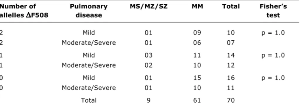
Table 3 - Distribution of alpha 1 antitrypsin alleles and pulmonary disease severity (patients separated according to presence or absence of the
∆
F508 mutation)Number of Pulmonary MS/MZ/SZ MM Total Fishers allelles
∆∆∆∆∆
F508 disease test2 Mild 01 09 10 p = 1.0
2 Moderate/Severe 01 06 07
1 Mild 03 11 14 p = 1.0
1 Moderate/Severe 02 10 12
0 Mild 01 15 16 p = 1.0
0 Moderate/Severe 01 10 11
Total 9 61 70
of the buffer recommended by the supplier and the final volume was made up with 29 µl of H2O.
After digestion samples were subjected to polyacrylamide gel electrophoresis at 14%.
Results
Seventy cystic fibrosis patients were analyzed. There were 36 males (51%) and 34 females (49%), with a mean age of 10.2 and standard deviation of 9.44 years. Ninety-seven percent of the patients were Caucasoids and 3% Negroids. Nine patients with cystic fibrosis (12.8%) were heterozygotes for the S or Z allele or composite heterozygotes (SZ). No patient was homozygous for either S or Z alleles . In Brazil, the normal frequency for S heterozygotes is 9.2% and for Z heterozygotes it is 5.5%.14 No statistically significant difference was found in clinical severity between A1AT genotypes (MS
χ
2(1) = 0.0487- 0.80 < p < 0.90; MZ
χ
2(1) = 2.1228-0.10 < p < 0.20; SZχ
2(1) = 2.9719, 0.05 < p < 0.10) (Table 2). No statistically significant difference was found when patients were separated according to presence or absence of the
∆
F508 mutation (Table 3). The age groups of the patients and the severity of pulmonary status can be observed in Table 4.Discussion
This study evaluated children and adults suffering from cystic fibrosis. It was confirmed that there was a great majority of Caucasoids, which was expected, despite the elevated number of people of mixed race in Brazil.
The Shwachman score was used to evaluate the severity of pulmonary status.12 In earlier studies carried out at Unicamp, it was confirmed that the score exhibited a statistically significant correlation with colonization by Pseudomonas aeruginosa, colonization by Pseudomonas aeruginosa mucosa, forced vital capacity, first second forced expiratory volume, transcutaneous hemoglobin saturation, number of infectious exacerbations in the previous year, indications for Dornase Alpha, indications for regular respiratory physiotherapy and indications for home oxygen therapy.15,16 The Shwachman score is therefore a good indicator of general severity.
Proteases have potentially deleterious effects such as the destruction of elastin in the lungs, stimulation of mucus production and cleavage of immunoglobulins and fibronectin.7,17
In cases with bacterial colonization, the deleterious effect of this imbalance has been well studied. Thus, with infectious processes, as dos neutrophils are lysed, elastase
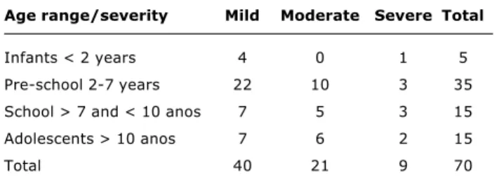
Age range/severity Mild Moderate Severe Total
Infants < 2 years 4 0 1 5
Pre-school 2-7 years 22 10 3 35
School > 7 and < 10 anos 7 5 3 15
Adolescents > 10 anos 7 6 2 15
Total 40 21 9 70
Table 4 - Age groups of the patients and the severity of pulmo-nary status
could be liberated into the extracellular medium. This elevated quantity of liberated protease could attack the elastin and cause pulmonary damage. In cases where patients have CF and DA1AT concomitantly, this imbalance would be exacerbated and pulmonary damage could be greater. Based on this, clinical trials of the use of neutrophilic elastase inhibitors, such as alpha 1 antitrypsin aerosol for the treatment of CF, have been performed and achieved some success.18
Nevertheless, published literature shows that the genotypes that result in mild to moderate A1AT deficiency, MS, SS and MZ were not associated with aggravation of the lung disease of cystic fibrosis patients.10,19,20 In contrast, they postulated that the association of these two alterations could exhibit a significant improvement in pulmonary function8,9 because the neutrophil elastase is a powerful proteolytic enzyme capable of lysing bacteria such as Pseudomonas aeruginosa7,20,21 . It was suggested that the neutrophil elastase and other proteases are not exclusively destructive, but could also exhibit beneficial effects and that they are important to lyse bacteria and could reduce the regulation of inflammation, by inhibiting neutrophil activation, by cleavage of immunoglobulin , neutrophil and complement receptors and by inducing apoptosis.19,22 Overall, it appears that the final result of the action of elastase on the pulmonary epithelium depends on the balance between the positive and negative effects of elastase. Furthermore, in the case of those with alpha 1 antitrypsin deficiency, this effect will depend on the variation in enzyme activity.
It is apt to note that the severity of the pulmonary condition involves other variables such as environmental factors: age, pancreatic status, nutritional status23,24 and genetic factors such as: variations in DNA in introns, unidentified genes distant from the CF locus, A second mutation on the same allele which attenuates the effect of the primary mutation.25-28 It is clear, therefore, that the pulmonary manifestation of CF are multifactorial.
In this study, the first performed in Brazil into the association between A1AT deficiency and CF did not find any significant relation between the pulmonary status of CF patients and the distribution pf alleles for alpha 1 antitrypsin deficiency. When the patients were split according to the presence of the two alleles, one allele or no alleles for the
∆
F508 mutation and compared them according to pulmonary status, no statistical difference was observed, proving thereis no relation between the presence of the
∆
F508, mutation, alpha 1 antitrypsin deficiency and pulmonary status.It is important to emphasize that only one patient with the SZ allele was detected, having a moderate enzyme deficiency; the remainder were all heterozygotes for the S or Z alleles. Therefore, since the frequency of homozygotes for the S and Z mutations and the composite mutation SZ was low in the population of cystic fibrosis patients studied, it was concluded that A1AT deficiency did not perform a significant role in the pulmonary manifestations of cystic fibrosis because this was a small sample.
It is worth pointing out that nothing can be stated on the subject of the SS, ZZ or SZ genotypes since none were identified in the group studied. Therefore the sample should be amplified for further analysis with multicenter studies.
Acknowledgements
This research was supported by the Fundação de Amparo à Pesquisa do Estado de São Paulo (Foundation for the Support of Research for the State of São Paulo -Fapesp).
References
1. Cystic fibrosis mutation database. www.genet.sickkids.on.ca/ cftr/.
2. Vankeerberghen A, Cuppens H, Cassiman JJ. The cystic fibrosis transmembrane conductance regulator: an intriguing protein with pleiotropic functions. J Cystic Fibr. 2002;1:13-29. 3. Pelmutter DH. Clinical manifestations of alpha-1-antitrypsin
deficiency. Gastroenterol Clin N Am. 1995;24:27-43.
4. Lai EC, Kao FT, Law ML, Woo SL. Assignment of the
alpha-1-antitrypsin gene and a sequence- related gene to human chromosome 14 by molecular hybridization. Am J Hum Genet. 1983;35:385-92.
5. Faber JP, Poller W, Weidinger S, Kirchgesser M, Schwaab R,
Bidlingmaier F. Identification and DNA sequence analysis of 15 new alpha-1-antitrypsin variants, including two PI*Q0 alleles and one deficient PI*M allele. Am JHum Genet. 1994;55:1113-21.
6. Pierce JA. Antitrypsin and emphysema: perspectives and
prospects. J Am Med Ass. 1988;259:2890-5.
7. Sommerhoff CP, Nadel JA, Basbaum CB, Caughey GH. Neutrophil elastase and cathepsin G stimulate secretion from cultured bovine airway gland serous cells. J Clin Invest. 1990;85:682-9. 8. Mahadeva R, Westerbeek RC, Perry DJ, Lovegrove JU, Whitehouse DB, Carroll NR, et al. Alpha1 antitrypsin deficiency alleles, the Taq- I G→A allele and cystic fibrosis lung disease. Eur Respir J. 1998;11:873-9.
9. Mahadeva R, Sharples L, Roos-Russell RI, Webb AK, Bilton D, Lomas DA. Association of Alpha1 antichimotrypsin deficiency with milder lung disease in patients with cystic fibrosis. Thorax. 2001;56:53-8.
10. Frangolias DD, Ruan J, Wilcox PJ, Davidson AG, Wong LT, Berthiaume Y, et al. Alpha-1-antitrypsin deficiency alleles in cystic fibrosis lung disease. Am J Respir Cell Mol Biol. 2003;29:390-6.
11. Gibson LE, Cooke RE. A test for concentration of electrolytes in sweat in cystic fibrosis of the pancreas utilizing pilocarpine by iontophoresis. Pediatrics. 1959;23:545-9.
12. Shwachman H, Kulczycki LL. Long term study of 105 patients with cystic fibrosis: studies made over a five to fourteen year period. Am J Dis Child. 1958;96:6-15.
Correspondence: Carmen Sílvia Bertuzzo
Shigeo Mori, 1710, Barão Geraldo CEP 13082-084 Campinas, SP, Brazil Tel.: +55 (19) 3788.8907
Fax: +55 (19) 3788.8909
E-mail: [email protected] or [email protected] 14. Pagotto RC. Polimorfismo da Alfa1-1- antitripsina humana em
populações brasileiras [dissertação]. São Paulo: Universidade de São Paulo; 1993.
15. Alvarez AE, Ribeiro AF, Hessel G, Bertuzzo CS, Ribeiro JD. Fibrose Cística em um centro de referência no Brasil: características clínicas e laboratoriais de 104 pacientes e sua associação com o genótipo e a gravidade da doença. J Pediatr (Rio J). 2004;80:371-9.
16. Domee Espinoza MD. Fibrose Cística em jovens e adultos do hospital das clínicas da UNICAMP [dissertação]. Campinas: Universidade Estadual de Campinas; 1998.
17. Suter S, Schaad UB, Morgenthaler JJ. Fibronectin-cleaving activity in bronchial secretions of patients with cystic fibrosis. J Infect Dis. 1988;158:89-100.
18. Allen ED. Opportunities for the use of aerosolized alpha 1 antitrypsin for the treatment of cystic fibrosis. Chest. 1996;110:S256S-60.
19. Doring G, Krogh-Johansen H, Weidinger S. Allotypes of alpha 1 antitrypsin in patients with cystic fibrosis, homozygous and heterozygous for delta F508. Pediatr Pulmon. 1994;18:3-7. 20. Meyer P, Braun A, Roscher AA. Analysis of the two common
alpha-1-antitrypsin deficiency alleles PiMS and PiMZ as modifiers of Pseudomonas aeruginosa susceptibility in cystic fibrosis. Clin Genet. 2002;62:325-7.
21. Belaaouaj A, McCarthy R, Baumann M. Mice lacking neutrophil elastase reveal impaired host defense against gram negative bacterial sepsis. Nature Med. 1998;4:615-8.
22. Mahadeva R, Stewart S, Bilton D, Lomas DA. Alpha1 antitrypsin deficiency alleles and severe cystic fibrosis lung disease. Thorax. 1998;53:1022-4.
23. Mahadeva R, Lomas DA. Secondary genetic factors in cystic fibrosis lung disease. Thorax. 2000;55:446.
24. McKone EF, Emerson SS, Edwards KL, Aitken ML. Effect of genotype on phenotype and mortality in cystic fibrosis: a retrospective cohort study. Lancet.2003;361:1671-6. 25. Kerem E, Kerem B. Genotype-phenotype correlations in cystic
fibrosis. Pediatr Pulmonol. 1996;22:387-95.
26. Dork T, Wulbrand U, Richter T. Cystic fibrosis with three mutations in the cystic fibrosis transmembrane conductance regulator gene. Hum Genet. 1991;87:441-6.
27. Bienvenu T. Les bases moléculaires de 1hétérogénéité phénotypique dans la muviscidose. Ann Biol Clin. 1997;55: 113-21.