Disease Is Not Associated with Worsening of Intestinal
Inflammatory Activity
Alexandre Medeiros do Carmo1*, Fabiana Maria Santos1, Carmen Lucia Ortiz-Agostinho1,
Ieˆda Nishitokukado1, Cintia S. Frota1, Flavia Ubeda Gomes1, Andre´ Zonetti de Arruda Leite1, Claudio Se´rgio Pannuti2, Lucy Santos Vilas Boas3, Magaly Gemio Teixeira4, Aytan Miranda Sipahi1
1Departamento de Gastroenterologia, Hospital das Clı´nicas da Faculdade de Medicina da Universidade de Sa˜o Paulo – LIM 07, Sa˜o Paulo, Sa˜o Paulo, Brazil,2Instituto de Medicina Tropical e Departamento de Doenc¸as Infecciosas e Parasitarias (LIM-HC) da Faculdade de Medicina da Universidade de Sa˜o Paulo, Sa˜o Paulo, Sa˜o Paulo, Brazil, 3Instituto de Medicina Tropical e Hospital das Clı´nicas da Faculdade de Medicina (LIM-HC), Universidade de Sa˜o Paulo, Sa˜o Paulo, Sa˜o Paulo, Brazil,4Departamento de Cirurgia do Servic¸o de Cirurgia do Co´lon Reto e Aˆnus, Hospital das Clı´nicas da Faculdade de Medicina da Universidade de Sa˜o Paulo, Sa˜o Paulo, Sa˜o Paulo, Brazil
Abstract
Background: Cytomegalovirus is highly prevalent virus and usually occurs in immunocompromised patients. The pathophysiology and treatment of inflammatory bowel disease often induce a state of immunosuppression. Because this, there are still doubts and controversies about the relationship between inflammatory bowel disease and cytomegalovirus.
Aim:Evaluate the frequency of cytomegalovirus in patients with inflammatory bowel disease and identify correlations.
Methods:Patients with inflammatory bowel disease underwent an interview, review of records and collection of blood and fecal samples. The search for cytomegalovirus was performed by IgG and IgM blood serology, by real-time PCR in the blood and by qualitative PCR in feces. Results were correlated with red blood cell levels, C-reactive protein levels, erythrocyte sedimentation rates and fecal calprotectin levels for each patient.
Results:Among the 400 eligible patients, 249 had Crohn’s disease, and 151 had ulcerative colitis. In the group of Crohn’s disease, 67 of the patients had moderate or severe disease, but 126 patients presented with active disease, based on the evaluation of the fecal calprotectin. In patients with ulcerative colitis, only 21 patients had moderate disease, but 76 patients presented with active disease, based on the evaluation of the fecal calprotectin. A large majority of patients had positive CMV IgG. Overall, 10 patients had positive CMV IgM, and 9 patients had a positive qualitative detection of CMV DNA by PCR in the feces. All 400 patients returned negative results after the quantitative detection of CMV DNA in blood by real-time PCR. Analyzing the 19 patients with active infections, we only found that such an association occurred with the use of combined therapy (anti-TNF-alpha+azathioprine)
Conclusion:The findings show that latent cytomegalovirus infections are frequent and active cytomegalovirus infection is rare. We did not find any association between an active infection of CMV and inflammatory bowel disease activity.
Citation:Carmo AMd, Santos FM, Ortiz-Agostinho CL, Nishitokukado I, Frota CS, et al. (2014) Cytomegalovirus Infection in Inflammatory Bowel Disease Is Not Associated with Worsening of Intestinal Inflammatory Activity. PLoS ONE 9(11): e111574. doi:10.1371/journal.pone.0111574
Editor:Juliet Spencer, University of San Francisco, United States of America
ReceivedJuly 6, 2014;AcceptedSeptember 20, 2014;PublishedNovember 11, 2014
Copyright:ß2014 Carmo et al. This is an open-access article distributed under the terms of the Creative Commons Attribution License, which permits unrestricted use, distribution, and reproduction in any medium, provided the original author and source are credited.
Data Availability:The authors confirm that all data underlying the findings are fully available without restriction. All relevant data are within the paper and its Supporting Information files.
Funding:This project received funding from FAPESP helping to buy products to experiments (project 2010/18275-8, http://www.fapesp.br/). The funders had no role in study design, data collection and analysis, decision to publish, or preparation of the manuscript.
Competing Interests:The authors have declared that no competing interests exist. * Email: [email protected]
Introduction
Cytomegalovirus (CMV) is a ß herpes-virus with double-stranded DNA and represents a common viral infection in humans, with infection levels ranging from 40% in developed countries to 100% in developing countries [1]. Primary CMV infection is subclinical in most patients and is often followed by CMV disease or latent CMV infection. CMV disease is rare in immunocompetent patients but not among the immunocompro-mised population. The organs affected by CMV disease include
the eyes, lungs, liver, urinary tract, pancreas, central nervous system, heart and gastrointestinal tract [2].
inflammation, it is necessary to use a combination of parameter settings, covering clinical, endoscopic, laboratory, endoscopic and radiological values. The CDAI (Crohn’s disease activity index) and the TrueLove-Witts are clinical indices that are widely used as a means to quantify the inflammatory activity of CD and UC, respectively [4,5,6]. In addition, Fecal Calprotectin (FC) is a very good marker of intestinal inflammation that has a close correlation with endoscopic and histological findings, as well as good sensitivity and specificity [7,8].
A clinical treatment is not curative and often relies on the use of immunosuppressive drugs. The pathophysiology of a disease may compromise the immune system. This observation, in combination with the fact that CMV presents a tropism for sites of inflammation, prompted us to hypothesize whether patients with IBD are more susceptible to CMV infection and disease [9,10].
Several authors have tried to correlate CMV and IBD, hypothesizing the following: possible triggering of CMV infection at the onset of IBD [11,12]; a worsening of the infection [13,14,15,16]; an association between the use of anti TNF-alpha and a higher risk of CMV disease [17,18]; or the higher prevalence of CMV disease in UC patients, compared with CD [19,20]. On the other hand, other authors have advocated that CMV is only a bystander or surrogate marker of severe colitis [21,22,23,24,25] or that the anti TNF-alpha therapies may reduce the risk of CMV reactivation in UC patients [26].
The prevalence of CMV infection in patients with IBD is highly variable. One reason for this wide variation is the difficulty and lack of standardization among indices for diagnosing the different grades of CMV infection [13,27]. The prevalence of CMV infection and its clinical significance in IBD, especially in outpatients, have yet to be determined.
In this context, the aim of this study was to explore the prevalence of CMV infection in 400 outpatients with IBD, using serology, a real-time quantitative PCR assay on blood samples, and a qualitative PCR assay on fecal samples. Another objective was to correlate CMV viral replication with various demographic, therapeutic and inflammatory activities in IBD patients.
Patients and Methods
Patients and Samples
The study was carried out from June 2011 to August 2012 with IBD outpatients from the Hospital das Clı´nicas, School of Medicine, University of Sa˜o Paulo (HC-FMUSP). The authors followed the Resolution number 196/96, the National Health Council/MS, respecting the basic principles of bioethics, auton-omy, non-maleficence, beneficence and justice. The study was submitted to the Ethics Committee of theHospital das Clı´nicas, Faculty of Medicine, University of Sa˜o Paulo (USP-HC) (Research Protocol: 0144/11) and accepted in session on 11/05/2011, before it starts. All patients or guardians, after reading, filled in and signed the consent form and agreed to participate in the study.
The diagnosis of inflammatory bowel disease, UC or CD, had been previously established by clinical, laboratory, endoscopic, radiological and histological findings, according to standard criteria.
Patients were asked to bring a fecal sample and were submitted to an interview on the day of consultation. Patients were evaluated to verify the levels of their disease activity, according to the pathology: CD (CDAI- Crohn’s disease activity index) [5] and UC (Truelove-Witts index) [6]. After data collection, all patients were referred to the laboratory for collection of venous blood samples. All of the data collected were analyzed and compared with data collected from these patients medical records. Those who did not meet the established criteria, did not sign the consent form, or failed to collect or deliver laboratory fecal materials were automatically excluded from the study.
Definition of active CMV infection
Active CMV infection was defined by a positive IgM test and/ or detection of CMV DNA in peripheral blood or fecal samples by qualitative or real time PCR
Laboratory Analysis
Peripheral blood samples were collected, and the tests performed included the following: complete blood count, eryth-rocyte sedimentation rate, C-reactive protein(CRP), and IgG and IgM antibodies against CMV.
Real Time Quantitative PCR (qPCR)
Extraction of viral DNA was performed by processing whole bloodusing a QIAamp DNA Mini-kit (Qiagen, Valencia, Calif.) according to the manufacturer’s instructions.
For Amplification and detection of viral DNA, the sequences of the PCR primers and probe were selected from the US17 region of CMV AD169, as previously described [28]. Summarizing, forward and reverse primers were 59 -GCGTGCT-TTTTAGCCTCTGCA-39 and 59 -AAAAGTTTGTGCCC-CAACGGTA-39, respectively, and the probe FAM -TG-ATCGGCGTTATCGCGTTCTTGATC - BHQ. PCR was performed using TaqMan Universal PCR master mix (Applied Biosystems), prepared as follows: 20mL of DNA from whole blood was mixed with 25ml of PCR master mix, 15 pmol of each of the primers, 10 pmol of the TaqMan probe, and DPOC water were added to final volume of 50ml.
Reactions were performed using the ABI PRISM 7300 Real Time PCR System (Applied Biosystems, Foster City, Calif.) with the following cycling conditions: 1 cycle at 50uC for 2 min and 95uC for 10 min to activate the Taq-Polymerase followed by 45 cycles at 95uC for 15 s and 61uC for 1 min.
To determine the sensitivity of the method, standard DNA at a concentration of 15.000 copies/ml (DNA from ADN169, Sellex, Table 1.The different grades of CMV infection.
CMV infection status Description
Not infected Not infected with the virus; negative IgG and IgM antibodies against CMV Latent infection Carrier of the CMV genome without active replication
Active infection Detectable viral replication in peripheral blood or organs or a significant rise in IgM antibodies against CMV Disease Clinical expression of active infection, that is, active CMV infection with end-organ involvement
Washington, DC, USA) was serially diluted until the concentration of 150 copies/ml and tested in triplicate.
For specificity, DNAs of the following pathogens: Epstein-Barr virus, Adenovirus, Mycoplasma pneumoniae, Herpes Simplex Virus Type 1, Herpes Simplex Virus Type 2, Varicella-Zoster Virus, Pneumocystis jiroveci, Toxoplasma gondii), obtained from commercial crops, acquired from SellexInc (Washington DC, USA), were subjected to PCR.
After performing the RT-PCR, DNA samples extracted from 300 of the 400 randomly selected patients) were analyzed by
conventional qualitative PCR. The same qualitative technique was used for stool samples[29].
Qualitative PCR
The fecal DNA was extracted using a Qiagen (QIAamp DNA stool Mini Kit) kit, and the detection of the cytomegalovirus genome was performed using the qualitative PCR technique (conventional) to amplify a specific segment of the CMV gB, with specific primers gB 1319 and gB 1604 of the glycoprotein B gene [29,30].
Table 2.Demographic and clinical profile of the inflammatory bowel disease patients.
Characteristics Crohn’s disease Ulcerative Colitis
Number of cases 249 151
Gender M:F 113:136 64:87
Age variation (mean) 15–82 (45,5) 23–80 (49,92)
Duration of disease, months (mean) 12–456 (132,5) 6–576 (118,77)
BMI, kg/m2(mean) 15,42–48,05 (24,3) 16,9–37,32 (25,51)
Disease location
ileal64 (25,7%) distal50 (33,1%)
colonic65 (26,1%) left colonic41 (27,1%) ileocolonic108 (43,3%) pancolonic60 (39,4%) upper GI12 (4,8%)
Clinical Index (CDAI/Truelove)
Remission 126 (50,8%)
Mild 55 (22,18%) 129 (86%)
Moderate 57 (22,98%) 21 (14%)
Severe 10 (4%) 0
FC$150, number (%) 126 (50,6%) 76 (50,8%)
Anemia, number (%) 56 (22,5%) 28 (18,5%)
C-reactive protein$5, number (%) 92 (37,7%) 56 (37,6%)
Previous surgery 100/247 (40,5%) 15/151 (9,9%)
doi:10.1371/journal.pone.0111574.t002
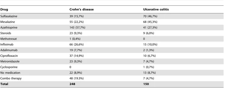
Table 3.Medications used by the patients enrolled in the study.
Drug Crohn’s disease Ulcerative colitis
Sulfasalazine 39 (15,7%) 70 (46,7%)
Mesalazine 55 (22,2%) 68 (45,3%)
Azathioprine 143 (57,7%) 41 (27,3%)
Steroids 23 (9,3%) 9 (6,0%)
Methotrexat 1 (0,4%) 0
Infliximab 66 (26,6%) 15 (10,0%)
Adalimumab 19 (7,7%) 2 (1,3%)
Ciprofloxacin 37 (14,9%) 10 (6,7%)
Metronidazole 23 (9,3%) 7 (4,7%)
Cyclosporine 0 1 (0,7%)
No medication 22 (8,9%) 13 (8,7%)
Combo therapy 48 (19.3%) 7 (4,7%)
Total 248 150
The result was revealed by the presence or absence of a band at the expected segment of DNA electrophoresis on an agarose gel marked with ethidium bromide. Internal, negative and positive controls were performed in parallel to assess the proper operation of the reaction.
Fecal Calprotectin
Stool samples were stored at220uC to be thawed later, and a survey of fecal calprotectin using ELISA, PhiCal (Caprotectin Elisa Kit) was conducted [8,31].
Statistical Analysis
A statistical analysis to determine the characteristics associated with CMV and IBDs patients was conducted. Descriptive analyses were performed first, and the Fisher’s exact test and Mann-Whitney test were used for categorical variables, with Student’s t test being used for continuous variables. In a more detailed study, a simple and multiple logistic regression was considered. A P value
,0.05 was considered significant.
Results
Clinical Characteristics
The clinical and demographic characteristics of the 400 patients involved in the study are shown in Table 2. Using clinical indices, including the Crohn’s disease activity index (CDAI) for CD and the Truelove, for UC, only 67 CD patients and 21 UC patients presented with moderate or severe disease. However, using fecal calprotectin (FC), over half of the patients had an FC above 149 ng/ml, suggesting active inflammatory bowel disease. Ana-lyzing these data, we found associations among FC$150, CRP$5 and anemia. The association between FC$150 and the clinical indices (CDAI and Truelove) did not achieve statistical signifi-cance.
The medications used are shown in Table 3. Immunosuppres-sive agents were widely used among these patients. Analyzing the combined therapy (anti TNF-aplus azathioprine), 37 patients with CD used infliximab and azathioprine, and 11 patients used adalimumab and azathioprine. Among UC patients, six used
infliximab and azathioprine, and one used adalimumab and azathioprine.
CMV Status
The serologic status of CMV in the blood of patients is shown in Table 4. Only 24 patients were not infected with CMV, and 90,9% of patients exhibited IgG antibodies against CMV in the blood, indicating previous CMV infection. Ten patients were IgG positive, whereas 28 patients did not complete the tests. The analysis of fecal samples from 394 study patients by a qualitative PCR revealed that nine of them exhibited CMV DNA (Table 5). The detection of CMV DNA by RT-PCR in the blood produced values under the inferior limit (150 copies/mL) in all 400 patients. As a result of these overall negative results, we randomly selected 300 patients and repeated the detection of CMV by PCR in blood using qualitative PCR, the same technique used on the feces. Again, all of the samples were negative.
The detection of CMV DNA by real time PCR in the blood produced values under the inferior limit (150 copies/mL) in all 400 patients. In order to double-check these overall negative results, we randomly selected 300 patients and repeated the detection of CMV in blood by using the same qualitative technique used on the feces. Again, all blood samples were negative.
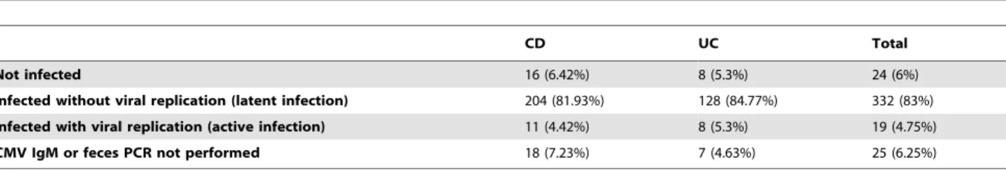
Using serology and the results from the CMV DNA PCR, it is possible to conclude that 24 patients were not infected with CMV, 332 patients were infected without viral replication, and 19 were infected with viral replication. (Table 6)
Clinical-Virological Correlations
After dividing the patients into three different groups, the not infected, the latent infection and the active infection groups, we looked for associations between these groups and the variables of interest.
Analyzing exclusively CD patients, we found an association between the use of infliximab and CMV viral replication (p = 0.0045) and between the use of combined therapy and viral replication (p = 0.005). In UC patients, no variable was signifi-cantly associated with the three different groups of CMV infection. Table 4.Serologic distribution for CMV in patients with CD and UC.
Serologic CD UC Total
IgG (-) IgM (-) 16 (7%) 8 (5,6%) 24 (6,5%)
IgG (+) IgM (-) 207 (90,4%) 131 (91,6%) 338 (90,9%)
IgG (+) IgM (+) 6 (2,6%) 4 (2,8%) 10 (2,6%)
IgG (-) IgM (+) 0 0 0
Not performed 20 8 28
doi:10.1371/journal.pone.0111574.t004
Table 5.Analysis of CMV DNA by qualitative PCR on the feces of study patients.
Feces PCR CD UC Total
(+) 5 (2,01%) 4 (2,65%) 9 (2,25%)
(-) 239 (95,98%) 146 (96,69%) 385 (96,25%)
Not performed 5 (2,01%) 1 (0,66%) 6 (1,5%)
Considering all patients with inflammatory bowel disease, the likelihood ratio test demonstrated a statistically significant association between the use of combined therapy and the CMV infection group (Table 7).
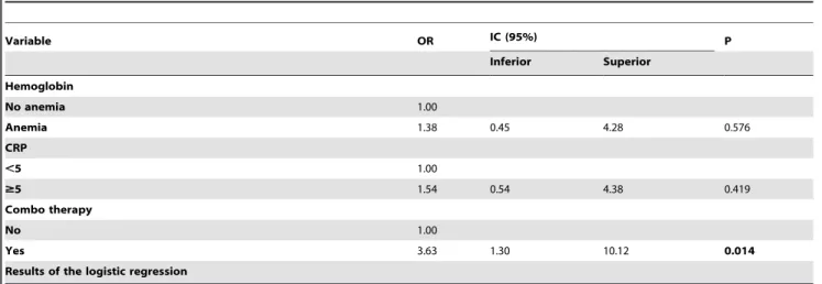
Utilizing the variables in univariate tests with descriptive levels below 0.2 (p,0.2), we verified whether the infection’s character-istics as whole could influence replication. We found that a patient using combined therapy had a chance of replication that was 3.63 times greater than that of patients not using this treatment (p = 0.014) (Table 8).
Discussion
This study examined patients with IBD. Without any previous selection, 400 outpatients were randomly selected in order to evaluate a group that would best represent the general population of patients with IBD from the HCFMUSP.
More than half of these patients had intestinal disease activity, as confirmed by fecal calprotectin. Based on the clinical index, most patients were categorized with quiescent/mild disease (50.8%/22.18%CD and 0/86% of UC). However, such clinical indices have been criticized due to their highly variable sensitivity and specificity, and we believe that fecal calprotectin is the better parameter to demonstrate that half of the patients had active IBD. These results corroborate the literature, which is quite critical of these indices but reports a good sensitivity and specificity of fecal calprotectin in relation to endoscopic findings [8,31,32].
The very high percentage of patients infected with CMV, as evidenced by the presence of IgG antibodies to CMV, can be justified by the socioeconomic status of the study group [19,33,34]. Using Fischer’s test and simple logistic regression, we did not find significant differences between the group of uninfected patients and the group with latent infections in relation to the activity index of IBD, CRP levels or fecal calprotectin. This contradicts the original hypothesis, which held that the latent CMV infection could worsen the IBD activity compared to the uninfected CMV patients [14,15,16,35]. It is worth noting that only 24 patients (6.45%) were not infected, instead having negative antibodies IgG and IgM for CMV.
Despite the large number of patients undergoing immunosup-pressive therapy, comprising more than half of patients with active IBD, viral replication was not detected by PCR in their blood in any case. These null results may have been obtained because our sample was composed of IBD outpatients, rather than a specifically selected population. Our study corroborates other studies arguing that the frequency of viral replication and CMV disease in patients with IBD is not as high as sometimes reported, questioning the virulence of CMV in patients with IBD [21,24,25,26,27]. The detection of CMV DNA in the fecal specimens by a qualitative PCR assay employing primers to the gB region, a highly variable region of HCMV, could underestimate the number of patients shedding CMV in feces. However, as this
assay was performed blindly in all samples regardless of the clinical category or activity index of inflammatory bowel disease, we believe that our result concerning the lack of association between CMV and worsening of intestinal inflammatory activity was not affected.
When we began our study, the association between IBD and CMV was controversial, with the literature pointing to the risk that the rates of CMV infection, replication and disease might be higher in IBD patients. As the present project has progressed, new studies have emerged to challenge these ideas, suggesting that CMV is an occasional finding, a bystander in IBD enterocolitis [21,24,25,26,27].
In our series of ulcerative colitis patients, we did not find any patients with a severe form of the disease, according to the Truelove-Witts classification, which affects the assessment of the role of CMV in this particular group of patients. CD patients were found in all categories of CMV infection, but we did not find any correlation between CMV and the severity of IBD. We found that, in patients with mild and moderate forms of CD and UC, there was no association between the severity of IBD and CMV viral replication in the blood or stool specimens.
Pillet et al. [26] suggest that immunosuppressive therapy does not appear to have an impact on CMV reactivation in CD patients. They state that TNF alpha enhances CMV viral replication and that anti-TNF drugs may reduce the risk of CMV reactivation. However, we detected CMV viral replication in UC and CD patients, indicating that anti-TNF alpha does not protect against viral replication. In CD patients, an association was found between the viral replication of CMV and anti-TNF alpha, contradicting the hypothesis put forth by Pillet et al.
In the present study, we found a statistically significant association between CMV viral replication and the use of combined therapy, consisting of biological therapy (infliximab or adalimumab) plus an immunosuppressive drug (azathioprine or methotrexat). We believe that higher immunosuppression favors viral replication. However, the increase in CMV replication was not accompanied by a greater intestinal inflammatory activity, as evidenced by fecal the calprotectin levels. The present findings further support the hypothesis that CMV is a spectator of the inflammatory process in IBD. The higher the degree of immunosuppressive state caused by the therapy and/or disease is, the greater the chance for CMV replication; however, this finding did not seem to worsen the IBD activity.
The methodological difficulties in achieving a clear categoriza-tion of the CMV infeccategoriza-tions in uninfected, infected non-replicating, and infected with replication and with CMV disease patients might jeopardize the critical evaluation of each work. This effect can be seen in the large variation among the rates of CMV disease that have been published in previous works, suggesting that many cases of the disease were most likely misdiagnosed as CMV infection or CMV replication.
Table 6.Patients distribution regarding CMV infection.
CD UC Total
Not infected 16 (6.42%) 8 (5.3%) 24 (6%)
Infected without viral replication (latent infection) 204 (81.93%) 128 (84.77%) 332 (83%)
Infected with viral replication (active infection) 11 (4.42%) 8 (5.3%) 19 (4.75%)
CMV IgM or feces PCR not performed 18 (7.23%) 7 (4.63%) 25 (6.25%)
Variable Not infected Infected without replication Infected with replication Total p
N % N % N %
Hemoglobin 0.195#
No anemia 17 5.8 264 90.1 12 4.1 293
Anemia 7 8.5 68 82.9 7 8.5 82
CRP 0.395
,5 14 6.1 207 90.0 9 3.9 230
$5 9 6.4 122 86.5 10 7.1 141
Fecal calprotectin 0.464
,150 10 5.6 163 90.6 7 3.9 180
$150 14 7.2 168 86.6 12 6.2 194
Sulfasalazine 0.587
No 19 7.0 237 87.5 15 5.5 271
Yes 5 4.9 94 91.3 4 3.9 103
Mesalazine 0.704
No 18 6.9 230 88.5 12 4.6 260
Yes 6 5.3 101 88.6 7 6.1 114
Azathioprine 0.165
No 10 5.1 181 91.4 7 3.5 198
Yes 14 8.0 150 85.2 12 6.8 176
Steroids 0.433#
No 21 6.1 305 89.2 16 4.7 342
Yes 3 9.4 26 81.3 3 9.4 32
Ifx 0.068#
No 22 7.3 266 88.7 12 4.0 300
Yes 2 2.7 65 87.8 7 9.5 74
Ada 0.066#
No 24 6.8 314 88.7 16 4.5 354
Yes 0 0.0 17 85.0 3 15.0 20
Combo therapy 0.001#
No 24 7.4 287 88.9 12 3.7 323
Yes 0 0.0 44 86.3 7 13.7 51
Result of the chi-square. #Result of the likelihood ratio doi:10.1371/journal.pone.0111574.t007
Inflammato
ry
Bowel
Disease
and
Cytomegalo
virus
ONE
|
www.ploson
e.org
6
November
2014
|
Volume
9
|
Issue
11
|
Based on the improved standardization of diagnostic methods and increasing understanding of the pathophysiology of CMV, studies should soon converge on lower rates of diagnosis for CMV disease in the IBD population.
In summary, our study has shown that latent infection by CMV was quite prevalent in a study group of patients with inflammatory bowel disease. We found no association between IBD activity and CMV infection in IBD patients, even among patients with viral replication using immunosuppressive therapy. Finally, although the use of combined therapies in patients with IBD was associated with the viral replication of CMV, such therapies bore no relation to inflammatory activity of IBD.
Author Contributions
Conceived and designed the experiments: AMC AZAL CSP MGT AMS. Performed the experiments: AMC FMS CLOA IN CSF FUG LSVB. Analyzed the data: AMC AZAL CSP MGT AMS. Wrote the paper: AMC FMS CLOA AZAL CSP MGT AMS.
References
1. Staras SA, Dollard SC, Radford KW, Flanders WD, Pass RF, et al. (2006) Seroprevalence of cytomegalovirus infection in the United States, 1988–1994. Clin Infect Dis 43: 1143–1151.
2. Goodgame RW (1993) Gastrointestinal cytomegalovirus disease. Ann Intern Med 119: 924–935.
3. Rowshani AT, Bemelman FJ, van Leeuwen EM, van Lier RA, ten Berge IJ (2005) Clinical and immunologic aspects of cytomegalovirus infection in solid organ transplant recipients. Transplantation 79: 381–386.
4. Vilela EG, Torres HO, Martins FP, Ferrari Mde L, Andrade MM, et al. (2012) Evaluation of inflammatory activity in Crohn’s disease and ulcerative colitis. World J Gastroenterol 18: 872–881.
5. Best WR, Becktel JM, Singleton JW, Kern F Jr (1976) Development of a Crohn’s disease activity index. National Cooperative Crohn’s Disease Study. Gastroen-terology 70: 439–444.
6. Truelove SC, Witts LJ (1955) Cortisone in ulcerative colitis; final report on a therapeutic trial. Br Med J 2: 1041–1048.
7. Konikoff MR, Denson LA (2006) Role of fecal calprotectin as a biomarker of intestinal inflammation in inflammatory bowel disease. Inflamm Bowel Dis 12: 524–534.
8. Vieira A, Fang CB, Rolim EG, Klug WA, Steinwurz F, et al. (2009) Inflammatory bowel disease activity assessed by fecal calprotectin and lactoferrin: correlation with laboratory parameters, clinical, endoscopic and histological indexes. BMC Res Notes 2: 221.
9. Papadakis KA, Tung JK, Binder SW, Kam LY, Abreu MT, et al. (2001) Outcome of cytomegalovirus infections in patients with inflammatory bowel disease. Am J Gastroenterol 96: 2137–2142.
10. Maher MM, Nassar MI (2009) Acute cytomegalovirus infection is a risk factor in refractory and complicated inflammatory bowel disease. Dig Dis Sci 54: 2456– 2462.
11. Orvar K, Murray J, Carmen G, Conklin J (1993) Cytomegalovirus infection associated with onset of inflammatory bowel disease. Dig Dis Sci 38: 2307–2310. 12. Lakatos PL (2009) Environmental factors affecting inflammatory bowel disease:
have we made progress? Dig Dis 27: 215–225.
13. Criscuoli V, Rizzuto MR, Cottone M (2006) Cytomegalovirus and inflammatory bowel disease: is there a link? World J Gastroenterol 12: 4813–4818. 14. Kambham N, Vij R, Cartwright CA, Longacre T (2004) Cytomegalovirus
infection in steroid-refractory ulcerative colitis: a case-control study. Am J Surg Pathol 28: 365–373.
15. Kishore J, Ghoshal U, Ghoshal UC, Krishnani N, Kumar S, et al. (2004) Infection with cytomegalovirus in patients with inflammatory bowel disease: prevalence, clinical significance and outcome. J Med Microbiol 53: 1155–1160. 16. Onyeagocha C, Hossain MS, Kumar A, Jones RM, Roback J, et al. (2009) Latent cytomegalovirus infection exacerbates experimental colitis. Am J Pathol 175: 2034–2042.
17. Helbling D, Breitbach TH, Krause M (2002) Disseminated cytomegalovirus infection in Crohn’s disease following anti-tumour necrosis factor therapy. Eur J Gastroenterol Hepatol 14: 1393–1395.
18. Pickering O, Weinstein T, Rubin LG (2009) Fatal disseminated cytomegalovirus infection associated with infliximab and 6-mercaptopurine therapy in a child with Crohn disease. Pediatr Infect Dis J 28: 556.
19. Domenech E, Vega R, Ojanguren I, Hernandez A, Garcia-Planella E, et al. (2008) Cytomegalovirus infection in ulcerative colitis: a prospective, comparative study on prevalence and diagnostic strategy. Inflamm Bowel Dis 14: 1373–1379. 20. Dimitroulia E, Spanakis N, Konstantinidou AE, Legakis NJ, Tsakris A (2006) Frequent detection of cytomegalovirus in the intestine of patients with inflammatory bowel disease. Inflamm Bowel Dis 12: 879–884.
21. Matsuoka K, Iwao Y, Mori T, Sakuraba A, Yajima T, et al. (2007) Cytomegalovirus is frequently reactivated and disappears without antiviral agents in ulcerative colitis patients. Am J Gastroenterol 102: 331–337. 22. Criscuoli V, Casa A, Orlando A, Pecoraro G, Oliva L, et al. (2004) Severe acute
colitis associated with CMV: a prevalence study. Dig Liver Dis 36: 818–820. 23. Maconi G, Colombo E, Zerbi P, Sampietro GM, Fociani P, et al. (2005)
Prevalence, detection rate and outcome of cytomegalovirus infection in ulcerative colitis patients requiring colonic resection. Dig Liver Dis 37: 418–423. 24. Lawlor G, Moss AC (2010) Cytomegalovirus in inflammatory bowel disease:
Pathogen or innocent bystander? Inflamm Bowel Dis.
25. Leveque N, Brixi-Benmansour H, Reig T, Renois F, Talmud D, et al. (2010) Low frequency of cytomegalovirus infection during exacerbations of inflamma-tory bowel diseases. J Med Virol 82: 1694–1700.
26. Pillet S, Pozzetto B, Jarlot C, Paul S, Roblin X (2012) Management of cytomegalovirus infection in inflammatory bowel diseases. Dig Liver Dis 44: 541–548.
27. Kim JJ, Simpson N, Klipfel N, Debose R, Barr N, et al. (2010) Cytomegalovirus infection in patients with active inflammatory bowel disease. Dig Dis Sci 55: 1059–1065.
Table 8.Results of the multiple logistic regression model to explain the replication of cytomegalovirus.
Variable OR IC (95%) P
Inferior Superior
Hemoglobin
No anemia 1.00
Anemia 1.38 0.45 4.28 0.576
CRP
,5 1.00
$5 1.54 0.54 4.38 0.419
Combo therapy
No 1.00
Yes 3.63 1.30 10.12 0.014
Results of the logistic regression
28. Bankier AT, Beck S, Bohni R, Brown CM, Cerny R, et al. (1991) The DNA sequence of the human cytomegalovirus genome. DNA Seq 2: 1–12. 29. Chou SW, Dennison KM (1991) Analysis of interstrain variation in
cytomegalovirus glycoprotein B sequences encoding neutralization-related epitopes. J Infect Dis 163: 1229–1234.
30. Boom R, Sol C, Weel J, Lettinga K, Gerrits Y, et al. (2000) Detection and quantitation of human cytomegalovirus DNA in faeces. J Virol Methods 84: 1– 14.
31. Gisbert JP, McNicholl AG (2009) Questions and answers on the role of faecal calprotectin as a biological marker in inflammatory bowel disease. Dig Liver Dis 41: 56–66.
32. Denis MA, Reenaers C, Fontaine F, Belaiche J, Louis E (2007) Assessment of endoscopic activity index and biological inflammatory markers in clinically
active Crohn’s disease with normal C-reactive protein serum level. Inflamm Bowel Dis 13: 1100–1105.
33. Pannuti CS, Vilas-Boas LS, Angelo MJ, Carvalho RP, Segre CM (1985) Congenital cytomegalovirus infection. Occurrence in two socioeconomically distinct populations of a developing country. Rev Inst Med Trop Sao Paulo 27: 105–107.
34. Suassuna JH, Leite LL, Villela LH (1995) Prevalence of cytomegalovirus infection in different patient groups of an urban university in Brazil. Rev Soc Bras Med Trop 28: 105–108.