Efficacy and safety of Elevate® system on apical and anterior
compartment prolapse repair with personal technique
modification
_______________________________________________
Daniele Castellani
1, Vikiela Galica
1, Pietro Saldutto
1, Giuseppe Paradiso Galatioto
1, Carlo Vicentini
11 Department of Life, Health & Environmental Sciences, University of L’Aquila, Urology Unit, “Giuseppe Mazzini” Hospital, Teramo, Italy
ABSTRACT
ARTICLE
INFO
______________________________________________________________ ______________________
Aim: To evaluate the effectiveness and safety of Anterior Elevate® mesh kit system (AES) in woman with symptomatic stage 3 or 4 anterior and/or apical pelvic organ prolapse (POP).
Materials and Methods: This retrospective, monocentric, single surgeon study enrolled between May 2010 and January 2013 fifty-six woman experiencing symptomatic an-terior vaginal prolapse with or without apical descent (POP-Q stage 3 or 4). All women received a AES and 7 (12.5%) received a concomitant transvaginal hysterectomy. Pri-mary endpoint was anatomic correction of prolapse; success was defined as POP-Q stage ≤ 1 or asymptomatic stage 2. Secondary endpoints were quality-of-life (QOL) re-sults and patients’ safety outcomes, which were assessed by 3 validated self-reporting questionnaires at baseline and annually: ICIQ-UI short form, ICIQ-VS and P-QOL. All patients completed 2-years and 28 women 3-years of follow-up. Surgical approach was modified in women with uterus, moving the two-propylene strips anteriorly around the cervix itself crossing one another, so the left will take place in the right side and the right on the opposite. This modification was made in order to better support the uterus.
Results: Vaginal mesh exposure was present in 3 (5,3%) patients. Very good anatomi-cal outcomes were seen, with one (1,8%) failure at 6-months, 4 (7,1%) at 1-year, 6 at 2-years (10,7%). Statistically significant improvements were seen in the ICIQ-VS and P-QOL questionnaires throughout follow-up.
Conclusion: Our data suggest that AES is a minimally-invasive transvaginal procedure to repair anterior and apical POP, with good evidence related to mid-term safety and efficacy.
Keywords:
Pelvic Organ Prolapse; Surgical Mesh; Vagina; Surgical Procedures, Operative
Int Braz J Urol. 2017; 43: 1115-21
_____________________ Submitted for publication: April 21, 2016
_____________________ Accepted after revision: September 11, 2016 _____________________ Published as Ahead of Print: January 24, 2017
INTRODUCTION
Pelvic organ prolapse (POP) occurs when there is a disruption of the natural supporting structures of the pelvic organs, often with impai-red function of the pelvic floor musculature. The loss of these normal attachments and the dynamic support of the pelvic floor result in the descent of one or more pelvic structures including the
the lifetime risk of needing surgery for prolapse or urinary incontinence by 80 years of age has been reported as high as 19% (3). Many women with clinically evident POP on physical exami-nation may be asymptomatic. When symptoma-tic, they complain of bothersome symptoms that can be divided in vaginal urinary, defecatory and sexual. Treatment for POP is based upon symp-tom bother, patient expectation, and quality of life impact. The direct costs of POP in the United States has been estimated at U.S. $1billion, with similar costs expected throughout the developed countries (4). Given the increasing time and re-sources that will be required for POP surgery in the future, it is paramount to perform effective, durable, cost-effective interventions with minimal morbidity. The failure of traditional repairs has led to the use of graft materials, particularly synthetic mesh, to augment prolapse repair in an attempt to improve success and durability. In the last de-cade, several mesh kits have been developed and commercialized to repair POP through a vaginal approach (5). All these procedures have gained popularity, because of their minimally-invasive approach and low morbidity rate. Despite that, the Food and Drug Administration issued a Pu-blic Health Notification in October 2008 to inform physicians and patients of adverse events related to vaginal reconstructive surgical use of synthetic mesh and to provide recommendations on how to mitigate risks and counsel patients appropriately (6). A wide spectrum of potential complications exists with the use of transvaginal mesh in POP surgery, even severe complications, such as fistula formation, mesh erosion into adjacent organs, and death. In 2010, after a 15-years of experience on transvaginal POP and stress urinary incontinen-ce (SUI) mesh surgery, we started using the Ele-vate® Anterior and Apical prolapse system (AES) kit (American Medical Systems, Minnetonka, MN) to repair anterior and apical compartment pro-lapse. In women with uterus we decide to make a change to the standard technique, moving the two-propylene strips anteriorly around the cervix itself crossing one another. This approach could give a better support to the uterus left in place. The aim of the study presented in this paper was to evaluate the effectiveness and safety of AES in
women with symptomatic stage 3 or 4 anterior and/or apical POP.
MATERIALS AND METHODS
and to The International Urogynecological Asso-ciation (IUGA)/ICS joint terminology and classifi-cation of the compliclassifi-cations related directly to the insertion of prostheses and grafts in female pelvic floor surgery (13). Between May 2010 and January 2013 fifty-seven women met the criteria and were enrolled in the study, held at Urology Unit, Uni-versity of L’Aquila, Teramo Hospital, Italy. One pa-tient was lost at follow-up. Fifty-six papa-tients were available for analysis. Slings were not performed at the same time in any patient with SUI, becau-se we prefer a staged mesh surgery. Mid-urethral slings were offered subsequently only in patients who needed. Statistical analysis was performed using the Student t-test. Qualitative date are sho-wn as mean±SD. Statistically significantly diffe-rence is considered as p<0.05.
Surgical Technique
AES kit contains a shaped mesh with two--self anchoring tips and two-propylene strips with a self-fixating tip at their top. Distally the mesh has to be fixed in the obturator foramen using the attached self-fixating tips. Proximally two strips have to be attached to the sacrospinous li-gament using the needle present in the kit, which are assembled with the mesh. All patients recei-ved antibiotics prophylaxis with ceftriaxone 1g intravenously 30 minutes prior surgery. All proce-dures were performed by single experienced pel-vic surgeon (C.V.), in a dorsal lithotomic position and under spinal anesthesia. Surgery is different in woman with uterus respect of those with post--hysterectomy vault prolapse. In latter group the AES is placed in the standard fashion, according to the instructions for use. Conversely in patients with uterus in place, surgery begins with an inci-sion at level of posterior vaginal wall, followed by a sharp and blunt dissection bilaterally towards the sacrospinous ligament, where the combined elements are placed in the standard fashion. After that, a second incision and dissection are perfor-med in the anterior vaginal wall. After a blunt dis-section of the cervix, the two-propylene strips are moved anteriorly around the cervix itself crossing one another, so the left will take place in the right side and the right on the opposite. This personal
technique modification can be watched in a video clip available online in the journal website. At this point, surgery is equal in both groups; the dis-tal part of the mesh is anchored using the needle, which drives the self-fixating tips to the obturator internal muscle. The mesh is distally fixed with two tension-free vicryl 2/0 sutures at the level of the bladder neck and proximally to the uterosacral ligaments or their residual part in patients without uterus. Finally, both propylene strips are inserted to the open eyelets and adjusted in a tension-free manner using the locking eyelets. Cystoscopy is performed to rule out any bladder injury. Surgery ends with closure of vaginal wall incisions with a double vicryl suture, 2/0 internally and 0/0 exter-nally, positioning a 16-Fr indwelling catheter and vaginal packing. In case of stage 2 posterior com-partment prolapse a simple colpoperineoplasty is performed.
RESULTS
month were grade I (intravenous analgesics and anti-emetics) in ten cases, and grade III-a (vaginal infected hematoma with wound dehiscence, re-quiring drainage in local anesthesia) in one. This complication can be classified as 3CbT2S1 accor-ding to IUGA/ICS terminology and classification of the complications related directly to the inser-tion of prostheses and grafts in female pelvic floor surgery (13). Transient buttock pain was reported in 4 (7.1%) of the patients in the first week post--operatively and it disappeared spontaneously. All patients were examined 6-weeks after surgery, 6 months and then annually with urine culture and pelvic examination according to POP-Q. ICIQ-UI--short form, ICIQ-VS and P-QOL questionnaires were self-administered annually. Post-operative results are shown in Table-2. All patients comple-ted a 2-years and 28 3-years of follow-up. Anato-mical results were excellent because failure (defi-ned as symptomatic POP stage 2 or stage ≥3) was seen in 1 (1.8%) woman at 6 months, in 4 (7.1%) at 1-year and in 6 (10.7%) at 2-years. At 3-years
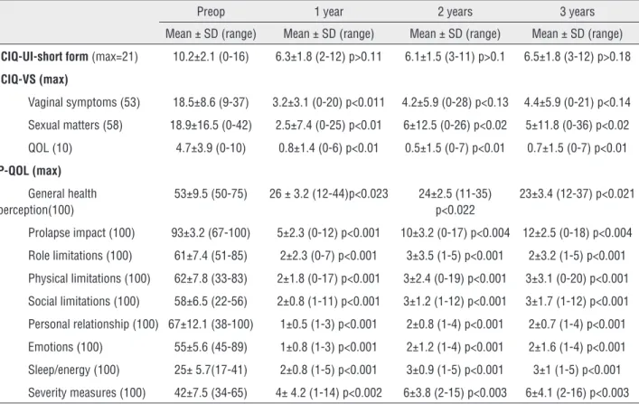
of follow-up only 3 patients out of 28 (10.7%) had POP ≥3 stage (data not showed in Table-2). Vagi-nal mesh exposure, defined as grade III-b (12) or 3BT3S1 (13) complication, was seen in 3 patients (5.3%) during the first year of follow-up. Ques-tionnaires outcomes showed statistical significant improvement of symptoms and QOL domain ex-cept for incontinence (Table-3).
DISCUSSION
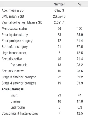
Traditional anterior colporrhaphy for re-pair of anterior prolapse has an estimate risk of recurrence between 30-50% (14, 15). Randomized controlled trials and recent meta-analysis sho-wed superior anatomical outcomes in mesh repair compared to anterior colporrhaphy (14, 16, 17). Nowadays more than 40 implants are available on the market (18) even with little evidence on their safety and efficacy related to mid- and long--term. Indeed, the FDA warned in 2011 regarding serious complications associated with transvagi-nal placement of surgical mesh and reinforced the basis that surgeons should perform prolapse repair only if they are adequately subspecialized in this area (19). The AES is a relative new kit compo-sed of a type I polypropylene mesh with bilateral anterior and posterior graft arms for anchoring them to the obturator foramen and sacro-spinous ligament, respectively. This kit has two major ad-vantages; first its fixation is easy to perform via self-fixating tips, avoiding blind trocar passage through the obturator and perirectal fossa seen with alternative mesh kit techniques. Secondly it seems to fit perfectly anterior and apical prolapse surgical repair, because it is well known that most anterior-compartment prolapse is associated with apical prolapse (20). Crossing of the strips of the mesh anteriorly to the cervix is different from the standard technique. We made this change because we believe that it could better support uterus. Our data show results of the AES for the repair of an-terior and apical vaginal wall prolapse with a mi-nimum of 2-years of follow-up in 56 patients and 3-years in 28. It is possible, hence, to find several important findings; first of all, the AES appears to be a safe and minimally invasive procedure with very low incidence of associated adverse events. Table 1 - Baseline demographics and characteristics.
Number % Age, mean ± SD 69±5.3
BMI, mean ± SD 26.5±4.5 Vaginal deliveries, Mean ± SD 2.6±1.4
Menopausal status 56 100 Prior hysterectomy 33 58.9 Prior prolapse surgery 12 21.4 SUI before surgery 21 37.5 Urge incontinence 7 12.5 Sexually active 40 71.4 Dyspareunia 13 23.2 Sexually inactive 16 28.6 Stage 3 anterior prolapse 22 39.2 Stage 4 anterior prolapse 19 33.9
Apical prolapse
Table 2 - Anatomical POP-Q results and complications at follow-up.
6 weeks 6 months 1 year 2 years n (%) n (%) n (%) n (%) Stage 1 POP 3 (5.3) 6 (10.7) 9 (16) 12 (21.4) Stage 2 POP 1 (1.8) 5 (8.9) 5 (8.9) 6 (10.7)
symptomatic 0 (0) 1 (1.8) 1 (1.8) 2 (3.5) Stage 3 POP 0 (0) 0 (0) 2 (3.5) 3 (5.3) Stage 4 POP 0 (0) 0 (0) 1 (1.8) 1 (1.8)
De novo SUI 5 (3) 4 (7.1) 5 (8.9) 5 (8.9)
Persistent SUI 10 (17.8) 10 (17.8) 10 (17.8) 11 (19.6)
De novo urgency 2 (3.5) 3 (5.3) 8 (14.3) 6 (10.7)
Urge incontinence 0 (0) 1 (1.8) 8 (14.3) 6 (10.7) Persistent dyspareunia 2 (3.5) 5 (8.9) 4 (7.1) 5 (8.9)
De novo dyspareunia 3 (5.3) 3 (5.3) 5 (8.9) 5 (8.9)
Vaginal mesh exposure 0 (0) 1 (1.8) 2 (3.5) 0 (0) Positive Urine Culture 8 (14,3) 10 (17,8) 7 (12,5) 9 (16)
Table 3 - Subjective ICIQ-UI short form, ICIQ-VS and P-QOL outcomes at baseline and after AES implant.
Preop 1 year 2 years 3 years Mean ± SD (range) Mean ± SD (range) Mean ± SD (range) Mean ± SD (range)
ICIQ-UI-short form (max=21) 10.2±2.1 (0-16) 6.3±1.8 (2-12) p>0.11 6.1±1.5 (3-11) p>0.1 6.5±1.8 (3-12) p>0.18
ICIQ-VS (max)
Vaginal symptoms (53) 18.5±8.6 (9-37) 3.2±3.1 (0-20) p<0.011 4.2±5.9 (0-28) p<0.13 4.4±5.9 (0-21) p<0.14 Sexual matters (58) 18.9±16.5 (0-42) 2.5±7.4 (0-25) p<0.01 6±12.5 (0-26) p<0.02 5±11.8 (0-36) p<0.02 QOL (10) 4.7±3.9 (0-10) 0.8±1.4 (0-6) p<0.01 0.5±1.5 (0-7) p<0.01 0.7±1.5 (0-7) p<0.01
P-QOL (max)
General health perception(100)
53±9.5 (50-75) 26 ± 3.2 (12-44)p<0.023 24±2.5 (11-35) p<0.022
23±3.4 (12-37) p<0.021
Prolapse impact (100) 93±3.2 (67-100) 5±2.3 (0-12) p<0.001 10±3.2 (0-17) p<0.004 12±2.5 (0-18) p<0.004 Role limitations (100) 61±7.4 (51-85) 2±2.3 (0-7) p<0.001 3±3.5 (1-5) p<0.001 2±3.2 (1-5) p<0.001 Physical limitations (100) 62±7.8 (33-83) 2±1.8 (0-17) p<0.001 3±2.4 (0-19) p<0.001 3±3.1 (0-20) p<0.001 Social limitations (100) 58±6.5 (22-56) 2±0.8 (1-11) p<0.001 3±1.2 (1-12) p<0.001 3±1.7 (1-12) p<0.001 Personal relationship (100) 67±12.1 (38-100) 1±0.5 (1-3) p<0.001 2±0.8 (1-4) p<0.001 2±0.7 (1-4) p<0.001 Emotions (100) 55±5.6 (45-89) 1±0.8 (1-3) p<0.001 2±1.2 (1-4) p<0.001 2±1.6 (1-4) p<0.001 Sleep/energy (100) 25± 5.7(17-41) 2±0.8 (1-5) p<0.001 3±0.9 (1-5) p<0.001 3±1 (1-5) p<0.001 Severity measures (100) 42±7.5 (34-65) 4± 4.2 (1-14) p<0.002 6±3.8 (2-15) p<0.003 6±4.1 (2-16) p<0.003
There was no bladder and rectal injuries. Only 3 vaginal mesh exposure were identified and were treated surgically with excision of the exposed vaginal area. Transient buttock pain was repor-ted in 4 (7.1%) of the patients in the first week post-operatively and it disappeared spontaneou-sly. The mechanism of this transient pain is like-ly due to local entrapment of pudendal branches such as the perforating cutaneous nerve (21). Secondly, our mean operative times is shorter (47.3 min) than reports of abdominal (221–225 min) and robotic (226–328 min) sacrocolpope-xy (22, 23). Third, our series demonstrate very good anatomical outcome, with one (1.8%) fai-lure at 6-months, 4 (7.1%) at 1-year, 6 at 2-ye-ars (10.7%). At 3-ye2-ye-ars follow-up only 3 patients out of 28 (10.7%) were POP ≥3 stage. Our 1-year anatomic results were similar to other transva-ginal mesh procedure; Vaiyapuri (24) reported in his series of Prolift® a cure rate of 92.1%, Jac-quetin (25) 81.6% of success rate in TVM tech-nique. The anatomic result remained stable for the next two years (89.2% at 2-year of follow-up and 87.4% at 3-year of follow-up). Our results are consistent with other AES series recently pu-blished (26-27). Both Rapp (26) and Huang (27) have 90% of anatomical success rate at 2-years follow-up. Anatomic failure is present in our se-ries at one-year follow-up and it remains almost the same during the next two years; no patients required a second surgery so far. Last but not least, the current series demonstrates excellent subjective outcomes; ICIQ-VS and P-QOL ques-tionnaires demonstrated statistically significant improvements not only in vaginal and sexual symptoms, but also in QOL at each follow-up visit. Regarding preoperative SUI we prefer, as previously mentioned, a staged surgery in these patients, because restoring pelvic organ support has cured SUI in 10 women out of 21 who had preoperative SUI as showed in Table-2 (11 pa-tients with persistent SUI after 2-years of follow--up). We performed a secondary sling procedure only in patients asking for it (8 women) after at least one year of follow-up. Our results sho-wed that AES is a minimally-invasive transvagi-nal procedure to repair anterior and apical POP, with good evidence related to mid-term safety
and efficacy. Further studies are indeed needed to confirm the long-term results.
ABBREVIATIONS
POP = Pelvic organ prolapse
AES = Elevate® Anterior and Apical prolapse sys-tem
SUI = stress urinary incontinence
POP-Q = pelvic organ prolapse quantitative ICS = International Continence Society QOL = quality-of-life
ICIQ-UI = International Consultation on Inconti-nence questionnaire on urinary incontiInconti-nence ICIQ-VS = International Consultation on Inconti-nence questionnaire on vaginal symptoms
P-QOL = prolapse-quality of life questionnaire
ACKNOWLEDGMENT
The authors would like to thank Dr. Ales-sandra Valloni and Dr. Davide Milardi for their outstanding contribution in editing surgical video.
CONFLICT OF INTEREST
None declared.
REFERENCES
1. Maher C, Baessler K, Barber M, et al. Surgical management of pelvic organ prolapse. In: Abrams C, Khoury W (eds), 5th International Consultation on Incontinence. Paris, Health Publication Ltd. 2013; pp. 1379-81.
2. Samuelsson EC, Victor FT, Tibblin G, Svärdsudd KF. Signs of genital prolapse in a Swedish population of women 20 to 59 years of age and possible related factors. Am J Obstet Gynecol. 1999;180:299-305.
3. Smith FJ, Holman CD, Moorin RE, Tsokos N. Lifetime risk of undergoing surgery for pelvic organ prolapse. Obstet Gynecol. 2010;116:1096-100.
4. Subak LL, Waetjen LE, van den Eeden S, Thom DH, Vittinghoff E, Brown JS. Cost of pelvic organ prolapse surgery in the United States. Obstet Gynecol. 2001;98:646-51.
6. Food and Drug Administration, FDA Safety Communication: UPDATE on Serious Complications Associated with Transvaginal Placement of Surgical Mesh for Pelvic Organ Prolapse, FDA, Silver Spring, Md, USA, 2011. available at <http://www.fda.gov/MedicalDevices/Safety/AlertsandNotices/ ucm262435.htm> (Accessed on August 08, 2011).
7. Bump RC, Mattiasson A, Bø K, Brubaker LP, DeLancey JO, Klarskov P, et al. The standardization of terminology of female pelvic organ prolapse and pelvic floor dysfunction. Am J Obstet Gynecol. 1996;175:10-7.
8. Abrams P, Cardozo L, Fall M, Griffiths D, Rosier P, Ulmsten U, et al. The standardisation of terminology of lower urinary tract function: report from the Standardisation Sub-committee of the International Continence Society. Neurourol Urodyn. 2002;21:167-78.
9. Avery K, Donovan J, Peters TJ, Shaw C, Gotoh M, Abrams P. ICIQ: a brief and robust measure for evaluating the symptoms and impact of urinary incontinence. Neurourol Urodyn. 2004;23:322-30.
10. Price N, Jackson SR, Avery K, Brookes ST, Abrams P. Development and psychometric evaluation of the ICIQ Vaginal Symptoms Questionnaire: the ICIQ-VS. BJOG. 2006;113:700-12.
11. Digesu GA, Khullar V, Cardozo L, Robinson D, Salvatore S. P-QOL: a validated questionnaire to assess the symptoms and quality of life of women with urogenital prolapse. Int Urogynecol J Pelvic Floor Dysfunct. 2005;16:176-81. 12. Dindo D, Demartines N, Clavien PA. Classification of
surgical complications: a new proposal with evaluation in a cohort of 6336 patients and results of a survey. Ann Surg. 2004;240:205-13.
13. Haylen BT, Freeman RM, Swift SE, Cosson M, Davila GW, Deprest J, et al. An International Urogynecological Association (IUGA)/International Continence Society (ICS) joint terminology and classification of the complications related directly to the insertion of prostheses (meshes, implants, tapes) and grafts in female pelvic floor surgery. Neurourol Urodyn. 2011;30:2-12.
14. Altman D, Väyrynen T, Engh ME, Axelsen S, Falconer C; Nordic Transvaginal Mesh Group. Anterior colporrhaphy versus transvaginal mesh for pelvic-organ prolapse. N Engl J Med. 2011;364:1826-36. Erratum in: N Engl J Med. 2013;368:394.
15. Kapoor DS, Nemcova M, Pantazis K, Brockman P, Bombieri L, Freeman RM. Reoperation rate for traditional anterior vaginal repair: analysis of 207 cases with a median 4-year follow-up. Int Urogynecol J. 2010;21:27-31.
16. Nguyen JN, Burchette RJ. Outcome after anterior vaginal prolapse repair: a randomized controlled trial. Obstet Gynecol. 2008;111:891-8.
17. Maher C, Feiner B, Baessler K, Adams EJ, Hagen S, Glazener CM. Surgical management of pelvic organ prolapse in women. Cochrane Database Syst Rev. 2010;4:CD004014.
18. Washington JL. Commercial products for pelvic repair. Female Pelvic Med Reconstr Surg. 2011;17:218-25. 19. FDA safety communication: update on serious
complications associated with transvaginal placement of surgical mesh for pelvic organ prolapse. available at. <http://www.fda.gov/medicaldevices/safety/ alertsandnotices/ucm262435.htm> (Accessed on July 13, 2011).
20. Rooney K, Kenton K, Mueller ER, FitzGerald MP, Brubaker L. Advanced anterior vaginal wall prolapse is highly correlated with apical prolapse. Am J Obstet Gynecol. 2006;195:1837-40.
21. Bohrer JC, Chen CC, Walters MD. Pudendal neuropathy involving the perforating cutaneous nerve after cystocele repair with graft. Obstet Gynecol. 2008;112:496-8. 22. Elliott CS, Hsieh MH, Sokol ER, Comiter CV, Payne CK,
Chen B. Robot-assisted versus open sacrocolpopexy: a cost-minimization analysis. J Urol. 2012;187:638-43. 23. Geller EJ, Siddiqui NY, Wu JM, Visco AG.
Short-term outcomes of robotic sacrocolpopexy compared with abdominal sacrocolpopexy. Obstet Gynecol. 2008;112:1201-6.
24. Vaiyapuri GR, Han HC, Lee LC, Tseng LA, Wong HF. Use of the Gynecare Prolift system in surgery for pelvic organ prolapse: 1-year outcome. Int Urogynecol J. 2011;22:869-77.
25. Jacquetin B, Fatton B, Rosenthal C, Clavé H, Debodinance P, Hinoul P, et al. Total transvaginal mesh (TVM) technique for treatment of pelvic organ prolapse: a 3-year prospective follow-up study. Int Urogynecol J. 2010;21:1455-62.
26. Rapp DE, King AB, Rowe B, Wolters JP. Comprehensive evaluation of anterior elevate system for the treatment of anterior and apical pelvic floor descent: 2-year followup. J Urol. 2014;191:389-94.
27. Huang KH, Huang LY, Chu LC, Chuang FC, Wu MP, Kung FT. Evaluation of the single-incision Elevate system to treat pelvic organ prolapse: follow-up from 15 to 45 months. Int Urogynecol J. 2015;26:1341-6.