Ureteral orifice involvement by urothelial carcinoma: long
term oncologic and functional outcomes
_______________________________________________
Muammer Altok
1, Ali F. Sahin
2, Mehmet I. Gokce
3, Gokhan R. Ekin
4, Rauf Taner Divrik
51 Department of Urology, MD Anderson Cancer Center, University of Texas, Houston, TX, USA; 2
Department of Urology, Sivas Numune Hospital, Sivas, Turkey; 3 Department of Urology, School of Medicine, Ankara University, Ankara, Turkey; 4 Department of Urology, Tepecik Education and Research Hospital, Izmir, Turkey; 5 Department of Urology, Private Ege City Hospital, Izmir, Turkey
ABSTRACT
ARTICLE
INFO
______________________________________________________________ ______________________
Purpose: Bladder cancer (BC) may involve the ureteral orifice, and the resection of the orifice has oncological and functional consequences such as development of up-per tract urothelial carcinoma (UTUC), vesicoureteral reflux or ureteral stenosis. The aim of this study was to investigate the oncological and functional outcomes of the ureteral orifice resection in BC patients and determine the predictive factors for UTUC development.
Materials and methods: A total of 1359 patients diagnosed with BC, between 1992 and 2012, were reviewed retrospectively. Patients were grouped with respect to orifice resection and compared for development of UTUC, survival and functional outcomes. Kaplan-Meier method was used to compare survival outcomes. Logistic regression analysis was performed to determine predictors of UTUC development.
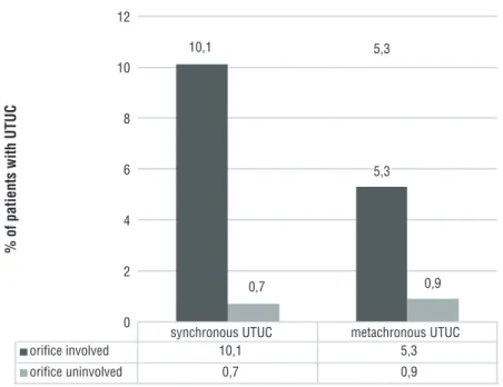
Results: Ureteral orifice involvement was detected in 138 (10.2%) patients. The rate of synchronous (10.1% vs. 0.7%, p=0.0001) and metachronous (5.3% vs. 0.9%, p=0.0001) UTUC development was found to be higher in patients with ureteral orifice involve-ment. Orifice involvement and tumor stage were found to be associated with develop-ment of UTUC in the regression analysis. Overall (p=0.963) and cancer specific survival rates (p=0.629) were found to be similar. Hydronephrosis was also significantly higher in patients with orifice involved BC, due to the orifice obstruction caused by the tumor (33.3% vs. 13.9%, p<0.05).
Conclusions: BC with ureteral orifice involvement has significantly increased the risk of having synchronous or metachronous UTUC. However, orifice involvement was not found to be associated with survival outcomes. Development of stricture due to resec-tion is a very rare complicaresec-tion.
INTRODUCTION
Urothelial carcinoma of the bladder is the most common malignancy of the urinary tract (1, 2). Bladder cancer (BC) may be localized anywhere in the bladder and involvement of ureteral
orifi-ce or its close environment has been reported in 5-35% of the cases (3-7). Involvement of ureteral orifice is a diagnostic and therapeutic dilemma as the disease location itself or the applied treat-ments may cause oncological and functional de-rangements in the upper urinary tract (5, 6, 8, 9).
Keywords:
Urinary Bladder Neoplasms; Hydronephrosis; Therapeutics
Int Braz J Urol. 2017; 43: 1052-9
_____________________
Submitted for publication: April 05, 2017
_____________________
Accepted after revision: May 22, 2017
_____________________
Transurethral resection (TUR) of the urete-ral orifice is necessary during treatment of these cases (3, 4, 8, 10) and TUR of the ureteral orifice is suggested to cause vesicoureteral reflux (VUR), due to the destruction of the muscle fibers, which leads to upper tract urothelial carcinoma (UTUC) development (5, 8, 9). Additionally, the electro--resection of the ureteral orifice may cause orifice stenosis, and secondary obstruction of the upper urinary tract as well (3, 6, 11).
In the current literature there are a num-ber of studies that report the treatment outcomes of patients with involvement of the ureteral orifice (3-12). These studies involve either relatively low number of patients (6, 8-11) or insufficient follow--up data (3, 5, 7, 8).
In this study, we investigated the data of 138 patients underwent orifice resection from a cohort of 1359 patients underwent TUR for uro-thelial carcinoma and aimed to report the oncolo-gical and physiolooncolo-gical outcomes of the patients underwent TUR of the ureteral orifice in compari-son with patients that have no evidence of urete-ral involvement.
MATERIALS AND METHODS
This study began after Local Ethics Com-mittee approval, and the medical records were based on the Oncologic Urology Clinics of Tepe-cik Research and Education Hospital in Izmir, in Turkey. All patients, diagnosed with BC between 1992 and 2012 were reviewed retrospectively, and 1359 patients with available data about tumor lo-calization were evaluated.
The tumors were staged and graded accor-ding to the International Union Against Cancer TNM classification and WHO 1973 grading sche-me (1, 13). The tumors were classified as <3cm or ≥3cm, and as solitary or multiple. An atrophic kidney was detected in some patients due to obs-truction; therefore, the development of hydrone-phrosis was described as hydronehydrone-phrosis±atrophic kidney. Tumors involving the ureteral orifice were treated with wide, deep resection, including the entire orifice area, as mentioned in the literature (7, 9, 10). During TUR, pure cutting current was used and selective coagulation was performed
to achieve hemostasis. According to our depart-mental policy, ureteral stenting was avoided. All patients were routinely evaluated via intravenous urography or ultrasound during the first visit and, if necessary, computed tomography and further imaging were performed. During the follow-up, adjuvant intravesical chemotherapy or immuno-therapy, re-TUR, second TUR, imaging, advanced therapy, etc. were performed according to the va-lid guidelines at the time (13, 14). Survival was calculated from the date of surgery, to either the last follow-up or death.
Statistical analysis was performed using the SPSS 22.0 software program for Windows (SPPS Inc., Chicago, IL, USA). Descriptive statistics for the clinical, pathological and treatment related data were provided. The Student t and Fisher exact tests were used to compare continuous and cate-gorical variables, respectively. Logistic regression analysis was performed to define factors associa-ted with the development of UTUC. Kaplan-Meier analysis was performed to evaluate cancer-speci-fic and overall survival rates of patients with and without ureteral orifice involvement. Cox regres-sion analysis was performed to define the factors associated with survival rates. For statistical signi-ficance p-value of 0.05 was accepted.
RESULTS
Among 1359 patients, 138 (10.2%) had BC involving the ureteral orifice. The two groups did not show significant difference in terms of de-mographic and cancer-related characteristics ex-cept, multiple tumors were significantly more fre-quent in patients without orifice involvement, and hydronephrosis at the initial diagnosis was more prevalent in the group of patients with orifice in-volvement. The patients and tumor characteristics are summarized in Table-1. One patient had a his-tory of nephrectomy for renal cell cancer (RCC) before the diagnosis of BC.
UTUC development
Table 1 - Patients and tumor characteristics.
Characteristics Orifice involved (n=138)
Non-Orifice involved (n=1221)
Total (n=1359) P
Age (Mean ± SD) 65.1±10.5 63.4±11.7 63.5±11.6 0.087
Follow-up, months (mean ± IQR)* 45.5 (9-68) 47.1 (9-70) 46.9 (9-69) 0.721
No. Gender (%)
M 119 (86.2) 1095 (89.7) 1214 (89.3)
0.214 F 19 (13.8) 126 (10.3) 145 (10.7)
No.TCC tumor grade (%)
G1 47 (34.1) 493 (40.4) 540 (39.7)
0.067 G2 36 (26.1) 286 (23.4) 322 (23.7)
G3 40 (29.0) 248 (20.3) 288 (21.2) Unspecified 15 (10.8) 194 (15.9) 209 (15.4)
No.TCC Tumor stage (%)
Ta 47 (34.1) 349 (28.6) 396 (29.1)
0.565 T1 59 (42.8) 520 (42.6) 579 (42.6)
≥T2 32 (23.2) 302 (24.7) 334 (24.6) Unspecified - 50 (4.1) 50 (3.7)
Carsinoma in situ (CIS)(%)
CIS at initial diagnosis 6 (4.3) 42 (3.4) 48 (3.5) 0.584 CIS progression 3 (2.2) 22 (1.8) 25 (1.8) 0.758 Total CIS 9 (6.5) 64 (5.2) 73 (5.3)
No. Tumor size (%)
Tumor < 3 cm 35 (25.4) 348 (28.5) 383 (28.2)
0.305 Tumor ≥ 3 cm 94 (68.1) 755 (61.8) 849 (62.5)
Unspecified 9 (6.5) 118 (9.7) 127 (9.3)
No.Tumor number (%)
Solitary 99 (71.7) 728 (59.6) 827 (60.9)
0.003 Multiple 36 (26.1) 479 (39.2) 515 (37.9)
Unspecified 3 (2.2) 14 (1.1) 17 (1.2)
Hydronephrosis (initial diagnosis)(%)
Hydronephrosis±Atrophic kidney 46 (33.3) 170 (13.9) 216 (15.9) 0.0001
Presence of UTUC (%)
Synchronous 14 (10.1) 8 (0.7) 22 (1.6)
0.0001 Metachronous* 7 (5.3) 11 (0.9) 18 (1.4)
metachronous UTUC could be evaluated in 1299 patients (132 orifice involved bladder cancer) and after a mean follow-up of 47 (IQR: 9-69) months, metachronous UTUC developed in 5.3% and 0.9% of the patients in the orifice involved and uninvolved groups of patients respectively (p=0.0001). The results of synchronous and me-tachronous UTUC are summarized in Figure-1. Logistic regression analysis was performed to determine factors associated with synchronous and metachronous UTUC development. Orifice involvement (OR: 16.044, 95% CI: 6.575-39.151, p=0.0001) and tumor stage (OR: 15.516, 95% CI:1.908-126.182, p=0.01) were identified as the parameters associated with synchronous UTUC development. For metachronous UTUC develo-pment, orifice involvement (OR: 9.141, 95% CI: 3.104-26.923, p=0.0001) and T stage (OR: 8.892, 95% CI: 1.163-67.978, p=0.035) were detected as significant. The results of logistic regression analysis are summarized in Table-2.
Survival analysis
Kaplan-Meier analysis was performed to determine the effect of orifice involvement on cancer-specific and overall survival. Both
cancer-specific and overall survival rates of the orifice involved and uninvolved groups were similar. The survival rates are summarized in Table-3 and Kaplan-Meier figures are given in Figure-2.
Functional outcomes
Development of hydronephrosis or renal failure could be evaluated in 132 of the 138 patients with ureteral orifice involvement. One patient underwent nephrectomy due to RCC and hydronephrosis was present in 44 of these pa-tients prior to resection. Seventeen of these 44 patients also had non-functional kidney and 15 of these patients underwent nephrectomy. Hydronephrosis reversed in 10 of the remaining 27 patients (with hydronephrosis and a func-tioning kidney) after orifice resection. Hydro-nephrosis at the ipsilateral kidney developed in 17 of the 87 remaining patients without ini-tial hydronephrosis. The underlying cause of hydronephrosis was vesicoureteral reflux in 8 (47%) patients, cancer progression and invol-vement of orifice in 5 (29%) patients, stone di-sease in 3 (18%) patients and orifice stenosis in 1 (6%) patient.
Figure 1 - UTUC Status.
orifice uninvolved
orifice involved 10,1
10,1 12
10
8
6
4
2
0
0,7 0,9
5,3 5,3
5,3 0,9 0,7
synchronous UTUC
% of patients with UTUC
Table 2 - Results of logistic regression analysis for development of synchronous and metachronous UTUC.
Synchronous UTUC development Metachronous UTUC development Parameter OR 95% CI P value OR 95% CI P value Age 1.006 0.964-1.051 0.780 0.964 0.922-1.009 0.113 Sex (male vs female) 0.774 0.200-2.994 0.710 0.572 0.069-4.721 0.604 Tumor grade 2.089 0.896-4.868 0.088 1.650 0.164-16.585 0.670 Tumor stage 15.516 1.908-126.182 0.01 8.892 1.163-67.978 0.035 Tumor multiplicity 0.523 0.166-4.648 0.269 0.443 0.158-1.240 0.121 Tumor size (<3 cm vs. ≥3 cm) 0.579 0.200-1.677 0.314 1.731 0.585-5.127 0.322 Orifice involvement 16.044 6.575-39.151 0.0001 9.141 3.104-26.923 0.0001
Table 3 - Survival rates of the ureter orifice involved and uninvolved patient groups.
Time Cancer specific survival rates (%) Overall survival rates (%) Orifice uninvolved Orifice involved P value Orifice
uninvolved
Orifice involved
P value
3 years 85.8 82.0
0.629
61.2 60.8
0.963
5 years 83.8 79.6 52.1 47.5
10 years 76.1 Not reached 33.4 34.2
Figure 2 - Kaplan-Meier curves for cancer specific (2A) and overall survival (2B).
A B
Survival Functions
Survival Functions Ureteral orifice
involvement Ureteral orifice
involvement No
Yes No-censored Yes-censored
No Yes No-censored Yes-censored
1.0
0.8
0.6
0.4
0.2
0.0
0 50 100 150 200 250 300
0 50 100 150 200 250 300
Time (months)
Cum Sur
vival
Cum Sur
vival
Time (months)
1.0
0.8
0.6
0.4
0.2
DISCUSSION
Involvement of ureteral orifice or its close environment by urothelial carcinoma is observed in up to 35% of the cases (4, 6, 7, 9, 12). Resection of the orifice is necessary in these cases and this has potential to result in loss of anti-reflux me-chanism and therefore seeding of malignant cells in the upper urinary tract or ureteral orifice steno-sis, which may lead to renal function impairment. In this study, we reported the long-term oncologi-cal and functional outcomes of 138 patients un-derwent ureteral orifice resection due to involve-ment by urothelial carcinoma and ureteral orifice involvement and resection was shown to increase the risk of UTUC development.
Results of resection of the ureteral orifice have been reported as early as 1936 and in a series of 5 patients, no cases of ureteral orifice stenosis were reported (15). Later on, Rees et al. reported their outcomes in 20 patients, which revealed re-flux in 12 of the 17 patients with follow-up data and no cases of stenosis was reported (4). In the-se two early the-series, no data for development of UTUC was available. The first study with evalua-tion of UTUC development was published by Got-tfries et al. and the authors reported their results of 19 patients with a 12 month mean follow-up. In this, no cases of UTUC or ureteral orifice steno-sis were reported, with 9 patients found to have reflux (9). Resection of ureteral orifice seems to provide favorable results based on the results of these very early studies which have either very low number of patients of very short duration of follow-up. However, De Torres Mateos et al. re-ported 26% rate of reflux following resection and they also found a 22-fold greater risk of UTUC development. Therefore, the authors concluded on close follow-up for UTUC development following resection of the ureteral orifice (5). Palou et al. reported the results of their 19 patients underwent resection of the ureter with a mean follow-up of 57 months and they reported UTUC development in 8 patients (42.1%), and nontumoral stenosis in 3 (16%) of the patients. Therefore, the authors also concluded in closer follow-up of the upper uri-nary tract (11). In a more recent series, Chou et al. reported the results of 31 patients underwent
ureteral orifice resection and UTUC was observed in 4 (12.9%) of the patients after a mean follow-up of 33.5 months. Orifice stenosis was reported in 3 (10%) patients as well (6). In another recent series, Mano et al. reported results from 79 patients and 89 renal units underwent ureteral orifice resec-tion. The median follow-up duration was 15 mon-ths and they reported 11 (13%) patients to develop hydronephrosis. However, orifice stricture was the cause of hydronephrosis in only 3 (4%) of these patients. UTUC development during the follow-up was reported in only one patient (3).
Our study included a high number of patients with ureteral orifice involvement and different from the previous studies we reported synchronous and metachronous UTUC develo-pment separately. Ureteral orifice involvement was found to be associated with 14.4 and 5.7 times increased risk of development of synchro-nous and metachrosynchro-nous UTUC, respectively. This increased rate of development of metachronous UTUC is parallel to the findings of De Torres Mateos et al. (5). But it is much higher compared to the results of Mano et al. (3), which reported UTUC development in only one patient. This di-fference may be associated with didi-fferences in the duration of follow-up.
Also, the logistic regression analysis reve-aled ureteral orifice involvement as a significant factor for the development of synchronous and metachronous UTUC. The risk factors for UTUC in primary BC are strongly related to the primary tumor risk stratification, where the incidence is as low as 0.7% in the low-risk group, to as high as 24% in high-risk groups (16). Tumor grade, the presence of carcinoma in situ (CIS), tumor stage, and tumor multiplicity were the factors identified to have an association with the development of UTUC (11, 16, 17). Our result revealed an evidence for the significance of ureteral orifice involvement for further development of UTUC.
Our data indicate that resection of the ureteral orifice resulted in resolution of hydro-nephrosis in 10 of the 27 patients that have hydronephrosis prior to resection. New develo-ped hydronephrosis was observed in 17 of the 87 patients without prior hydronephrosis and orifice stenosis was the cause in only one pa-tient. This result is consistent with the results of the study by Mano et al. (3). In our series, ureteral catheterization following resection was not performed in any of the patients and urete-ral stricture developed in only one patient. The-refore, we support the idea of ureteral stenting unnecessary, contrary to the results of the study by Chou et al. which reported 10% obstruction rate (6). Ureteral stenting may be beneficial to prevent the consequences related to ureteral ori-fice edema, but fibrotic changes were shown to develop after about a month following surgery, which corresponds to the time for extraction of the ureteral stent (18). Therefore, we recommend against routine ureteral stenting following ure-teral orifice resection and any symptom related to ureteral orifice edema should be tried to be managed conservatively in the first step.
Our study has some limitations. First of all, retrospective nature and inclusion of patients from a 20 years of time interval limits the ho-mogeneity of follow-up and imaging protocols. Additionally, treatment guidelines showed signi-ficant changes during the study period, therefore patients received different adjuvant treatments for urothelial cancer, which has an effect on the survival rates as well.
CONCLUSIONS
The involvement of the ureteral orifice seems to be an important risk factor for both synchronous metachronous UTUC development. However, ureteral orifice involvement was not found to be associated with overall and cancer specific survival outcomes. Resection of ureteral orifice seems to ahieve acceptable functional ou-tcome results. Clinicians should suspect UTUC in patients with BC involving the ureteral orifice, especially when associated with hydronephrosis.
ABBREVIATIONS
BC = Bladder Cancer CIS = Carcinoma in situ TUR = Transurethral resection
UTUC = Upper tract urothelial carcinoma VUR = Vesicoureteral reflux
RCC = Renal Cell Cancer
CONFLICT OF INTEREST
None declared.
REFERENCES
1. Rouprêt M, Babjuk M, Compérat E, Zigeuner R, Sylvester R, Burger M, et al. European Association of Urology. European guidelines on upper tract urothelial carcinomas: 2013 update. Eur Urol. 2013;63:1059-71.
2. Ploeg M, Aben KK, Kiemeney LA. The present and future burden of urinary bladder cancer in the world. World J Urol. 2009;27:289-93.
3. Mano R, Shoshany O, Baniel J, Yossepowitch O. Resection of ureteral orifice during transurethral resection of bladder tumor: functional and oncologic implications. J Urol. 2012;188:2129-33.
4. Rees RW. The effect of transurethral resection of the intravesical ureter during the removal of bladder tumours. Br J Urol. 1969;41:2-5.
5. De Torres Mateos JA, Banús Gassol JM, Palou Redorta J, Morote Robles J. Vesicorenal reflux and upper urinary tract transitional cell carcinoma after transurethral resection of recurrent superficial bladder carcinoma. J Urol. 1987;138:49-51.
6. Chou EC, Lin AT, Chen KK, Chang LS. Superficial transitional cell carcinoma of the ureteral orifice: higher risk of developing subsequent upper urinary tract tumors. Int J Urol. 2006;13:682-5.
7. Kisbenedek L, Szeldeli P, Biró G, Balogh F. Vesicoureteral reflux following transurethral resection of bladder tumours at the ureteral orifice. Eur Urol. 1982;8:9-10.
8. Freed SZ. Vesicoureteral reflux following transurethral resection of bladder tumors. J Urol. 1976;116:184-7. 9. Gottfries A, Nilsson S, Sundin T, Viklund LG. Late effects of
transurethral resection of bladder tumours at the ureteric orifice. Scand J Urol Nephrol. 1975;9:32-5.
11. Palou J, Salvador J, Millán F, Collado A, Algaba F, Vicente J. Management of superficial transitional cell carcinoma in the intramural ureter: what to do? J Urol. 2000;163:744-7. 12. See WA. Distal ureteral regeneration after radical transurethral
bladder tumor resection. Urology. 2000;55:212-5; discussion 215-6.
13. Babjuk M, Burger M, Zigeuner R, Shariat SF, van Rhijn BW, Compérat E, et al. EAU guidelines on non-muscle-invasive urothelial carcinoma of the bladder: update 2013. Eur Urol. 2013;64:639-53.
14. Witjes JA, Compérat E, Cowan NC, De Santis M, Gakis G, Lebret T, et al. EAU guidelines on muscle-invasive and metastatic bladder cancer: summary of the 2013 guidelines. Eur Urol. 2014;65:778-92.
15. Counseller VS, Braasch WF. Diathermy for carcinoma of the bladder. Ann Surg. 1935;101:1418-25.
16. Kirkali Z, Tuzel E. Transitional cell carcinoma of the ureter and renal pelvis. Crit Rev Oncol Hematol. 2003;47:155-69.
17. Ayyathurai R, Soloway MS. Monitoring of the upper urinary tract in patients with bladder cancer. Indian J Urol. 2011;27:238-44.
18. Graham JB. Electroresection injury of the ureteral orifice. J Urol. 1961;86:539-47.
_______________________ Correspondence address:
Muammer Altok, MD Assistant Professor in Urology Department of Urology MD Anderson Cancer Center University of Texas 1515 Holcombe Blvd, Unit 1373