Diagnostic relevance of metastatic renal cell carcinoma in
the head and neck: An evaluation of 22 cases in 671 patients
_______________________________________________
Anja Lieder
1, Thomas Guenzel
2, Steffen Lebentrau
3, Constanze Schneider
4, Achim Franzen
11 Department of Otorhinolaryngology, Ruppiner Kliniken and Brandenburg Medical School
Theodor-Fontane, Neuruppin, Germany; 2 Department of Otorhinolaryngology, Head and Neck Surgery, Borromaeus-Hospital Leer Germany; 3 Department of Urology and Pediatric Urology, Ruppiner Kliniken and Brandenburg Medical School Theodor-Fontane, Neuruppin, Germany; 4 Clinical Cancer Registry Brandenburg, Neuruppin, Germany
ABSTRACT
ARTICLE
INFO
______________________________________________________________ ______________________
Purpose: Renal cell carcinoma (RCC) is a malignant tumor that metastasizes early, and patients often present with metastatic disease at the time of diagnosis. The aim of our evaluation was to assess the diagnostic and differential diagnostic relevance of meta-static renal cell carcinoma (RCC) with particular emphasis on head and neck manifesta-tions in a large patient series.
Patients and methods: We retrospectively evaluated 671 consecutive patients with RCC who were treated in our urology practice between 2000 and 2013.
Results: Twenty-four months after diagnosis, 200/671 (30%) of RCC had metastasized. Distant metastases were found in 172 cases, with 22 metastases (3.3%) in the head and neck. Cervical and cranial metastases were located in the lymph nodes (n=13) and in the parotid and the thyroid gland, tongue, the forehead skin, skull, and paranasal si-nuses (n=9). All head and neck metastases were treated by surgical excision, with 14 patients receiving adjuvant radiotherapy and 9 patients receiving chemotherapy or targeted therapy at some point during the course of the disease. Five patients (23%) survived. The mean time of survival from diagnosis of a head and neck metastasis was 38 months, the shortest period of observation being 12 months and the longest 83 months.
Discussion and conclusion: Our findings show that while RCC metastases are rarely found in the neck, their proportion among distantly metastasized RCC amounts to 13%. Therefore, the neck should be included in staging investigations for RCC with distant metastases, and surgical management of neck disease considered in case of re-sectable metastatic disease. Similarly, in patients presenting with a neck mass with no corresponding tumor of the head and neck, a primary tumor below the clavicle should be considered and the appropriate staging investigations initiated.
Keywords:
Carcinoma, Renal Cell; Neoplasm Metastasis; Carcinoma,
squamous cell of head and neck [Supplementary Concept]
Int Braz J Urol. 2017; 43: 202-8
_____________________ Submitted for publication: December 22, 2015 _____________________ Accepted after revision: June 29, 2016
________________________ Published as Ahead of Print: September 20, 2016
INTRODUCTION
Renal cell carcinoma (RCC) is a malignant tumour of the kidney that metastasizes early. Most
Numerous single case reports and small series of metastasis of RCC into the head and neck region are available in the literature. These case reports focus mainly on particular, unusual, and especially extranodal location of the metas-tases as well as unusual clinical courses (2-8). The aim of this study was to assess the differen-tial diagnostic and also the therapeutic relevance of metastatic RCC in a large series of RCC and to evaluate if a systematic examination of the head and neck is appropriate in the context of staging RCC. We present an analysis on RCC metastasi-zing into the head and neck region based on a large group of 671 consecutive patients with an RCC treated in our unit.
PATIENTS AND METHODS
Medical records of 671 consecutive pa-tients who were diagnosed with an RCC in the De-partment of Urology of Ruppiner Kliniken, a large District General Hospital, between 2000 and 2013 were evaluated. All patients were followed-up un-til the time of their death; surviving patients were followed-up for at least 24 months from the date of diagnosis. All data were collected from case notes,anonymized and maintained in an Apache OpenOffice4 database and analyzed using a sta-tistical software package (Apache OpenOffice4 Calc with R4Calc R extension). As this study was a retrospective case notes study, formal ethical approval was not required. Written consent was obtained from all patients prior to undertaking any procedures but for this retrospective case note audit, formal written consent was not required. All investigations and treatments were carried out according to accepted clinical practice and were compliant with the medical principles of the Dec-laration of Helsinki.
RESULTS
Of 671 consecutive patients diagnosed with RCC, 200 (30%) had distant or regional lymph node metastases either at the time of diagnosis or within 24 months of diagnosis. The overall metastatic rate, including locoregional metastases, was 17% (111/671) at the time of diagnosis of
RCC, with an additional 13% (89/671) diagnosed following treatment.
Distant metastases were found in 172 pa-tients (26%), and regional lymph node metastases in 22 patients (3%). In 92 patients (14%), metasta-ses were identified at the time of diagnosis of the primary tumour, and in the remaining 80 patients (12%) metastasis occurred over the course of the following 24 months despite curative intent treat-ment (Table-1).
Metastases of RCC in the head and neck were found in 22 patients (3%). Sixteen patients were male and six were female. The mean age of these patients at the time of diagnosis was 66 years (32-81 years). In 10 patients (45%), head and neck metastases appeared simultaneously to the primary tumour, or the metastasis was the first manifestation of the RCC. In 12 patients (55%), metastases were detected at follow-up following curative intent treatment after 24 months on average. The longest period between treatment of the primary RCC and the detection of metastases in the Head and Neck was 87 months.
The histological type of renal cell carci-noma was clear cell renal carcicarci-noma in 14 cases, poorly differentiated or undifferentiated carcino-ma in six cases, nephroblastocarcino-ma in one case and small cell renal carcinoma in one case. Tumour Grade was G2 in 8 cases, G3 in 9 cases, and un-determined in 5 cases. Initial TNM stages ranged from T1N0M0 to T3N2M1 at the time of diagnosis.
Table 1 - Patient and tumour characteristics.
No. Age * Histology Grade TNM at
diagnosis
primary
management
Time to
Metastasis **
Location
Metastasis
Other metastases Management of
metastatic disease
Survival after Metastasis
(H&N) in months
1 70 clear cell 2 pT2bpNxM1 nephrectomy 0 Level IV Lymph
node
Lung, bones Excision 2
2 58 clear cell 2 pT3apN0M1 nephrectomy 5 Thyroid Gland Retroperitoneum, pancreas Excision
(Thyroidectomy),
Sunitimib
24 (patient alive)
3 74 clear cell *** unknown nephrectomy 78 Parotid Gland none Excision
(Parotidectomy),
56 (patient alive)
4 52 clear cell/poly 3 pT3pN2 M1 nephrectomy 5 Level IV Lymph
node
Lung, bones Excision, radiotherapy,
chemotherapy
14
5 78 clear cell 2 pT2NxM1 nephrectomy 65 Frontal Sinus Brain, lung, bones, liver Radiotherapy 2
6 69 undifferentiated 3 cT3cN1cM1 chemotherapy 0 Level IV Lymph
node
Bone, liver, peritoneum Excision, radiotherapy,
chemotherapy
14
7 73 clear cell 2 pT1bNxMx nephrectomy 94 Level IV Lymph
node
Lung, bones Excision, radiotherapy,
chemotherapy,
Sorafenib
7
8 69 clear cell 3 pT1cN0cM0 nephrectomy 14 Level IV Lymph
node
Peritoneum Excision, radiotherapy 4
9 70 undifferentiated *** TxN1M1 chemotherapy 0 Level IV Lymph
node
Lung, mediastinum Chemotherapy,
Sunitimib
28
10 69 undifferentiated *** pT3cN0cM1 nephrectomy 24 Level IV Lymph
node
Lung, bones, liver, adrenal
glands
Excision,
chemotherapy,
Sunitimib
12
11 32 Nephroblastoma 3 pT3bpN0cM1 nephrectomy 0 Level IV Lymph
node
Lung, liver Chemotherapy,
radiotherapy
(other centre)
24
12 56 clear cell 3 pT3acN1cM1 nephrectomy 5 Tongue Lung, bones, mediastinal
nodes, soft tissue finger
Excision (glossectomy),
radiotherapy
3
13 66 polymorph 3 pT1bNxMx nephrectomy 75 Frontal Skull
bone
Lung, bones, soft tissue
back
Sunitimib,
chemotherapy
radiotherapy
13
14 81 undifferentiated *** unknown declined treatment 0 Level IV Lymph
node
none Declined treatment 0
15 69 clear cell 2 pT1acNxM0 nephrectomy 69 Level IV Lymph
node
Retroperitoneal lymph
nodes (paraaortal)
Excision, chemotherapy 19
16 68 clear cell 2 pT1bpN0cM0 nephrectomy 36 Facial Skin
(forehead)
Lung, adrenal glands,
jejunum
Excision 19 (patient alive)
17 48 clear cell 2 pT1bN0M0 nephrectomy
adrenalectomy
87 Thyroid Gland Lung, bones, mediastinum Excision, laminectomy,
chemotherapy,
Sunitimib
27 (patient alive)
18 72 small cell *** cT4cN1M1 resection
metastasis
0 Frontal Skull
bone and
mandible
Lung, brain, mediastinal
lymph nodes
Excision, radiotherapy 13
19 73 clear cell 2 pT1a cN0 M1 nephrectomy 40 Thyroid Gland Lung, bones, mediastinum Excision, radiotherapy 86 (patient alive)
20 78 clear cell 3 pT2bNxM0 nephrectomy 6 Level IV Lymph
node
Lung, retroperitoneum Excision, Sunitimib 7
21 63 undifferentiated *** unknown nephrectomy 0 Level IV Lymph
node
Lung Excision, none to lung 2
22 65 undifferentiated *** unknown unknown unknown unknown unknown unknown Lost to follow up
*Patient age at the time of head and neck metastasis
All 22 patients received curative intent tre-atment at the time of diagnosis, except for one pa-tient, who declined treatment. Eighteen patients, all of whom had the primary tumour diagnosed first or synchronous with the head and neck me-tastasis, received a nephrectomy.
In the 18 cases where metastases in the head and neck were found after diagnosis of the primary tumour or at staging of the primary tu-mour, patients received curative intent treatment at the time of initial diagnosis and were then followed-up by either a hospital or community urology tumour surveillance programme.
Nephrectomy was performed in 17 pa-tients, total nephrectomy in 13 patients and par-tial nephrectomy in 4 patients. When metastases of the head and neck occurred, they were treated by surgical resection and adjuvant radiotherapy. In the 10 patients where diagnosis of the RCC head and neck metastasis preceded (4 patients) or coincided (6 patients) with diagnosis of the primary tumour, patients received surgical treat-ment of the head and neck metastasis first follo-wed by surgery to the primary tumour follofollo-wed by adjuvant radiotherapy.
Radiotherapy was performed in 14 cases. Radiotherapy after primary tumour resection was
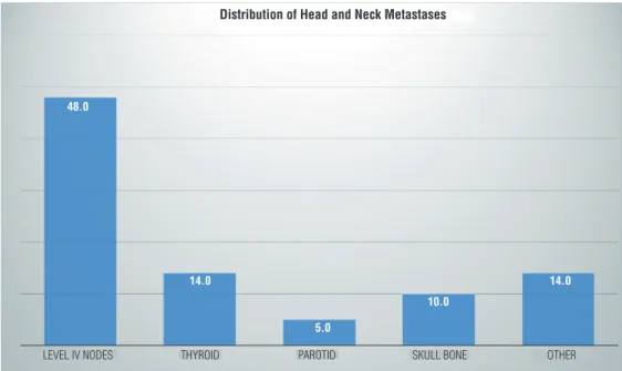
Figure 1 - Distribution of head and neck metastases by location (in % of n=22 patients).
LEVEL IV NODES 48.0
14.0
5.0
10.0
14.0
THYROID PAROTID SKULL BONE OTHER
Distribution of Head and Neck Metastases
performed in one patient, following resection of the head and neck metastasis in 4 cases, and follo-wing detection of other metastases in 9 cases. The dose of radiotherapy was 40 Gray except in four patients who requested palliative treatment; 25 Gray were administered in such cases.
Chemotherapy was performed in 9 pa-tients, usually following the diagnosis of dissemi-nated metastatic disease. Due to the long observa-tion period, chemotherapy regimens changed over time and included both standard chemotherapy, chemoimmunotherapy and targeted therapy. In particular, targeted therapy with either Sunitinibe or Sorafenibe was given to 6 patients.
Five patients (23%) survived and one patient was lost to follow-up. The mean time of survival from diagnosis of a head and neck metastasis was 38 months, the shortest period of observation being 12 months and the lon-gest 83 months (standard deviation 30 months) (Figure-2).
DISCUSSION
RCC are slowly growing, capsule-forming tumours and most frequently metastasize into lung and the lymph nodes, followed by the ske-letal system and the liver – in the majority of the cases, several organ systems are affected simulta-neously (1). The metastatic rate of 17% (111/671) in our patient group at the time of diagnosis, and an additional 13% (89/671) in the further course of the disease, is lower than described in other studies (2). This could be explained by the fact that 66% of our cases had been diagnosed in an
early stage (61% in stage I) and had been well differentiated (Grade 1 + Grade 2: 68%).
RCC are considered to be the third most frequent infraclavicular tumour metastasizing to the head and neck. Supraclavicular metastases were found in 3% (22/671) of all our patients with RCC. In the literature, there are reports of metasta-tic rates of up to 15% (3-6). Whether these results can be compared to those presented here, remains to be discussed with regards to size of study, stage of disease, duration of follow-up and whether all patients were staged specifically for metastases of the head and neck.
While the proportion of RCC metastasizing to the head and neck was low at 3%, we observed head and neck metastases in 11% (22/200) of all metastasized RCC and in 13% (22/172) of all dis-tantly metastasized RCC.
According to our results, RCC metastasizing into the head and neck area primarily metastasize into the cervical lymph nodes. In the literature,
Figure 2 - Survival after diagnosis of a head and neck metastasis, shown in months with standard deviation.
1
0.9
0.8
0.7
0.6
0.5
0.4
0.3
0.2
0.1
0
0 6 12 18 24 30 36
there are several reports about unusual manifestations of RCC in different organs of the head and neck. Single case observations refer to the parotid gland, the skull, the skin, the oral cavity, and the paranasal sinuses (3, 5-10), which were also seen in our patient group. Two large multicentre studies also reported metastatic spread of RCC into the thyroid gland, a phenomenon that was also observed in two of our patients (10, 11).
Our observations also show the variable pat-tern of cervical metastasis of RCC. In some cases, a cervical metastasis may represent the first mani-festation of an RCC, in other cases, cervical me-tastases may occur months or years after curative intent treatment of an RCC (5, 6, 12). In 3 out of 22 patients, a solitary cervical lymph node metas-tasis was the first manifestation of a previously unknown RCC. At the other end of the spectrum, a solitary metastasis appeared in the parotid gland 6 years after diagnosis of the primary tumour. In the other 19 patients, the metastatic spread of the RCC into the head and neck occurred at the same time as metastasis into other organ systems.
Lymph node metastases and metastases of the parotid gland generally occur as painless, re-latively slowly growing tumours (7, 9, 13). Facial nerve palsies in conjunction with parotid metas-tasis of a RCC are rare (3). Metastases within the upper aerodigestive tract such as the oral cavi-ty and the pharynx are often painful. They are usually diagnosed when patients present with sore throats or oral pain, and grow nearly always sub-mucosally, show signs of increasing vasculariza-tion and are often distinguished from mucosa by their red discolouration. Such lesions will bleed profusely when biopsied or haemorrhage spon-taneously, and life-threatening haemorrhage has been reported. Metastases in the supraglottic la-rynx may cause narrowing of the upper airway, stridor and dyspnoea. Manifestations of the nasal cavity or the paranasal sinuses lead to nasal obs-truction, sinusitis-like complaints, or significant haemorrhage (14).
According to our observations, the head and neck were involved in 13% of distantly me-tastasized RCC. This must be considered in pa-tients who are due to undergo extensive surgery of either the primary tumour or metastases in
other locations. Appropriate staging procedu-res would include imaging of the neck by either computed tomography (CT) or magnetic resonan-ce imaging (MRI) with contrast and, if upper ae-rodigestive tract symptoms are present, a laryn-go-pharyngoscopy.
Surgery as therapeutic option of metasta-sized RCC has an great significance. Good onco-logic clearance is achieved in particular if metas-tases occur more than two years after treatment of the primary tumour, and where there is good surgical access. This applies to large case series on treatment outcomes of lung and liver metas-tases of RCC (15-17), and international guidelines recommend surgical therapy of metastases despi-te improvements of chemotherapy outcomes (2). Larger series of surgical therapy of supraclavicular metastases have only been published for patients with thyroid gland metastases. The five-year sur-vival rate of those patients amounted to 51% (10, 11). Only case reports only exist about the surgical therapy of RCC metastases in other supraclavicu-lar locations. Curative therapeutic options exist in cases of single metastasis into the head and neck (7, 8), but surgical management of head and neck metastases can also be appropriate for symptom control in cases of airway obstruction, haemor-rhage, or pain (13). We observed survival of 23% of patients with a head and neck metastasis follo-wing treatment, and would therefore have no he-sitation in recommending curative intent manage-ment of head and neck metastases in all patients fit for surgery.
CONCLUSIONS
sore throat, dysphagia or foreign body sensation should prompt an otolaryngologist examination and an ultrasound examination of the neck and thyroid at the very least, bearing in mind that while most metastases occur in supraclavicular lymph nodes, they may also present in an unusual location. Surgical management of such metastases should be considered in all patients fit for surgery for both curative intent and palliative treatment.
CONFLICT OF INTEREST
None declared.
REFERENCES
1. Deeb R, Zhang Z, Kini S, Ghanem T. Metastatic renal cell carcinoma to the parotid gland presenting 19 years after nephrectomy: case report and review of literature. Laryngoscope. 2010;120:S128.
2. Gnepp DR. Secondary Tumors. In: Ellies GL, Auclair PL. Tumors of the salivary glands. Armed Forces Institute of Pathology, Washington D.C. Chapt. 8. 2008; pp. 471-9. 3. Kundu S, Eynon-Lewis NJ, Radcliffe GJ. Extensive metastatic
renal cell carcinoma presenting as facial nerve palsy. J Laryngol Otol. 2001;115:488-90.
4. Langille G, Taylor SM, Bullock MJ. Metastatic renal cell carcinoma to the head and neck: summary of 21 cases. J Otolaryngol Head Neck Surg. 2008;37:515-21.
5. Ozkiriş M, Kubilay U, Sezen OS. Cervical lymph node metastasis in renal cell carcinoma. J Oral Maxillofac Pathol. 2011;15:211-3.
6. Park YW, Hlivko TJ. Parotid gland metastasis from renal cell carcinoma. Laryngoscope. 2002;112:453-6.
7. Pritchyk KM, Schiff BA, Newkirk KA, Krowiak E, Deeb ZE. Metastatic renal cell carcinoma to the head and neck. Laryngoscope. 2002;112:1598-602.
8. Spreafico R, Nicoletti G, Ferrario F, Scanziani R, Grasso M. Parotid metastasis from renal cell carcinoma: a case report and review of the literature. Acta Otorhinolaryngol Ital. 2008;28:266-8.
9. Torres-Carranza E, Garcia-Perla A, Infante-Cossio P, Belmonte-Caro R, Loizaga-Iriondo JM, Gutierrez-Perez JL. Airway obstruction due to metastatic renal cell carcinoma to the tongue. Oral Surg Oral Med Oral Pathol Oral Radiol Endod. 2006;101:e76-8.
10. Döme B, Hendrix MJ, Paku S, Tóvári J, Tímár J. Alternative vascularization mechanisms in cancer: Pathology and therapeutic implications. Am J Pathol. 2007;170:1-15.
11. Kroeger N, Seligson DB, Klatte T, Rampersaud EN, Birkhäuser FD, Rao PN, et al. Clinical, molecular, and genetic correlates of lymphatic spread in clear cell renal cell carcinoma. Eur Urol. 2012;61:888-95.
12. Ljungberg B, Cowan NC, Hanbury DC, Hora M, Kuczyk MA, Merseburger AS, et al. European Association of Urology Guideline Group. EAU guidelines on renal cell carcinoma: the 2010 update. Eur Urol. 2010;58:398-406.
13. Serouya SM, Dultz LA, Concors SJ, Wang B, Patel KN. Late solitary metastasis of renal cell carcinoma to the submandibular gland. J Oral Maxillofac Surg. 2012;70:2356-9.
14. Mrena R, Leivo I, Passador-Santos F, Hagström J, Mäkitie AA. Histopathological findings in parotid gland metastases from renal cell carcinoma. Eur Arch Otorhinolaryngol. 2008;265:1005-9.
15. Heffess CS, Wenig BM, Thompson LD. Metastatic renal cell carcinoma to the thyroid gland: a clinicopathologic study of 36 cases. Cancer. 2002;95:1869-78.
16. Iesalnieks I, Winter H, Bareck E, Sotiropoulos GC, Goretzki PE, Klinkhammer-Schalke M, et al. Thyroid metastases of renal cell carcinoma: clinical course in 45 patients undergoing surgery. Assessment of factors affecting patients’ survival. Thyroid. 2008;18:615-24.
17. Firek P, Richter S, Jaekel J, Brehmer B, Heidenreich A. Metastasectomy in renal cell cancer after neoadjuvant therapy with multi-tyrosine kinase inhibitors. Urologe A. 2012;51:398-402.
18. Assouad J, Petkova B, Berna P, Dujon A, Foucault C, Riquet M. Renal cell carcinoma lung metastases surgery: pathologic findings and prognostic factors. Ann Thorac Surg. 2007;84:1114-20.
19. Pfannschmidt J, Klode J, Muley T, Dienemann H, Hoffmann H. Nodal involvement at the time of pulmonary metastasectomy: experiences in 245 patients. Ann Thorac Surg. 2006;81:448-54.
20. Staehler MD, Kruse J, Haseke N, Stadler T, Roosen A, Karl A, et al. Liver resection for metastatic disease prolongs survival in renal cell carcinoma: 12-year results from a retrospective comparative analysis. World J Urol. 2010;28:543-7.