Inspiratory muscle training in quadriplegic patients*
,**
Treinamento de músculos inspiratórios em pacientes com quadriplegia
Janne Marques Silveira, Ada Clarice Gastaldi, Cristina de Matos Boaventura, Hugo Celso Souza
Abstract
Objective: To determine whether inspiratory muscle training can increase strength and endurance of these muscles in quadriplegic patients. Methods: Eight quadriplegic patients (7 males and 1 female) with injury to the lower cervical spine (segments C4-C7) were submitted to inspiratory muscle training with a threshold inspiratory muscle trainer adjusted to 30% of MIP. The training sessions were carried out with the patients in a sitting position, 5 days a week for 8 weeks. Endurance time, MIP, MEP and FVC were determined at baseline, week 4 and week 8. Results: In comparison with the mean baseline value, there was an increase in MIP, measured in the sitting position, at weeks 4 and 8 (−83.0 ± 18.9 cmH2O vs. −104.0 ± 19.4 cmH2O and −111.3 ± 22.7 cmH2O). There was an increase in MEP, also in the sitting position, at week 4 (from 36.8 ± 8.1 to 42.6 ± 8.8 cmH2O). There was an improvement in FVC at week 4 (from 2.1 ± 0.8 to 2.5 ± 0.6 L, representing an increase of 24 ± 22%). Although there was an increase in endurance (sitting) at week 8, the difference was not significant in comparison with the baseline value (29.8 ± 21.0 vs. 35.9 ± 15.5 min, an increase of 173 ± 233%). Conclusions: Quadriplegic patients can benefit from training at low loads (30% of MIP), which can improve inspiratory muscle strength, FVC and expiratory muscle performance.
Keywords: Respiratory mechanics; Quadriplegia; Breathing exercises.
Resumo
Objetivo: Determinar se o treinamento de músculos inspiratórios pode aumentar a força e endurance desses músculos em pacientes com quadriplegia. Métodos: Oito pacientes quadriplégicos (7 homens e 1 mulher) com lesão medular cervical entre C4 e C7 foram submetidos ao treinamento de músculos inspiratórios utilizando-se um resistor de carga linear ajustado em 30% da PImáx. As sessões de treinamento foram realizadas com os pacientes sentados 5 vezes por semana por 8 semanas. Tempo de endurance, PImáx, PEmáx e CVF foram medidos antes do treinamento e nas semanas 4 e 8. Resultados: Em comparação ao valor basal médio, houve um aumento da PImáx, mensurada na posição sentada, nas semanas 4 e 8 (−83,0 ± 18,9 cmH2O vs. −104,0 ± 19,4 e −111,3 ± 22,7 cmH2O). Houve aumento da PEmáx, também na posição sentada, na semana 4 (de 36,8 ± 8,1 a 42,6 ± 8,8 cmH2O). Houve uma melhora na FVC na 4ª semana (de 2,1 ± 0,8 a 2,5 ± 0,6 L, representando um incremento de 24 ± 22%). O tempo de endurance (sentado) não apresentou um aumento significativo entre o momento basal e a semana 8 (29,8 ± 21,0 min vs. 35,9 ± 15,5 min; aumento de 173 ± 233%). Conclusões: Pacientes com quadriplegia podem se beneficiar com o treinamento com baixas cargas (30% da PImáx), com melhora da força dos músculos inspiratórios, CVF e efetividade dos músculos expiratórios.
Descritores: Mecânica respiratória; Quadriplegia; Exercícios respiratórios.
* Study carried out in the Triângulo University Center School of Physiotherapy, Uberlândia, Brasil.
Correspondence to: . Departamento de Fisioterapia e Medicina, Rua Rio de Janeiro entre as ruas 9 e 10, Campus II, CEP 77400-000, Gurupi, TO, Brasil.
Tel 55 63 3612-7608 or 3612-7684. E-mail: [email protected] Financial support: None.
Submitted: 9 Sep 2009. Accepted, after review: 12 Feb 2010.
** A versão completa em português deste artigo está disponível em www.jornaldepneumologia.com.br
Introduction
Quadriplegia results from injury to the spinal cord, which can cause severe respiratory impairment depending on the level at which the lesion occurs.(1,2) Lesions above C3 are
life-threatening, causing paralysis of the diaphragmatic, intercostal, scalene and abdominal muscles, thereby leading to a lack of
ventilatory support and the need for mechanical ventilation. If the injury occurs below C3, the diaphragm remains totally or partially innervated.
(3) However, diaphragmatic function is impaired
MIP was enough to produce training effects.(6,7)
However, there have been no studies evaluating the benefits of this protocol in quadriplegic patients. Therefore, the objective of the present study was to determine whether inspiratory muscle training with threshold trainers at low loads (30% of MIP) can increase the strength and endurance of inspiratory muscles in quadriplegic patients.
Methods
Our sample comprised eight quadriplegic patients (7 males and 1 female), 19-52 years of age, with injury to the lower cervical spine (segments C4-C7) and classified as grade A on the American Spinal Injury Association Impairment Scale.(20)
All of the patients in the sample were being treated at the Triângulo University Center (UNITRI) School of Physiotherapy, in the city of Uberlândia, Brazil. The inclusion criteria were as follows: having complete lesion of the cervical spine for more than 12 months; being clinically stable; being a nonsmoker; and having no history of chronic respiratory disease.
The study was approved by the UNITRI Human Research Ethics Committee. All of the patients or their legal representatives gave written informed consent.
Before the study, four assistants were selected and trained in the techniques applied during the tests (strength and respiratory muscle endurance, FVC and inspiratory muscle training).
In addition, MIP, MEP, FVC and endurance were measured prior to the first recorded measurement in order to avoid any learning effect. These measurements were then randomly determined, in the sitting and supine positions, in the pretraining period and at the end of weeks 4 and 8 of training. The study was conducted for 8 consecutive weeks.
The patients received inspiratory muscle training, in a sitting position, with their natural breathing pattern, for 30 min/day, five days a week, for 8 consecutive weeks. The threshold inspiratory muscle trainer (HealthScan Products Inc., Cedar Grove, NJ, USA) was always adjusted to 30% of MIP or until the load of 41 cmH2O was reached, which is the maximum load for the device. All of the training sessions were supervised by one of the study team members, Quadriplegic patients have restrictive
ventilatory impairment, together with decreases in VC, TLC, MIP, MEP, inspiratory capacity and endurance time, as well as in the ability to cough, especially in a sitting position. Therefore, these patients are highly susceptible to inspiratory muscle fatigue and pulmonary complications.(4,5)
Specific training in which inspiratory muscles are overloaded through the use of resistive or threshold inspiratory muscle trainers can improve MIP. When a resistive trainer is employed, the inspiratory load depends on the diameter of the orifice and the breathing pattern of the patient, thus not allowing constant loads to be achieved.(6,7) However, when inspiratory muscle
training is carried out with a threshold trainer, it is possible to establish a constant inspiratory load (graduated in cmH2O) since the inspiratory
flow occurs only if the patient produces a pressure greater than the adjusted one, which opens the valve. Once the threshold is achieved, the resistance remains constant regardless of the inspiratory flow.(1,8)
Several studies have demonstrated that the inspiratory muscles of quadriplegic patients can be trained by increasing MIP, inspiratory muscle endurance, lung volume and capacity.(6,8-16)
Those studies have used different methods for overloading inspiratory muscles, such as the use of threshold trainers, resistive trainers or abdominal weights, as well as normocapnic hyperpneaand training protocols involving high or unknown loads.(11-13)
Some studies have included a control group,(2,11,16,17) whereas others have included only
a training group or compared the type of training used with that of another protocol. Although two recent systematic reviews have pointed to a few “trends”, no evidence of the effectiveness of respiratory muscle training in spinal cord-injury patients has been established.(18,19)
The benefits resulting from such training, which is usually associated with high loads, depend on both intensity and length. However, inspiratory muscle training at high loads is difficult to perform and can cause muscle fatigue.(6)
interrupted whenever the patient presented insufficient inspiratory flow to produce the minimum pressure necessary to open the resistance trainer during three consecutive inhalations or whenever the patient achieved the maximum time of 45 min.(22)
Student’s t-test was used in order to draw comparisons between measured and predicted values, as well as between the values obtained and the exercise loads were adjusted at the
beginning of each week.
Plateau maximal respiratory pressures were measured with a vacuum manometer (OEM Medical, Marshalltown, IA, USA) connected to a plastic mouthpiece with a 2-cm internal diameter and a 1-mm orifice at the top in order to avoid any interference that could be caused by the extra pressure from the buccinator muscles in the final measurement.(21,22)
Overall, MIP and MEP were measured, based on RV and TLC, respectively, ten times each, with the patient wearing a nose clip. There was a 2-min rest interval between the measurements.
Only pressures sustained for at least 2 s were recorded, and MIP was set at the highest value obtained.
The results were analyzed as absolute values and percentages, in accordance with the equation established by Neder et al.(20)
We used a Wright spirometer (Mark 8; Ferraris Medical Ltd., Enfield, UK) to measure FVC with the patients in sitting and supine positions.
The patients were instructed to inhale until TLC was reached and then to exhale into the mouthpiece of a 21-cm plastic resistance trainer with a 2-cm internal diameter. In accordance with the American Thoracic Society (ATS) recommendations,(21) at least three measurements
were performed, and the highest value was chosen for the statistical analysis. The results were analyzed as absolute values.
The endurance test was a steady-state test adjusted according to the data graphically presented by Bellemare & Grassino(22) and
based on the tension-time index. Those authors used transdiaphragmatic pressure (Pdi) as the maximal tension produced by the diaphragm. In the present study, in order to determine the inspiratory muscle strength, we employed MIP rather than Pdi.
The respiratory pattern was controlled by a sound signal based on the ratio between inspiratory time and total respiratory time (TI/Ttot) at 0.6, and the load was applied to the respiratory muscles with the threshold muscle trainer (%MIP). The tests were carried out with the patients in a sitting position.
The load was kept constant throughout the test period by controlling TI and the inspiratory pressure for each respiratory cycle, and the result is expressed in minutes. The respiratory test was
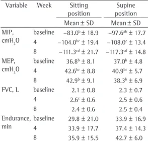
Table 1 - MIP, MEP, FVC and endurance time determined in the studied patients in the sitting and supine positions at baseline, week 4 and week 8 of training.
Variable Week Sitting position
Supine position Mean± SD Mean± SD MIP,
cmH2O
baseline −83.0b ± 18.9 −97.6ab± 17.7 4 −104.0bc± 19.4 −108.0c± 13.4 8 −111.3cd± 21.7 −117.3cd± 14.8 MEP,
cmH2O
baseline 36.8b± 8.1 37.0b± 4.8 4 42.6bc± 8.8 40.9bc± 5.7 8 42.9b± 9.1 38.3b± 6.9 FVC, L baseline 2.1 ± 0.8 2.3 ± 0.7 4 2.6c± 0.6 2.5 ± 0.6 8 2.4 ± 0.6 2.5 ± 0.4 Endurance,
min
baseline 29.8 ± 21.0 33.9 ± 16.9 4 33.9 ± 17.7 37.4 ± 14.3 8 35.9 ± 15.5 42.7 ± 6.0 aSupine > seated (p < 0.05). bObtained values < predictive
values (p < 0.05). cWeeks 4 and 8 > baseline (p < 0.05). dWeek 4 vs. week 8(p < 0.05).
supine (−83.0 ± 18.9 vs. −97.6 ± 17.7 cmH2O; p < 0.05). There were no significant differences between the two positions for baseline MEP, FVC and endurance.
Baseline MIP, baseline MEP, and MEP measured at weeks 4 and 8 were significantly lower than the predicted values published by
Neder et al. for MIP (−120 ± 11 cmH2O) and MEP (129 ± 12 cmH2O),(20) and the differences were
significant (p < 0.05 for all).(20) No significant
difference was found between MIP measured in weeks 4 and 8.
With patients in the sitting position, MIP was significantly higher at weeks 4 and
8 than at baseline (−104.0 ± 19.4 and
−111.3 ± 21.7 cmH2O, respectively, vs. −83.0 ± 18.9 cmH2O; p < 0.05 for both). With patients in the supine position, the MIP values obtained at weeks 4 and 8 were also significantly higher
than was that obtained at baseline (−108.0 ±
13.4 and −117.3 ± 14.8 cmH2O, respectively, vs.
−97.6 ± 17.7 cmH2O; p < 0.05 for both). After 8 weeks of training, MIP had increased by 39 ± 12% in the sitting position and by 23 ± 14% in the supine position (Figure 1).
Compared with baseline MEP, there was a significant increase in MEP at week 4 for the sitting and supine positions (36.8 ± 8.1 and 37.0 ± 4.8 cmH2O, respectively, vs. 42.6 ± 8.8 and 40.9 ± 5.7 cmH2O; p < 0.05 for both), representing, respectively, an 18 ± 22% and an 11 ± 11% increase in MEP. There were no significant increases in MEP at week 8 (Figure 2).
in the sitting position and those obtained in the supine position. In order to compare the pretraining values to those taken on weeks 4 and 8, we conducted ANOVA with repeated measures, followed by a paired t-test. The level of significance was set at 5%.
Results
Seven of the eight patients completed the study. One abandoned the training sessions after 4 weeks, for no apparent reason. The values obtained for MIP, MEP, FVC and endurance time are presented in Table 1.
Mean baseline MIP was lower when the patients were sitting than when they were Figure 2 - MEP determined in the studied patients in the sitting and supine positions at baseline, week 4 of training and week 8 of training.
Figure 3 - FVC determined in the studied patients in the sitting and supine positions at baseline, week 4 of training and week 8 of training.
inspiratory muscle training with a threshold trainer.
In the present study, MIP, MEP, FVC, and endurance obtained in the sitting position were compared with those in the supine position. A difference between the two positions was found for MIP at baseline, suggesting that the sitting position is disadvantageous for these patients.(24-26) However, this difference between
the sitting and supine positions did not persist after 4 and 8 weeks of training, possibly due to the fact that the sitting position was adopted during the training sessions, thus minimizing any disadvantages for these patients.
After 4 and 8 weeks of training, MIP improved. Several other studies have also found such an increase in MIP, although those studies involved inspiratory muscle training with high loads or resistive trainers.(9,11,27) It is possible to achieve
the training effect even without knowing the load. Nevertheless, according to one study,(5) the
advantages of the inspiratory muscle training with threshold trainers are that it allows accurate determination of the training load and thereby avoids fatigue of the inspiratory muscles.
Another interesting aspect relates to the duration of quadriplegia. According to one group of authors,(4) the tonus of the abdominal
muscles increases by approximately 3 months after the lesion, and that, at one year after the injury, pulmonary function can improve in relation to the initial phase and tends to become stable.(3,28) In our study, none of the patients
was in the acute phase, since the duration of the lesion ranged from 35 to 318 months, and, although there was no control group, spontaneous improvement is not probable in this phase.
A recent study evaluated the effect of normocapnic hyperpnea training, showing improvement in respiratory muscle strength and endurance in the trained group and no improvement in the control group.(16)
In the present study, MEP improved, in the sitting and supine position, after 4 weeks of training. This improvement can be due to an increased elastic recoil of the lungs, resulting from a high FVC. The increase in MEP observed by other authors is related to specific expiratory muscle intervention,(4,9,29) which did not occur in
our study, because the protocol was applied to the inspiratory muscles only.
There was also a significant increase in FVC at week 4 compared with the baseline value, for patients in the sitting position (2.5 ± 0.6 L vs. 2.0 ± 0.7 L; p < 0.05), translating to a 24 ± 22% increase. There was no significant increase in FVC at week 8 (Figure 3).
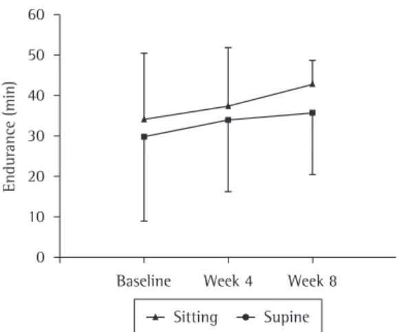
After 8 weeks of training, endurance did not improve significantly over the baseline value in either the sitting or the supine position (35.9 ± 15.5 vs. 29.8 ± 21.0 min and 42.7 ± 6.0 vs. 33.9 ± 16.9 min, respectively), with an increase of 173 ± 233% and 116 ± 189%, respectively (Figure 4).
Discussion
The results of this study suggests that inspiratory muscle training with a threshold trainer at low loads increases the strength of the respiratory muscles in quadriplegic patients.
The efficiency of inspiratory training at a low load (30% of MIP) has previously been reported in individuals with COPD(6,7) but not in
quadriplegic patients. Our study suggests that inspiratory muscle training at low loads has a positive effect in quadriplegic patients, since the initial values were lower than those predicted, whereas those obtained after 8 weeks of training were higher than the baseline values and showed no significant differences in relation to the predicted values, suggesting a trend towards normality after the specific training. These lower baseline values are in accordance with the findings of a study(23) showing that quadriplegic
patients with injuries to the upper cervical spine (at C4 or C5) presented with the lowest values of MIP, MEP and FVC. It is important to note that the protocol that we used (low load muscle training) is an easy-to-perform exercise, which favors adherence to treatment.
The training protocols for quadriplegic patients described in the literature are to be applied once or twice a day in 15- to 30-min sessions for 5-7 days a week over 6, 7, 8 or 16 weeks, or even one year.(4,8,11-13) Based on
the studies previously cited, our protocol used 30-min training sessions at 30% of MIP once a day for 5 days a week over 8 consecutive weeks. One group of authors,(1) working on the Spinal
quadriplegic patients were submitted to respiratory muscle training. Nevertheless, some protocols do not involve the determination of respiratory patterns during the test, which precludes any comparisons of the results.(7,8,10,11)
The small number of patients and the lack of a control group are major limitations to our study. However, other studies have presented the same limitations, probably due to specific difficulties related to this population of patients.(2,8,9,11,13)
In two systematic reviews,(16,17) three and six
studies were selected, respectively. According to one of the reviews,(16) there have been very few
studies showing evidence of the effectiveness of inspiratory muscle training in patients with cervical spinal cord injury. However, the authors of the other review suggested that there is a trend toward improvement of both expiratory muscle strength and VC, as well as a decrease in the RV, after respiratory muscle training. Nevertheless, the available data are still insufficient to draw conclusions, and further studies involving larger samples, control groups, the use of threshold trainers and different variables are needed.(1,16,17)
In conclusion, the present study showed that inspiratory muscle training at a 30% muscle training load is associated with improvements in inspiratory muscle strength and FVC, as well as in expiratory muscle effectiveness, for quadriplegic patients.
References
1. SCIRE Spinal Cord Injury Rehabilitation Evidence [homepage on the Internet]. Vancouver: SCIRE [cited 2009 Jul 20]. Sheel AW, Reid WD, Townson AF, Ayas N. Respiratory Management Following Spinal Cord Injury. In: Eng JJ, Teasell RW, Miller WC, Wolfe DL, Townson AF, Hsieh JTC, et al, editors. Spinal Cord Injury Rehabilitation Evidence. Version 2.0. [Adobe Acrobat document, 40p.] Available from: http://www. scireproject.com/pdf/SCIRE_II_CH8.pdf
2. Zupan A, Savrin R, Erjavec T, Kralj A, Karcnik T, Skorjanc T, et al. Effects of respiratory muscle training and electrical stimulation of abdominal muscles on respiratory capabilities in tetraplegic patients. Spinal Cord. 1997;35(8):540-5.
3. McMichan JC, Michel L, Westbrook PR. Pulmonary
dysfunction following traumatic quadriplegia.
Recognition, prevention, and treatment. JAMA. 1980;243(6):528-31.
4. Loveridge B, Sanii R, Dubo HI. Breathing pattern adjustments during the first year following cervical spinal cord injury. Paraplegia. 1992;30(7):479-88. 5. Larson JL, Kim MJ, Sharp JT, Larson DA. Inspiratory
muscle training with a pressure threshold breathing device in patients with chronic obstructive pulmonary disease. Am Rev Respir Dis. 1988;138(3):689-96.
At week 4, FVC increased only in the sitting position. Prior to the inspiratory muscle training, the mean FVC in the sitting position was 2.1 ± 0.8 L, increasing to 2.6 ± 0.6 L by week 4, equivalent to an increase of 24 ± 22%. This increase in FVC resulting from the increased inspiratory muscle strength can also be attributed to the sitting position adopted during training. In our study, the parameter used was FVC, whereas other authors found improvements in VC, FVC, TLC, maximal voluntary ventilation, FEV1, and PEF after muscle training.(2,12,13)
One group of authors(29) established that FVC
values above 1.8 ± 0.8 L can lower the risk of pulmonary complications in quadriplegic patients and therefore constitute a major predictor of morbidity. Since our patients had higher FVC values, they also had greater RV. Higher FVC means a better ability to cough, which reduces the accumulation of secretion and prevents a number of respiratory infections commonly seen in this group of patients.(13) Another group of
authors,(26) in a one-year study on respiratory
training with threshold trainers, demonstrated a decrease not only in the number of respiratory infections, but also in the frequency of secretion aspiration and of hospitalizations for the treatment of infections.
Endurance is the capacity of the inspiratory muscles to sustain a given load for a given time.(30) Our endurance results did not show an
improvement after 8 weeks of training in the sitting position only, our patients did show any significant improvement in terms of endurance, probably due to the variability of the obtained data, with an increase of 173 ± 233% in the sitting position and 116 ± 189% in the supine position.
According to the ATS/European Respiratory Society recommendations,(30) there are various
ways of assessing and quantifying the endurance properties of the inspiratory muscles, due to a lack of a standardized system for measuring these variables. A more precise technique for the measurement of diaphragm endurance was developed by Bellemare & Grassino.(22) The
authors stated that it is necessary to control the respiratory pattern (TI/Ttot) and to know the load (inspiratory pressure/MIP) to be applied to the muscles during the endurance tests.
18. Barros Filho TE. Avaliação padronizada nos traumatismos raquimedulares. Rev Bras Ortop. 1994;29(3):99-106. 19. Black LF, Hyatt RE. Maximal respiratory pressures:
normal values and relationship to age and sex. Am Rev Respir Dis. 1969;99(5):696-702.
20. Neder JA, Andreoni S, Lerario MC, Nery LE. Reference values for lung function tests. II. Maximal respiratory pressures and voluntary ventilation. Braz J Med Biol Res. 1999;32(6):719-27.
21. Standardization of Spirometry, 1994 Update. American Thoracic Society. Am J Respir Crit Care Med. 1995;152(3):1107-36.
22. Bellemare F, Grassino A. Effect of pressure and timing of contraction on human diaphragm fatigue. J Appl Physiol. 1982;53(5):1190-5.
23. Mateus SR, Beraldo PS, Horan TA. Maximal static mouth respiratory pressure in spinal cord injured patients: correlation with motor level. Spinal Cord. 2007;45(8):569-75.
24. Boaventura CM, Silveira JM, Santos PR, Gastaldi AC. Força da musculatura respiratória de pacientes tetraplégicos sentados e em supino. Rev Fisioter Univ Sao Paulo. 2004;11(6):70-6.
25. Ali J, Qi W. Pulmonary function and posture in traumatic quadriplegia. J Trauma. 1995;39(2):334-7.
26. Ehrlich M, Manns PJ, Poulin C. Respiratory training for a person with C3-C4 tetraplegia. Aust J Physiother. 1999;45(4):301-7.
27. Ledsome JR, Sharp JM. Pulmonary function in acute cervical cord injury. Am Rev Respir Dis. 1981;124(1):41-4.
28. Boaventura CM, Gastaldi AC, Silveira JM, Santos PR, Guimarães RC, De Lima LC. Effect of an abdominal binder on the efficacy of respiratory muscles in seated and supine tetraplegic patients. Physiotherapy. 2003;89(5):290-5.
29. Crane L, Klerk K, Ruhl A, Warner P, Ruhl C, Roach KE. The effect of exercise training on pulmonary function in persons with quadriplegia. Paraplegia. 1994;32(7):435 41.
30. American Thoracic Society/European Respiratory Society. ATS/ERS Statement on respiratory muscle testing. Am J Respir Crit Care Med. 2002;166(4):518-624.
6. Lisboa C, Muñoz V, Beroiza T, Leiva A, Cruz E. Inspiratory muscle training in chronic airflow limitation: comparison of two different training loads with a threshold device. Eur Respir J. 1994;7(7):1266-74.
7. Gross D, Ladd HW, Riley EJ, Macklem PT, Grassino A. The effect of training on strength and endurance of the diaphragm in quadriplegia. Am J Med. 1980;68(1):27-35.
8. Leith DE, Bradley M. Ventilatory muscle strength and endurance training. J Appl Physiol. 1976;41(4):508-16. 9. Loveridge B, Badour M, Dubo H. Ventilatory muscle
endurance training in quadriplegia: effects on breathing pattern. Paraplegia. 1989;27(5):329-39
10. Derrickson J, Ciesla N, Simpson N, Imle PC. A comparison of two breathing exercise programs for patients with quadriplegia. Phys Ther. 1992;72(11):763-9.
11. Rutchik A, Weissman AR, Almenoff PL, Spungen AM, Bauman WA, Grimm DR. Resistive inspiratory muscle training in subjects with chronic cervical spinal cord injury. Arch Phys Med Rehabil. 1998;79(3):293-7. 12. Lin KH, Chuang CC, Wu HD, Chang CW, Kou YR.
Abdominal weight and inspiratory resistance: their immediate effects on inspiratory muscle functions during maximal voluntary breathing in chronic tetraplegic patients. Arch Phys Med Rehabil. 1999;80(7):741-5. 13. Stiller K, Huff N. Respiratory muscle training for
tetraplegic patients: A literature review. Aust J Physiother. 1999;45(4):291-9.
14. Van Houtte S, Vanlandewijck Y, Kiekens C, Spengler CM, Gosselink R. Patients with acute spinal cord injury benefit from normocapnic hyperpnoea training. J Rehabil Med. 2008;40(2):119-25.
15. Uijl SG, Houtman S, Folgering HT, Hopman MT. Training of the respiratory muscles in individuals with tetraplegia. Spinal Cord. 1999;37(8):575-9.
16. Brooks D, O’Brien K, Geddes EL, Crowe J, Reid WD. Is inspiratory muscle training effective for individuals with cervical spinal cord injury? A qualitative systematic review. Clin Rehabil. 2005;19(3):237-46.
17. Van Houtte S, Vanlandewijck Y, Gosselink R. Respiratory muscle training in persons with spinal cord injury: a systematic review. Respir Med. 2006;100(11):1886-95.
About the authors
Janne Marques Silveira
Professor of Physical Therapy and Physiology. Department of Physiology, Medicine and Physical Therapy, Centro Universitário Unirg, Gurupi, Brazil.
Ada Clarice Gastaldi
Professor of Physical Therapy. Department of Biomechanics, Medicine and Rehabilitation, University of São Paulo at Ribeirão Preto School of Medicine, Ribeirão Preto, Brazil.
Cristina de Matos Boaventura
Professor. Triângulo University Center School of Physiotherapy, Uberlândia, Brazil.
Hugo Celso Souza