Necrotizing pneumonia in children submitted to
thoracoscopy due to pleural empyema:
incidence, treatment and clinical evolution*
,**
Pneumonia necrosante em crianças submetidas à toracoscopia por empiema pleural: incidência, tratamento e evolução clínica
Maurício Macedo, Karine Furtado Meyer, Tatiana Cristina Miranda Oliveira
Abstract
Objective: To assess the incidence of necrotizing pneumonia (NP) in children submitted to thoracoscopy, comparing patients with and without NP in terms of the presentation and clinical evolution. Methods: A retrospective study of
children with pleural empyema submitted to thoracoscopy. Thoracoscopy was performed in patients not previously submitted to thoracic drainage and in whom there was evidence of loculated effusion or pneumothorax, as well as in those previously submitted to thoracic drainage and in whom there was persistent pneumothorax or fever with purulent discharge. On the basis of the thoracoscopy findings, patients were distributed into two groups: those with NP (NP group) and those without (no-NP group). Results: The study sample comprised 52 patients. Of the 24 patients with NP, 19 (79%) had undergone thoracic drainage prior to thoracoscopy, 11 (46%) presented with pneumothorax, and 16 (67%) developed bronchopleural fistula. In the NP group, the median drainage time and the median length of hospital stay were 18 and 19 days, respectively. Of the 28 patients without NP, 10 (36%) had undergone thoracic drainage prior to thoracoscopy, 9 (32%) presented pneumothorax, and 5 (18%) developed bronchopleural fistula. In the no-NP group, the median drainage time and the median length of hospital stay were 6 and 10 days, respectively. Conclusions: Pneumothorax should raise the suspicion of NP. Early thoracoscopy
can be a valuable treatment option for NP in children because it hastens recovery in comparison with the medical treatment alone and avoids extensive late thoracotomy lung resections.
Keywords: Empyema, pleural; Thoracoscopy; Pneumonia; Child.
Resumo
Objetivo: Analisar a incidência de pneumonia necrosante (PN) em crianças submetidas a toracoscopia e comparar
pacientes com e sem PN em relação às diferentes apresentações e evolução clínica. Métodos: Estudo retrospectivo de crianças portadoras de empiema e submetidas a toracoscopia. A toracoscopia foi realizada em pacientes não submetidos a drenagem torácica prévia e evidência de derrame septado ou pneumotórax, assim como naqueles submetidos previamente a drenagem torácica e pneumotórax persistente ou febre e secreção purulenta. Baseado na presença de PN durante a toracoscopia, os pacientes foram divididos em dois grupos: com PN e sem PN.
Resultados: Participaram do estudo 52 pacientes. Dos 24 pacientes com PN, 19 (79%) foram submetidos a
drenagem torácica anterior à toracoscopia, 11 (46%) apresentaram pneumotórax, e 16 (67%) evoluíram com fístula broncopleural. Neste grupo, as medianas do tempo de drenagem e de hospitalização foram, respectivamente, 18 e 19 dias. Dos 28 pacientes sem PN, 10 (36%) foram submetidos a drenagem torácica anterior à toracoscopia, 9 (32%) apresentaram pneumotórax, e 5 (18%) evoluíram com fístula broncopleural. Neste grupo, as medianas do tempo médio de drenagem e de hospitalização foram, respectivamente, 6 e 10 dias. Conclusões: A PN deve ser
suspeitada na presença de pneumotórax. A toracoscopia precoce pode ser uma opção terapêutica de grande valor na PN da infância, pois acelera a recuperação quando comparada ao tratamento médico isolado e evita ressecções pulmonares extensas da toracotomia tardia.
Descritores: Empiema pleural; Toracoscopia; Pneumonia; Criança.
* Study carried out at the Hospital Estadual Infantil Darcy Vargas, São Paulo, Brazil.
Correspondence to: Mauricio Macedo. Rua Comandante Garcia D’Ávila, 37, CEP 05654-040, São Paulo, SP, Brasil. Tel 55 11 7152-2726. Fax: 55 11 3747-3113. E-mail: [email protected]
Financial support: None.
Submitted: 23 November 2009. Accepted, after review: 19 January 2010.
Vargas, located in the city of São Paulo, Brazil, between July of 2002 and June of 2008.
In patients who had not previously been submitted to thoracic drainage, thoracoscopy was performed when chest ultrasound demonstrated the presence of loculated effusion or when a chest X-ray revealed pneumothorax.
In patients who had undergone thoracic drainage, thoracoscopy was performed when a chest X-ray, ultrasound or CT scan revealed loculated effusion or persistent pneumothorax, or when purulent discharge and fever persisted.
All procedures were performed under general anesthesia, with orotracheal intubation and with the patient in the lateral decubitus position. Three access ports were used: a 10-mm port to introduce the scope and insufflate with CO2 (at 4-6 mmHg); and two 5-mm ports for forceps. Pleural adherences were sectioned and fibrin layers were removed until the pleural cavity was fully cleaned and the lungs expanded.
A diagnosis of NP was made when a cavity containing necrotic debris was found in the parenchyma (Figure 1). In such cases, debridement of all necrotic tissue and cleaning were performed, and, in order to provide better drainage, the opening of the pulmonary cavity was broadened.
At the end of the procedure, a chest drain was placed through an orifice of the posteroinferior port and was removed when there was complete lung expansion, without purulent discharge or bronchopleural fistula.
On the basis of the thoracoscopy findings, patients were distributed into two groups: those with NP (NP group) and those without (no-NP group). The following parameters were assessed in both groups: gender; age; radiological findings; pleural fluid culture; presence of bronchopleural fistula in the postoperative period; drainage time and length of hospital stay.
The groups were compared for qualitative variables using the chi-square test or Fisher’s exact test, as indicated. The quantitative variables were analyzed using the Student’s t-test for independent samples with normal distribution or, otherwise, with the nonparametric Mann-Whitney test. The level of significance was set at 0.05 (α = 5%).
Introduction
Despite advances in antimicrobial therapy, complications due to pulmonary infections continue to occur and have been associated with significant morbidity. The main complication is pleural empyema, which is seen in 10% of cases.(1,2)
Treatments for empyema previously included only antibiotics, thoracentesis, thoracostomy (with open or closed drainage) and thoracotomy.(1,3) With the advent of minimally invasive surgery, thoracoscopy was added to the therapeutic armamentarium.(4,5) Thoracoscopy provides a broad view of the pleural cavity, as well as allowing the sectioning of pleural adherences, the suction of collections and the removal of excess fibrin deposited on the parietal and visceral pleurae.(4,5) In some cases, thoracoscopy has revealed focal pulmonary necrosis, also known as pulmonary gangrene or necrotizing pneumonia (NP).(6)
It is known that NP is a severe complication of pneumonia,(6) and that it more often affects adults. (6,7) It is believed that NP is caused by proteolytic enzymes that are released by microorganisms and destroy the pulmonary parenchyma(7-9) or by an exacerbated inflammatory response, mediated by cytokines, resulting from the interaction between the pathogenic agent and the host. (7) The agent that is most often involved in the pathogenesis of NP is Streptococcus pneumoniae, followed by Aspergillus spp., Legionella spp. and Staphylococcus aureus.(6)
Although NP is responsible for high morbidity in child populations,(7) there have been few studies of its incidence, its clinical profile and the best practice management strategies.
The objective of this study was to assess the incidence of NP in children submitted to thoracoscopy due to pleural empyema and to compare patients with and without NP in terms of presentation and clinical evolution, as well as to determine the efficacy of thoracoscopy as a treatment option.
Methods
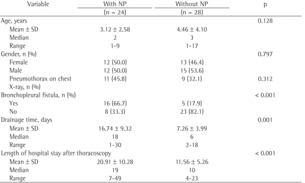
thoracic drainage prior to thoracoscopy. Pneumothorax was observed on plain film radiography in 9 patients (32%). Culture of pleural fluid was positive for Streptococcus pneumoniae in 5 patients and for Staphylococcus aureus in 3. Five patients (18%) developed bronchopleural fistula. The median drainage time and the median length of hospital stay were 6 and 10 days, respectively (Table 1).
No statistically significant differences were found between the groups in terms of age, gender, presence of pneumothorax on chest X-ray, result of pleural fluid culture or need to repeat the thoracoscopy.
Statistically significant differences were found between the groups regarding the development of bronchopleural fistula in the postoperative period (p < 0.001), drainage time (p = 0.001) and length of hospital stay after thoracoscopy (p < 0.001; Table 1). None of the patients required a second procedure (thoracoscopy or thoracotomy).
Discussion
The epidemiological pattern of pneumonia appears to be changing, especially in Europe(10) and North America,(11) where the incidence of necrotizing forms is on the rise.(12) In adults, such forms have been linked with alcohol abuse, diabetes mellitus and nutritional deficiencies,(8) whereas, in children, this pathology is being described in previously healthy young children, even those without predisposing factors.
One group of authors(13) evaluated 107 adult patients submitted to video-assisted thoracoscopy due to empyema and identified NP in 3 (2.8%). In another study,(14) involving 39 children who did not respond to drainage alone and presented loculations on chest CT scans, 2 (5.1%) were found to have NP. In yet another study,(15) the incidence of NP was reported to be 12.5% in a group of 16 children. In our study, NP was diagnosed in 46% of the cases. We believe that this high incidence is related to the fact that our hospital is a referral hospital for pediatric thoracic surgery and receives patients who do not respond to the conventional therapy— drainage and antibiotic therapy.
Pneumothorax on the initial chest X-ray should raise the suspicion of NP, as should pyopneumothorax that persists after chest drainage. Air in the pleural space implies
Results
Necrosis of the pulmonary parenchyma was found in 24 patients (46%). In this group (the NP group), 12 patients were female, the mean age was 3.12 years, and 19 patients (79%) had undergone thoracic drainage prior to the thoracoscopy being ordered. Pneumothorax was observed on plain film radiography in 11 patients (46%). Culture of pleural fluid was positive for Streptococcus pneumoniae in 6 patients, for Staphylococcus aureus in 3 and for Pseudomonas aeruginosa in 1. Sixteen patients (67%) developed bronchopleural fistula. The median drainage time and the median length of hospital stay were 18 and 19 days, respectively (Table 1).
In the no-NP group, there were 28 patients, 13 of whom were female, the mean age was 4.46 years, and 10 patients (36%) had undergone
Figure 1 - Images obtained during thoracoscopy
decortication. Therefore, conservative treatment can prolong the course of the disease, leading to prolonged hospitalization, and can promote the emergence of drug-resistant bacteria.
Radical and aggressive surgery has been considered to be the optimal treatment of NP in adults. The idea that early surgical intervention is necessary was first put forth in the 1970s.(17,18) In a more recent study,(19) pulmonary resection (from wedge resection to pneumonectomy) was advocated. However, in that study, lobectomy or pneumonectomy was typically performed only after antibiotic therapy had been ineffective for a long period of time. In addition, the decision to proceed to lung resection in children is highly controversial because it can impair future pulmonary function.(8,20)
The logical question is whether less aggressive but earlier surgery can shorten the progression of the disease and avoid later salvage surgery.(21) We believe that thoracoscopy should be considered as an intermediate option for treating NP of bacterial etiology. In fact, thoracoscopy is a semiconservative surgical procedure because it avoids extensive resection of the lung. Ablation of the necrotic debris, efficient drainage of empyema and closure of air leaks can hasten recovery. Therefore, when damage to the lung parenchyma. In our study,
pneumothorax was diagnosed in 20 patients, and 11 had NP. Of the 9 patients presenting pneumothorax without necrosis, 5 presented bronchopleural fistula in the immediate postoperative period, indicating the presence of a pulmonary lesion not observed during thoracoscopy. Failure to identify a necrotic lesion during thoracoscopy can occur when the lesion is covered by fibrin, is blocked by other structures or is quite small.
Of the 24 patients in the NP group, 16 (66.7%) developed bronchopleural fistula. This finding is in agreement with the literature, in which the incidence of bronchopleural fistula in patients NP is reported to be 63-70%.(6,7)
The classical strategies for the management of NP include conservative and radical treatment.
Conservative management with high-dose antibiotic therapy and pleural drainage can result in parenchymal preservation and re-expansion, especially in children.(16) One group of authors(16) showed unexpectedly satisfying recuperation of the lung parenchyma. However, patients receiving conservative treatment often require multiple drainages and occasionally require open thoracic drainage. In rare cases, such patients can require thoracotomy for pulmonary resections or
Table 1 - Statistical analysis of clinical and radiological data for patients with and without necrotizing
pneumonia.
Variable With NP Without NP p
(n = 24) (n = 28)
Age, years 0.128
Mean ± SD 3.12 ± 2.58 4.46 ± 4.10
Median 2 3
Range 1-9 1-17
Gender, n (%) 0.797
Female 12 (50.0) 13 (46.4)
Male 12 (50.0) 15 (53.6)
Pneumothorax on chest X-ray, n (%)
11 (45.8) 9 (32.1) 0.312
Bronchopleural fistula, n (%) < 0.001
Yes 16 (66.7) 5 (17.9)
No 8 (33.3) 23 (82.1)
Drainage time, days 0.001
Mean ± SD 16.74 ± 9.32 7.26 ± 3.99
Median 18 6
Range 1-30 2-18
Length of hospital stay after thoracoscopy < 0.001
Mean ± SD 20.91 ± 10.28 11.56 ± 5.26
Median 19 10
Range 7-49 4-23
7. Hacimustafaoglu M, Celebi S, Sarimehmet H, Gurpinar A, Ercan I. Necrotizing pneumonia in children. Acta Paediatr. 2004;93(9):1172-7.
8. Penner C, Maycher B, Long R. Pulmonary gangrene. A complication of bacterial pneumonia. Chest. 1994;105(2):567-73.
9. Refaely Y, Weissberg D. Gangrene of the lung: treatment in two stages. Ann Thorac Surg. 1997;64(4):970-3; discussion 973-4.
10. Gillet Y, Issartel B, Vanhems P, Lina G, Vandenesch F, Etienne J, et al. Severe staphylococcal pneumonia in children [Article in French]. Arch Pediatr. 2001;8 Suppl 4:742s-746s.
11. Hausdorff WP. Invasive pneumococcal disease in children: geographic and temporal variations in incidence and serotype distribution. Eur J Pediatr. 2002;161 Suppl 2:S135-9.
12. Rees JH, Spencer DA, Parikh D, Weller P. Increase in incidence of childhood empyema in West Midlands, UK. Lancet. 1997;349(9049):402.
13. Weissberg D, Refaely Y. Pleural empyema: 24-year experience. Ann Thorac Surg. 1996;62(4):1026-9. 14. Subramaniam R, Joseph VT, Tan GM, Goh A, Chay OM.
Experience with video-assisted thoracoscopic surgery in the management of complicated pneumonia in children. J Pediatr Surg. 2001;36(2):316-9.
15. Kercher KW, Attorri RJ, Hoover JD, Morton D Jr. Thoracoscopic decortication as first-line therapy for pediatric parapneumonic empyema. A case series. Chest. 2000;118(1):24-7.
16. Kerem E, Bar Ziv Y, Rudenski B, Katz S, Kleid D, Branski D. Bacteremic necrotizing pneumococcal pneumonia in children. Am J Respir Crit Care Med. 1994;149(1):242-4.
17. Knight L, Fraser RG, Robson HG. Massive pulmonary gangrene: a severe complication of Klebsiella pneumoniae. Can Med Assoc J. 1975;112(2):196-8. 18. Proctor RJ, Griffin JP, Eastridge CE. Massive pulmonary
gangrene. South Med J. 1977;70(9):1144-6.
19. Krishnadasan B, Sherbin VL, Vallières E, Karmy-Jones R. Surgical management of lung gangrene. Can Respir J. 2000;7(5):401-4.
20. Cowles RA, Lelli JL Jr, Takayasu J, Coran AG. Lung resection in infants and children with pulmonary infections refractory to medical therapy. J Pediatr Surg. 2002;37(4):643-7.
21. McCarthy VP, Patamasucon P, Gaines T, Lucas MA. Necrotizing pneumococcal pneumonia in childhood. Pediatr Pulmonol. 1999;28(3):217-21.
22. Kalfa N, Allal H, Lopez M, Counil FO, Forgues D, Guibal MP, et al. An early thoracoscopic approach in necrotizing pneumonia in children: a report of three cases. J Laparoendosc Adv Surg Tech A. 2005;15(1):18-22.
compared with thoracotomy, thoracoscopy plus conservative therapy can be beneficial, not only by shortening the length of hospital stay but by saving a potentially functional lung. This prognostic consideration is of great importance in such young children and might justify the longer drainage time.
In severe cases of NP, pulmonary resection can be necessary. Prior to pulmonary resection, thoracoscopy is used in order to perform the preliminary procedures (fenestration and local mechanical cleaning) so that patients can recover from the infectious condition.(7) None of the patients in this study required thoracotomy, a finding that corroborates the data in the literature on thoracoscopy for NP.(22)
Early thoracoscopy can be a valuable treatment option for NP in children because it hastens recovery in comparison with medical treatment alone and avoids extensive late thoracotomy lung resections.
References
1. Mocelin HT, Fischer GB. Fatores preditivos para drenagem de derrames pleurais parapneumônicos em crianças. J Pneumol. 2001;27(4):177-84.
2. Moir CR, Telander RL. Complications of lower respiratory tract infection: Empyema complicating pneumonia, pneumatoceles, and respiratory embarrassment. In: Fallis JC, Filler RM, Lemoine G, editors. Pediatric Thoracic Surgery. New York: Elsevier; 1991. p. 299-305. 3. Béchamps GJ, Lynn HB, Wenzl JE. Empyema in children:
review of mayo clinic experience. Mayo Clin Proc. 1970;45(1):43-50.
4. Kang DW, Campos JR, Andrade Filho Lde O, Engel FC, Xavier AM, Macedo M, et al. Thoracoscopy in the treatment of pleural empyema in pediatric patients. J Bras Pneumol. 2008;34(4):205-11.
5. Silen ML, Weber TR. Thoracoscopic debridement of loculated empyema thoracis in children. Ann Thorac Surg. 1995;59(5):1166-8.
6. Hoffer FA, Bloom DA, Colin AA, Fishman SJ. Lung abscess versus necrotizing pneumonia: implications for interventional therapy. Pediatr Radiol. 1999;29(2):87-91
About the authors
Maurício Macedo
Director of the Department of Surgery. Hospital Estadual Infantil Darcy Vargas, São Paulo, Brazil.
Karine Furtado Meyer
Surgeon. Hospital Estadual Infantil Darcy Vargas, São Paulo, Brazil.
Tatiana Cristina Miranda Oliveira