J Bras Pneumol. 2011;37(4):556-559
associated with a worse prognosis.(6,7) Physicians who deal with this pathology should be on the alert for the possibility of cutaneous metastasis as the initial manifestation of lung cancer.(8)
Case report
A 58-year-old waiter who had quit smoking 9 years prior (smoking history, 40 pack-years) was referred for medical oncology consultation because of an epigastric mass. The patient presented with a history of COPD with severe
Introduction
Annually, metastatic disease accounts for over half of the new cases of lung cancer in Europe.(1) In Portugal, the estimated annual incidence is approximately 2,500 cases, and mortality among such cases is nearly 100%, lung cancer being the leading cause of cancer death.(1,2) To date, there has been no evidence of an effective method for population screening,(3) smoking cessation being fundamental to reducing the number of cases.(4)
It is relatively common for lung tumors to metastasize to the skin,(5) and this is typically
Cutaneous metastasis as the initial
manifestation of lung adenocarcinoma*
Metástase cutânea como primeira manifestação de adenocarcinoma pulmonar
Marcos Pantarotto, Liliana Lombo, Helena Pereira, Antonio Araújo
Abstract
We report the case of a 58-year-old male patient who was referred for oncology consultation due to an epigastric mass that had been growing rapidly for three months. Diagnostic investigation revealed that the mass was a metastasis of stage IV lung adenocarcinoma. The patient received five cycles of chemotherapy with cisplatin and gemcitabine as a first-line treatment, which was interrupted due to major adverse events. Although the pulmonary disease stabilized, the cutaneous disease progressed. The patient then received pemetrexed as a second-line chemotherapy, together with concurrent external radiotherapy, which was well tolerated. There was complete remission of the epigastric mass. However, the patient died three months after the treatment. Here, we emphasize the importance of a multidisciplinary approach and of its role in individualizing the treatment.
Keywords: Lung neoplasms; Neoplasm metastasis; Antineoplastic combined chemotherapy protocols;
Radiotherapy, computer-assisted.
Resumo
Relatamos o caso de um paciente do sexo masculino de 58 anos de idade, que foi encaminhado para a consulta de oncologia por apresentar uma massa epigástrica de crescimento rápido em três meses de evolução. A investigação diagnóstica revelou tratar-se de um adenocarcinoma pulmonar metastático estádio IV. Recebeu cinco ciclos de cisplatina e gemcitabina como tratamento de primeira linha, que foi interrompido devido a efeitos adversos. Houve estabilidade da doença pulmonar e progressão cutânea. Recebeu pemetrexed como tratamento de segunda linha e radioterapia externa concomitante, com boa tolerância e regressão completa da massa epigástrica. Entretanto, o paciente faleceu três meses após o tratamento. Destacamos aqui importância da multidisciplinaridade e do seu papel na individualização do tratamento.
Descritores: Neoplasias pulmonares; Metástase neoplásica; Protocolos de quimioterapia combinada
antineoplásica; Radioterapia assistida por computador.
* Study carried out at the Francisco Gentil Portuguese Oncology Institute, Porto, Portugal.
Correspondence to: Marcos Pantarotto. Instituto Português de Oncologia, Rua Dr. António Bernardino de Almeida, 4200-072, Porto, Portugal.
Tel. 351 22 508-4000 extension 7685. Fax: 351 22 508-4001. E-mail: [email protected] Financial support: None.
Cutaneous metastasis as the initial manifestation of lung adenocarcinoma
J Bras Pneumol. 2011;37(4):556-559 557
wasting, and the Eastern Cooperative Oncology Group performance status scale score was 1. Palpation revealed a firm, painful mass in the epigastric region. The mass, approximately 15 cm in diameter, was accompanied by yellowish exudation and adjacent erythema. Pulmonary auscultation revealed absent breath sounds in the upper third of the left hemithorax.
A thoracoabdominal CT scan revealed the presence of a mass with ill-defined borders in the apical posterior segment of the upper lobe of the left lung (Figure 2a), measuring approximately 2.2 cm at its largest diameter, as well as the epigastric lesion itself, which infiltrated the sternum. Both images coincided with intense uptake of the radiotracer fluorine-18 fluorodeoxyglucose on positron emission tomography/CT scans. A biopsy of the abdominal mass was performed and revealed that it was a metastasis of an adenocarcinoma. Immunohistochemistry showed diffuse positivity for cytokeratin 7 and thyroid transcription factor-1, as well as negativity for cytokeratin 20, contributing to a diagnosis of primary lung tumor,(9,10) which was staged as cT1bN0M1b (stage IV) in accordance with the tumor-node-metastasis classification.
The patient was started on the first-line treatment of 100 mg/m2 of cisplatin on day 1 + 1,250 mg/m2 of gemcitabine on days 1 and 8 of a 21-day cycle. The treatment regimen was poorly tolerated, and there were multiple omissions of the day-8 gemcitabine cycle because of hematologic toxicity, with an episode of febrile neutropenia after the second cycle and the occurrence of grade 2 thrombocytopenia after the fifth cycle. A follow-up CT scan of the obstructive ventilatory changes and, 7 years
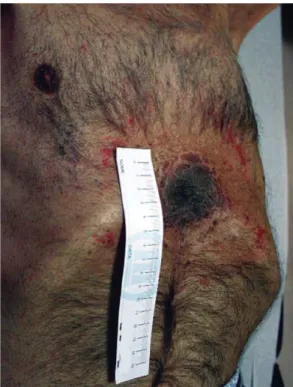
prior, had undergone partial gastrectomy for a perforated gastric ulcer. He had been under treatment with a long-acting inhaled β2 agonist. Three months before the consultation, the patient had noticed a firm, painful nodule that was growing rapidly on the abdominal surgical scar in the epigastric region (Figure 1). He complained of anorexia (grade 3) and weight loss (of approximately 10 kg, or 16% of his normal weight, in the 3 months preceding the consultation) and was quite distressed with the restriction that the mass, which was visible under his clothes, had imposed on his social life. He reported that the severity of his dyspnea had not changed. Physical examination revealed
Figure 1 - Initial appearance of the cutaneous
metastasis.
Figure 2 - In a, initial CT scan showing a spiculated pulmonary nodule in the apical posterior segment of the
558 Pantarotto M, Lombo L, Pereira H, Araújo A
J Bras Pneumol. 2011;37(4):556-559
Discussion
Cutaneous metastases have been described in association with most of the thoracic and abdominal tumors that cause distant metastases. Although there is no consensus in the literature regarding the frequency of cutaneous metastases, they are most commonly related to primary breast and lung tumors, the chest and abdomen being the sites that are most commonly affected by this type of dissemination.(5,11,12)
Skin lesions can present in different forms, the most common being hard, painless, rapidly growing nodules that adhere to deep planes and are skin-colored or slightly erythematous, with or without ulcerations. As a manifestation of breast and oral cavity cancers, skin lesions can present as nodules of sclerotic and inflammatory appearance; they can also present as a direct extension of the primary tumor.(13) Some lesions are intradermal and invade subcutaneous cellular tissue, whereas others are superficial, with no specific color or location that would indicate their nature.(11,13) They generally present as late manifestations of disseminated neoplastic disease. However, they can be the first clinical manifestation of thoracic or abdominal tumors.
In the clinical case in question, because of the location, size, and appearance of the skin lesion, as well as because of its implications for the autonomy of the patient, we considered it relevant to adjust the treatment in order to reduce the physical and emotional impact of the lesion. To reach such an objective, a multidisciplinary approach is fundamental.
Although the results obtained with the first-line pharmacological treatment were unsatisfactory, radiation therapy in association with second-line chemotherapy (pemetrexed) was well tolerated by the patient and provided better symptom control. The authors believe that the use of combined treatment provided the patient with better quality of life.
It should be highlighted that there is little evidence to support the combined use of radiation therapy and pemetrexed. However, an article describing the results of two recently conducted phase I studies(14) demonstrated that this treatment modality is well tolerated, pointing the way to a new line of research into the association between radiation therapy and therapies aimed at molecular targets(4) in lung cancer.
chest revealed that, although the pulmonary disease had stabilized, the cutaneous disease had progressed.
The treatment strategy was changed to second-line chemotherapy, with 500 mg/m2 of pemetrexed every 21 days, together with concurrent external radiation therapy, which consisted of palliative irradiation of the epigastric mass, at a total dose of 30 Gy (in 10 fractions), with 60Co (Figure 2b).
The patient received 6 cycles without any interruptions or significant adverse effects, and there was complete remission of the epigastric mass (Figure 3).
There was no recurrence of the skin lesion after the treatment with radiation therapy, and there were no new occurrences of such lesions.
A CT scan taken 2 months after the sixth cycle of pemetrexed revealed that the lung, liver, and bone diseases had progressed. The patient died 3 months after the end of the second-line chemotherapy, i.e., approximately 14 months after the initial diagnosis.
Figure 3 - Final appearance of the lesion three
Cutaneous metastasis as the initial manifestation of lung adenocarcinoma
J Bras Pneumol. 2011;37(4):556-559 559
7. Hoang T, Xu R, Schiller JH, Bonomi P, Johnson DH. Clinical model to predict survival in chemonaive patients with advanced non-small-cell lung cancer treated with third-generation chemotherapy regimens based on eastern cooperative oncology group data. J Clin Oncol. 2005;23(1):175-83.
8. Kamble R, Kumar L, Kochupillai V, Sharma A, Sandhoo MS, Mohanti BK. Cutaneous metastases of lung cancer. Postgrad Med J. 1995;71(842):741-3.
9. Capelozzi VL. Role of immunohistochemistry in the diagnosis of lung cancer. J Bras Pneumol. 2009;35(4):375-82.
10. Leonard N. Cutaneous metastases: Where do they come from and what can they mimic? Curr Diagn Pathol. 2007;13(4):320-30.
11. Krathen RA, Orengo IF, Rosen T. Cutaneous metastasis: a meta-analysis of data. South Med J. 2003;96(2):164-7. 12. Lookingbill DP, Spangler N, Sexton FM. Skin involvement
as the presenting sign of internal carcinoma. A retrospective study of 7316 cancer patients. J Am Acad Dermatol. 1990;22(1):19-26.
13. Molina Garrido MJ, Guillén Ponce C, Soto Martínez JL, Martínez Y Sevila C, Carrato Mena A. Cutaneous metastases of lung cancer. Clin Transl Oncol. 2006;8(5):330-3.
14. Surmont V, Smit EF, de Jonge M, Aerts JG, Nackaerts K, Vernhout R, et al. Pemetrexed and cisplatin with concurrent radiotherapy for locally advanced non-small cell and limited disease small cell lung cancer: results from 2 phase I studies. Lung Cancer. 2010;69(3):302-6.
Acknowledgments
The authors are sincerely grateful to Dr. Marta Soares and Dr. Isabel Azevedo, as well as to the nurses at the Francisco Gentil Portuguese Oncology Institute Lung Clinic, in the city of Porto, Portugal, for their unconditional support during the preparation of this report.
References
1. Ferlay J, Parkin DM, Steliarova-Foucher E. Estimates of cancer incidence and mortality in Europe in 2008. Eur J Cancer. 2010;46(4):765-81.
2. Ferlay J, Shin HR, Bray F, Forman D, Mathers C, Parkin DM. Estimates of worldwide burden of cancer in 2008: GLOBOCAN 2008. Int J Cancer. 2010;127(12):2893-917. 3. Gopal M, Abdullah SE, Grady JJ, Goodwin JS. Screening
for lung cancer with low-dose computed tomography: a systematic review and meta-analysis of the baseline findings of randomized controlled trials. J Thorac Oncol. 2010;5(8):1233-9.
4. Baumann M, Zips D, Appold S. Radiotherapy of lung cancer: technology meets biology meets multidisciplinarity. Radiother Oncol. 2009;91(3):279-81. 5. Coslett LM, Katlic MR. Lung cancer with skin metastasis.
Chest. 1990;97(3):757-9.
6. Terashima T, Kanazawa M. Lung cancer with skin metastasis. Chest. 1994;106(5):1448-50.
About the authors
Marcos Pantarotto
Resident in Medical Oncology. Francisco Gentil Portuguese Oncology Institute, Porto, Portugal.
Liliana Lombo
Resident in Radiology. Francisco Gentil Portuguese Oncology Institute, Porto, Portugal.
Helena Pereira
Director. Department of Radiology, Francisco Gentil Portuguese Oncology Institute, Porto, Portugal.
Antonio Araújo