Mouth breathing and forward head posture: effects on
respiratory biomechanics and exercise capacity in children*
Respiração bucal e anteriorização da cabeça: efeitos na biomecânica respiratória e na capacidade de exercício em crianças
Renata Tiemi Okuro, André Moreno Morcillo, Maria Ângela Gonçalves Oliveira Ribeiro, Eulália Sakano,
Patrícia Blau Margosian Conti, José Dirceu Ribeiro
Abstract
Objective: To evaluate submaximal exercise tolerance and respiratory muscle strength in relation to forward head posture (FHP) and respiratory mode in children, comparing mouth-breathing (MB) children with nasal-breathing (NB) children. Methods: This was a controlled, analytical cross-sectional study involving children in the 8-12 year age bracket with a clinical otorhinolaryngology diagnosis of MB, recruited between October of 2010 and January of 2011 from the Mouth Breather Clinic at the State University of Campinas Hospital de Clínicas, located in the city of Campinas, Brazil. The exclusion criteria were obesity, asthma, chronic respiratory diseases, heart disease, and neurological or orthopedic disorders. All of the participants underwent postural assessment and the six-minute walk test (6MWT), together with determination of MIP and MEP. Results: Of the 92 children in the study, 30 presented with MB and 62 presented with NB. In the MB group, the differences between those with moderate or severe FHP and those with normal head posture, in terms of the mean MIP, MEP and six-minute walk distance (6MWD), were not significant (p = 0.079, p = 0.622, and p = 0.957, respectively). In the NB group, the mean values of MIP and MEP were higher in the children with moderate FHP than in those with normal head posture (p = 0.003 and p = 0.004, respectively). The mean MIP, MEP, and 6MWD were lower in the MB group than in the NB group. Values of MIP and MEP were highest in the children with moderate FHP. Conclusions: Respiratory biomechanics and exercise capacity were negatively affected by MB. The presence of moderate FHP acted as a compensatory mechanism in order to improve respiratory muscle function.
Keywords: Mouth breathing; Posture; Exercise tolerance; Respiratory mechanics.
Resumo
Objetivo: Avaliar a tolerância ao exercício submáximo e a força muscular respiratória em relação à anteriorização da cabeça (AC) e ao tipo respiratório em crianças com respiração bucal (RB) ou nasal (RN). Métodos: Estudo analítico transversal com um grupo controle no qual foram incluídas crianças de 8 a 12 anos com diagnóstico clínico otorrinolaringológico de RB, recrutadas do Ambulatório do Respirador Bucal do Hospital de Clínicas da Universidade Estadual de Campinas, Campinas (SP), entre outubro de 2010 e janeiro de 2011. Os critérios de exclusão foram obesidade, asma, doenças respiratórias crônicas, cardiopatias e distúrbios neurológicos ou ortopédicos. Todos os participantes foram submetidos a avaliação postural, teste de caminhada de seis minutos (TC6) e determinação de PImáx e PEmáx. Resultados: Das 92 crianças do estudo, 30 tinham RB e 62 tinham RN. No grupo RB, não houve diferenças nas médias de PImáx, PEmáx e distância percorrida pelo TC6 (DTC6) entre o grupo com AC classificada como grave ou moderada e aquele com AC normal (p = 0,622; p = 0,957; e p = 0,079, respectivamente). No grupo RN, as médias de PImáx e PEmáx foram maiores no grupo com AC moderada do que naquele com AC normal (p = 0,003 e p = 0,004, respectivamente). Os valores de PImáx, PEmáx e DTC6 foram menores no grupo RB do que no grupo RN. A presença de AC moderada determinou maiores valores de PImáx e PEmáx. Conclusões: A RB afetou negativamente a biomecânica respiratória e a capacidade de exercício. A presença de AC moderada atuou como um mecanismo de compensação para uma melhor função da musculatura respiratória.
Descritores: Respiração bucal; Postura; Tolerância ao exercício; Mecânica respiratória.
* Study carried out at the Universidade Estadual de Campinas – Unicamp, State University at Campinas – Campinas, Brazil. Correspondence to: Renata Tiemi Okuro. Rua Francisco de Barros Filho, 52, casa 6, Barão Geraldo, CEP 13084-215, Campinas, SP, Brasil.
Tel. 55 19 3521-8983. E-mail: [email protected] or [email protected]
Financial support: This study received financial support from the Coordenação de Aperfeiçoamento de Pessoal de Nível Superior (CAPES, Office for the Advancement of Higher Education).
mouth-breathing children with nasal-breathing children.
Methods
This was a controlled, cross-sectional, descriptive, analytical study. The sample comprised male and female children in the 8-12 year age bracket with a diagnosis of mouth breathing confirmed by history, clinical examination, and rhinoscopy, by which we determined the degree of airway obstruction, as well as the presence of mechanical and anatomical changes. The children were recruited from the Mouth Breather Clinic of the Otolaryngology Department of the State University of Campinas School of Medical Sciences Hospital de Clínicas, located in the city of Campinas, Brazil. All children with a diagnosis of CMB who were treated at the Mouth Breather Clinic between October of 2010 and January of 2011 were invited to participate in the study. Healthy (control group) children were recruited from the D. Ana José Bodini Januário Municipal Elementary School, located in the city of Hortolândia, Brazil. Control group children underwent screening, which included a questionnaire sent to parents and otolaryngological examination in accordance with the criteria suggested by Yi et al.(5)
The questionnaire addressed comorbidities; medications in use; history of surgery; previous and ongoing treatments; signs and symptoms characteristic of mouth breathing (night time snoring and drooling, sleeping with the mouth open, frequent complaints of nasal obstruction, and restless sleep); and allergic rhinitis. Clinical examination of the ear, nose, and throat consisted of otoscopy, rhinoscopy, and oral endoscopy, in order to analyze the presence of factors causing obstruction of the nasal or oral cavities, or both, as described by Yi et al.
(5) Children presenting with one or more signs
or symptoms of mouth breathing were excluded the sympathetic trigeminal nerve, all of which
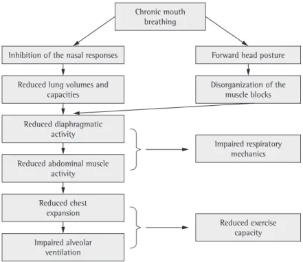
act in regulating the depth of breathing and airway patency. Nasal blockage results in an increase in lung resistance and a decrease in lung compliance, affecting chest expansion, with inadequate alveolar ventilation.(2)
It has also been demonstrated that the respiratory pattern imposed by CMB implies the need for postural adaptations. In order to facilitate the flow of air through the oral cavity, individuals bend the head forward and extend the neck. By doing so, they increase the amount of air passing through the pharynx, reducing airway resistance.(3)
Various studies have assessed body posture in mouth-breathing subjects, and the consensus is that forward head posture is the major change.
(4-7) This forward head posture will lead to
disorganization of the muscle blocks (anterior, posterior, and transverse muscles), impairing diaphragm muscle mobility and, consequently, diaphragmatic function. This postural change also leads to accessory muscle recruitment, with increased sternocleidomastoid muscle activity, causing rib cage elevation, reducing thoracoabdominal mobility, and compromising the ventilatory efficacy of the diaphragm. This mechanical disadvantage intensifies the inspiratory effort and increases the work of breathing.(3,8,9) Inefficient respiratory muscle
function decreases respiratory muscle strength, resulting in reduced chest expansion, which impairs pulmonary ventilation during physical activity.(9) Therefore, mouth breathing and
forward head posture, also present in nasal-breathing children, can affect the organization of the muscle blocks, resulting in reduced diaphragmatic activity and abdominal muscle hypoactivity, thus hindering the synergy between these two muscles.(8) These adaptations impair
pharyngeal tonsil hyperplasia in relation to the right and left choanae.
We used a 2.7-mm diameter flexible endoscope (Machida, Tokyo, Japan). The endoscope was introduced into the nasal cavity up to the region of the nasopharynx, where the presence of pharyngeal tonsils (adenoids) was assessed. The endoscope was removed backwards, and the size and aspect of the nasal conchae on the lateral wall of the nasal cavity were assessed. Adenoid size was classified in accordance with the study conducted by Modrzynski & Zawisza.
(10) Adenoids were defined as hyperplastic when
they occupied an area equal to or greater than 70% of the nasopharynx in the endoscopic assessment. The size of the palatine tonsils was defined by oral endoscopy, in accordance with the parameters recommended by Brodsky.
(11) In the assessment of the palatine tonsils,
obstruction was graded as follows: grade I, oropharyngeal obstruction ≤ 25%; grade II, oropharyngeal obstruction of 25-50%; grade III, oropharyngeal obstruction of 51-75%; and grade IV, oropharyngeal obstruction > 75%. A diagnosis of hyperplasia was made when the palatine tonsils were classified as having grade III or IV obstruction.
from the control group, as were those presenting with mechanical obstruction of the oral or nasal cavity. For both groups, the exclusion criteria were as follows: having a body mass index above the 95th percentile; having asthma, chronic respiratory diseases, neurological disorders, orthopedic disorders, or heart disease; and having undergone adenotonsillectomy. In the study group, the exclusion criteria were investigated by analyzing medical charts and interviewing parents, whereas, in the control group, they were investigated by analyzing the parental questionnaire.
The otolaryngological examination assessed the nasal fossae, paranasal sinuses, pharynx, larynx, and ears. Obstruction of the nasal cavities was investigated by rhinoscopy. Otoscopy consisted of examining the external auditory meatus and assessing the presence of tympanic membrane retraction. Changes in the oral cavity and palatine tonsil hyperplasia were assessed by oral endoscopy.
Rhinoscopy was performed for the assessment of the nasal cavities, septal deviation, turbinate hypertrophy, and nasopharyngeal hypertrophy, as well as for determining the degree of
Sciences (Protocol no. 849/2008). Prior to the beginning of the study, the parents or legal guardian of all children gave written informed consent.
Results
The study sample included 92 children. Of those, 30 (32.6%) presented with mouth breathing (MB group) and 62 (67.4%) presented with nasal breathing (NB group), with a mean age of 9.8 ± 0.9 years and 9.6 ± 0.9 years, respectively (p = 0.365). In the MB group, 23 (76.7%) were male and 7 (23.3%) were female, whereas, in the NB group, 23 (37.1%) were male and 39 (62.9%) were female (p < 0.001). There were no differences between the two groups in terms of race (p = 0.336), weight (p = 0.133), or height (p = 0.337).
Forward head posture, determined by the New York test, was detected in 29 children (96.7%) in the MB group, being considered severe in 12 (40.0%) and moderate in 17 (56.7%). In the NB group, moderately forward head posture was detected in 30 children (48.4%), and there was no severely forward head posture (p < 0.001).
The comparison between the MB group and the NB group in terms of the mean values of MIP, MEP, and 6MWD revealed that all values were lower in the MB group—MIP: 20.0 ± 7.1 cmH2O vs. 62.5 ± 21.9 cmH2O (p < 0.001); MEP: 25.3 ± 11.7 cmH2O vs. 58.8 ± 22.3 cmH2O
(p < 0.001); and 6MWD: 568.1 ± 47.4 m vs. 629.8 ± 47.6 m (p < 0.001).
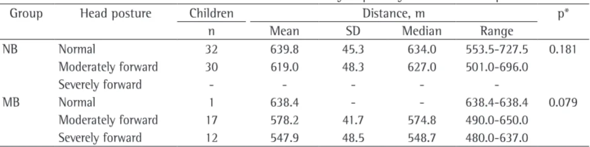
Tables 1 and 2 show that, in the MB group, the differences between those with (moderate or severe) forward head posture and those with normal head posture, in terms of the mean values of MIP and MEP, were not significant. However, in the NB group, the mean values of MIP and MEP were higher in those with forward head posture than in those with normal head posture (70.8 ± 19.1 cmH2O vs. 54.7 ± 21.7 cmH2O; The children underwent postural assessment
by the New York test,(13) an objective method for
postural assessment that contemplates thirteen body segments.(13) It has a scoring system that
allows a quantitative analysis with the power to classify the postural disorder assessed. Posture is classified as severely abnormal, moderately abnormal, or normal.(14) The classification of
head posture was specifically analyzed.
Respiratory muscle strength was assessed by determining MIP and MEP. Subsequently, the six-minute walk test (6MWT) was performed. These evaluations were performed by previously trained physical therapists, and each test was performed by a professional, always the same professional, who was blinded to the results of the other tests.
Measurements of MIP and MEP were obtained with a manometer (MV-120; Ger-Ar-SP Com. Equip. Ltda., São Paulo, Brazil) attached to a Y connector, with an air outlet (diameter, 1 mm) at its proximal end, and to a plastic mouthpiece (internal diameter, 2 cm).(15) Three
measurements were performed, and the highest value was considered the final result. After a 15-min rest period, the 6MWT was performed, in accordance with the American Thoracic Society recommendations.(16)
Before performing the tests, the children were given a demonstration. The tester verbally encouraged the children to make their best efforts.
there have been no studies involving all of these variables.
Our study showed that there was a predominance of mouth breathing in males, a fact that has also been observed by other authors.(1,17) Boys have smaller airway caliber
and a higher prevalence of allergic rhinitis and obstructive sleep apnea syndrome, major entities associated with CMB.(18)
Forward head posture was observed in 96.7% of the children in the MB group. It has been reported that this postural change, combined with flexion of the lower cervical spine and extension of the upper cervical spine, with decreased cervical lordosis, is the first postural compensation adopted by mouth-breathing subjects in order to decrease airflow resistance.
(5,6,19)
Cuccia et al.(6) assessed head posture in 35
mouth-breathing children who were compared with a control group, by means of cephalometric measurements, and found an increase in the extension of the upper cervical spine (atlanto-occipital joint) with decreased cervical lordosis, this being the principal finding. Another analysis by the same method showed that the extension of the cervical spine was greater in 56 mouth-breathing children with asthma than in normal-breathing children without asthma.(4)
Yi et al.(5) also observed extension of the head
p = 0.003; and 67.7 ± 22.1 cmH2O vs. 50.5 ± 19.5 cmH2O; p = 0.004, respectively). In terms of the 6MWD, the differences between those with forward head posture and those with normal head posture were not significant in the MB or NB group (p = 0.079 and p = 0.181, respectively; Table 3).
After multivariate analysis by multiple linear regression of MIP and MEP in relation to gender, age, respiratory mode, and forward head posture, the best adjusted model for MIP (adjusted R2 = 60.4%) included only the variables
respiratory mode and forward head posture. Mouth breathing was associated with lower MIP, whereas postural change was associated with higher MIP. The same was observed for MEP (adjusted R2 = 44.2%; Table 4). The same
adjustment was applied to the 6MWD. In this case, only the variable respiratory mode remained in the model, and the standard deviations were lower in the MB group (adjusted R2 = 26.6%;
Table 4).
Discussion
In the present study, we assessed the influence of respiratory mode and forward head posture on exercise capacity and respiratory muscle strength in children with CMB. To date,
Table 1 - Distribution of the MIP values by respiratory mode and head posture.a
Group Head
posture
Children MIP, cmH2O p*
n Mean SD Median Range
NB Normal 32 54.7 21.7 55.0 25.0-110.0 0.003
Moderately forward 30 70.8 19.1 72.5 35.0-110.0
Severely forward - - - -
-MB Normal 1 20.0 - - 20.0-20.0 0.622
Moderately forward 17 21.8 8.3 20.0 10.0-40.0
Severely forward 12 17.5 4.5 20.0 10.0-20.0
NB: nasal breathing; and MB: mouth breathing. aDetermined by the New York test. *Kruskal-Wallis test.
Table 2- Distribution of the MEP values by respiratory mode and head posture.a
Group Head
posture
Children MEP, cmH2O p*
n Mean SD Median Range
NB Normal 32 50.5 19.5 53.0 20.0-80.0 0.004
Moderately forward 30 67.7 22.1 67.5 20.0-120.0
Severely forward - - - -
-MB Normal 1 25.0 - - 25.0-25.0 0.957
Moderately forward 17 25.6 13.2 20.0 10.0-60.0
Severely forward 12 25.0 10.4 20.0 10.0-40.0
excursion found in the study conducted by Yi et al.(5) is also a finding that emphasizes the
change in respiratory mechanics in CMB.
In view of the changes in respiratory mechanics in CMB, we hypothesized the possibility of investigating, by using the 6MWT, the repercussion of such changes on exercise capacity, a subject that has not been studied in this type of population.
Reduced respiratory muscle strength can be caused by postural disorganization or by inhibition of the nasal responses, both of which result in lower lung volumes and capacities, affecting chest expansion and alveolar ventilation, with a decrease in PaO2, thereby reducing exercise tolerance.(21,22) In cases
that are more severe, this can be accompanied by obstructive sleep apnea syndrome or cor pulmonale.(18)
According to another hypothesis, known as the one-airway hypothesis, the effect of CMB can extend to the lung region and interfere with the physiological response to exercise. Individuals with CMB show changes in the muscular, circulatory, and respiratory systems, and such changes can affect the physiological response to exercise.(24)
Some studies have assessed cardiorespiratory function in subjects under conditions that induce mouth breathing.(25-28) Ribeiro & Soares(25)
observed that some spirometric indices (FEF25-75% and maximal voluntary ventilation) were below and decreased cervical lordosis in 30
mouth-breathing children. The results of those studies corroborate those of our study.
Forward head posture causes increased sternocleidomastoid muscle activity and leads to rib cage elevation, reducing thoracoabdominal mobility and compromising the ventilatory efficacy of the diaphragm.(3) This mechanical
disadvantage intensifies the inspiratory effort and generates a vicious cycle of muscle tension, postural change, and increased work of breathing.(3,20) Therefore, the disorganization of
the muscle blocks, which results in ineffective diaphragmatic contraction and, consequently, in ineffective abdominal muscle contraction, alters the respiratory dynamics completely, translating to reduced respiratory muscle strength. Another factor that might affect respiratory biomechanics is the lower respiratory effort required by mouth breathing, as well as the inhibition of the nasal afferent nerves, responsible for regulating lung capacity and lung volumes, resulting in poor use of the respiratory muscles and progressive muscle weakening.(8,21,22)
We found reduced respiratory muscle strength in the MB group. In a study evaluating the thoracic perimeter of mouth breathing children, lower values were found in relation to the control group.(23) This finding is explained
by the reduced expandability, with respiratory muscle weakness, leading to a smaller thoracic perimeter.(23) The reduced diaphragmatic
Table 4 - Multiple linear regression equations for the variables MIP, MEP, and six-minute walk distance.
Variable Adjusted R2 (%) Equation
MIP* 60.4 X = 0.323 – 1.585 × RM + 0.387 × FHPm
MEP* 44.2 Y = 0.243 – 1.360 × RM + 0.397 × FHPm
6MWD 26.6 Z = 629.7 – 61.674 × RM
assessing functional capacity because it is simple, inexpensive, and easily applied, providing an overall analysis of the respiratory, cardiac, and metabolic systems.(30)
In the present study, mouth breathing seemed to be the factor having the greatest impact the variables studied. It is therefore suggested that cervical repositioning is another of the changes triggered by mouth breathing, with a lesser effect on respiratory muscle strength and exercise capacity. Another consideration is that moderately forward head posture can act as a compensatory mechanism in order to improve respiratory muscle function, regardless of respiratory mode.
Although there is no evidence that forward head posture has an impact on respiratory biomechanics and exercise capacity, mouth breathing, with or without cervical change, compromises the musculoskeletal and cardiorespiratory systems. Therefore, a global intervention is essential to preventing pathological compensatory mechanisms.
A limitation of the present study was the fact that, although all of the children at the specialized clinic were invited to participate in the study, it was not possible to recruit all of the intended population. We therefore suggest that studies involving larger samples, as well as longitudinal studies involving older age groups, be conducted. In addition, we recommend the use of a more accurate postural assessment tool, the measurement of pulmonary function variables, and the use of maximal cardiopulmonary exercise testing. These future studies might clarify these relationships, which remain unexplored in the literature.
In the present study, mouth breathing negatively affected respiratory biomechanics and exercise capacity. Moderately forward head posture acted as a compensatory mechanism in order to improve respiratory muscle function.
Acknowledgments
We would like to thank Ester Piacentini Côrrea for her assistance in the data collection.
References
1. Barros JR, Becker HM, Pinto JA. Evaluation of atopy among mouth-breathing pediatric patients referred for treatment to a tertiary care center. J Pediatr (Rio J). 2006;82(6):458-64.
the predicted values, characterizing obstructive lung disease, mostly mild to moderate in severity, in mouth-breathing subjects. This brings us to the extensive impairment in the bronchial tree, the decreased nasal resistance changing intrathoracic pressure and reducing lung volume.
In subjects with asthma who were submitted to nasal occlusion with a nose clip during exercise, FEV1 decreased by 20%, compared with a decrease of 5% during spontaneous breathing, predominantly through the nose.(26) In subjects
with asthma, less nasal resistance is required in order to induce mouth breathing, and, when mouth breathing occurs, there is a decrease in pulmonary function and a greater predisposition to bronchial obstruction.(27) Melissant et al.(28)
induced upper airway obstruction during exercise and found that there were decreases in minute ventilation and CO2 production. The responses of the subjects included hypoventilation, hypoxia, and hypercapnia. Data similar to those reported in the aforementioned studies corroborate our finding of reduced cardiorespiratory capacity.
In our study, MIP and MEP were not found to be associated with head posture in the MB group children. In contrast, in the NB group, we observed that a more severely forward head posture translated to higher MIP and MEP values. This suggests that nose-breathing children use this as a compensatory mechanism, thus achieving higher MIP and MEP values than do those who retain a normal head posture. In contrast, mouth-breathing children seem to have more severe postural impairment, lacking compensatory resources to perform the maneuvers. Because we found no studies assessing these associations, further studies are needed in order to confirm and explain changes in respiratory muscle strength in relation to respiratory mode and forward head posture. Although we did not find a relationship between musculoskeletal change and lung disease in mouth-breathing subjects, Silveira et al.(29)
observed that forward head posture correlated negatively with spirometric variables in mouth-breathing subjects.
mechanics on orofacial pain. Dent Clin North Am. 1997;41(2):211-27.
21. Pires MG, Di Francesco RC, Grumach AS, Mello JF Jr. Evaluation of inspiratory pressure in children with enlarged tonsils and adenoids. Braz J Otorhinolaryngol. 2005;71(5):598-601.
22. Canning BJ. Neurology of allergic inflammation and rhinitis. Curr Allergy Asthma Rep. 2002;2(3):210-5. 23. Pires MG, Di Fracesco RC, Junior JF, Grumach AS.
Alterações Torácicas Secundárias ao Aumento de Volume de Tonsilas Palatinas e Faríngeas. Int Arch Otorhinolaryngol. 2007;11(2):99-105.
24. Bennett WD, Zeman KL, Jarabek AM. Nasal contribution to breathing with exercise: effect of race and gender. J Appl Physiol. 2003;95(2):497-503.
25. Ribeiro EC, Soares LM. Avaliação espirométrica de crianças portadoras de respiração bucal antes e após intervenção fisioterapêutica. Fisioter Bras. 2003;4(3):163-7.
26. Shturman-Ellstein R, Zeballos RJ, Buckley JM, Souhrada JF. The beneficial effect of nasal breathing on exercise-induced bronchoconstriction. Am Rev Respir Dis. 1978;118(1):65-73.
27. Hallani M, Wheatley JR, Amis TC. Initiating oral breathing in response to nasal loading: asthmatics versus healthy subjects. Eur Respir J. 2008;31(4):800-6. 28. Melissant CF, Lammers JW, Demedts M. Relationship
between external resistances, lung function changes and maximal exercise capacity. Eur Respir J. 1998;11(6):1369-75.
29. Silveira W, Mello FC, Guimarães FS, Menezes SL. Postural alterations and pulmonary function of mouth-breathing children. Braz J Otorhinolaryngol. 2010;76(6):683-6. 30. Morales-Blanhir JE, Palafox Vidal CD, Rosas Romero
Mde J, García Castro MM, Londoño Villegas A, Zamboni M. Six-minute walk test: a valuable tool for assessing pulmonary impairment. J Bras Pneumol. 2011;37(1):110-7.
between excursion of the diaphragm and curvatures of the spinal column in mouth breathing children. J Pediatr (Rio J). 2008;84(2):171-7.
6. Cuccia AM, Lotti M, Caradonna D. Oral breathing and head posture. Angle Orthod. 2008;78(1):77-82. 7. Neiva PD, Kirkwood RN, Godinho R. Orientation
and position of head posture, scapula and thoracic spine in mouth-breathing children. Int J Pediatr Otorhinolaryngol. 2009;73(2):227-36.
8. Lima LC, Baraúna MA, Sologurem MJ, Canto RS, Gastaldi AC. Postural alterations in children with mouth breathing assessed by computerized biophotogrammetry. J Appl Oral Sci. 2004;12(3):232-7.
9. Corrêa EC, Bérzin F. Mouth Breathing Syndrome: cervical muscles recruitment during nasal inspiration before and after respiratory and postural exercises on Swiss Ball. Int J Pediatr Otorhinolaryngol. 2008;72(9):1335-43. 10. Modrzynski M, Zawisza E. An analysis of the incidence
of adenoid hypertrophy in allergic children. Int J Pediatr Otorhinolaryngol. 2007;71(5):713-9.
11. Brodsky L. Modern assessment of tonsils and adenoids. Pediatr Clin North Am. 1989;36(6):1551-69.
12. Abreu RR, Rocha RL, Lamounier JA, Guerra AF. Etiology, clinical manifestations and concurrent findings in mouth-breathing children. J Pediatr (Rio J). 2008;84(6):529-35.
13. Althoff SA, Heyden SM, Robertson LD. Posture screening: a program that works. J Phys Educ Recreat. 1988;59(8):26-32.
14. Santos JB, Moro AR, César MR, Reis PF, Luz JD, Reis DC. Descrição do método de avaliação postural de Portland State University. Fisioter Brasil. 2005;6(5):392-5. 15. American Thoracic Society/European Respiratory Society.
ATS/ERS Statement on respiratory muscle testing. Am J Respir Crit Care Med. 2002;166(4):518-624.
About the authors
Renata Tiemi Okuro
Master’s Student. Graduate Program in Child and Adolescent Health, Faculdade de Ciências Médicas, Universidade Estadual de Campinas – FCM-Unicamp, State University at Campinas School of Medical Sciences – Campinas, Brazil.
André Moreno Morcillo
Tenured Associate Professor. Department of Pediatrics, Faculdade de Ciências Médicas, Universidade Estadual de Campinas – FCM-Unicamp, State University at Campinas School of Medical Sciences – Campinas, Brazil.
Maria Ângela Gonçalves Oliveira Ribeiro
Physical Therapist. Faculdade de Ciências Médicas, Universidade Estadual de Campinas – FCM-Unicamp, State University at Campinas School of Medical Sciences – Campinas, Brazil.
Eulália Sakano
Assistant Professor. Faculdade de Ciências Médicas, Universidade Estadual de Campinas – FCM-Unicamp, State University at Campinas School of Medical Sciences – Campinas, Brazil.
Patrícia Blau Margosian Conti
Physical Therapist. Faculdade de Ciências Médicas, Universidade Estadual de Campinas – FCM-Unicamp, State University at Campinas School of Medical Sciences – Campinas, Brazil.
José Dirceu Ribeiro