ABSTRACT
Objective: To evaluate the preventive effects of alkaline citrate on stone recurrence as well as stone growth post-ESWL or PCNL in patients with calcium-containing stones.
Materials and Methods: A total of 76 patients with calcium calculi who were stone-free or had residual stones less than 4 mm following ESWL and PCNL were enrolled. All patients were independently randomized into two groups. The treated group (N = 39) was given 81 mEq per day of oral potassium-sodium citrate (27 mEq three times a day), and the untreated group (N = 37) serving as controls. Blood, twenty-four hour urine analysis, and plain KUB were measured and compared at the baseline and after 12 months.
Results: At baseline, hypocitraturia was found in 20 of 39 patients (46.05%) of Group I and 15 of 37 patients (40.5%) of Group II. At 12 months, hypocitraturia was found in 3 of 39 (7.69%) and 14 of 37 (37.83%) of Group I and Group II, respectively (p = 0.007). At the 12 month follow-up, of the stone-free group, 92.3% of the treated group and 57.7% of the control group were still stone free. Of the residual stone group, 30.8% and 9.1 % of treated and control group were stone-free, respectively. The increased stone size found in 7.7% and 54.5% of treated and control groups, respectively. Conclusion: Sodium-potassium citrate provides positive effects on stone-forming activities in calcium stone patients suffering from urolithiasis following treatment with ESWL and PCNL procedures at the 12-month follow-up.
Key words: kidney; calculi; lithotripsy; citrates; urolithiasis Int Braz J Urol. 2011; 37: 611-616
With the advantage of high eficiency and
low morbidity rates, extracorporeal shock wave litho-tripsy (ESWL) has become the therapy of choice for small renal stones. Percutaneous nephrolithotomy (PCNL) is also becoming the therapy of choice for large renal stones due to the less invasive procedure as compared to open nephrolithotomy. These therapies provide good results, associated with an acceptable rate of complications, but unfortunately they do not change the underlying metabolic abnormality. Stone
recurrence is usually found after either treatment, even in those with a stone-free post-therapy status. In addition, retained stone fragments following those therapies may reaggregate or constitute a nucleus for new stone formation, thereby causing a high rate of
stone growth (1-4). The patient’s chances of irst epi -sode recurrent stone formation range between 27% and 50% (5,6). Medical treatment should therefore be considered following these interventions, in order to prevent further secondary treatments and hospitaliza-tions. Among the metabolic disorders usually found in recurrent stone forming patients, hypocitraturia
Alkaline citrate reduces stone recurrence and regrowth after
shockwave lithotripsy and percutaneous nephrolithotomy
Lojanapiwat B, Tanthanuch M, Pripathanont C, Ratchanon S, Srinualnad S,
Taweemonkong-sap T, Kanyok S, Lammongkolkul S
Division of Urology, Department of surgery (BL), Chiangmai University; Division of Urology,
De-partment of surgery (MT, CP), Prince Songkla University; Division of urology, DeDe-partment of
sur-gery (RS), Chulalongkorn University; Division of urology, Department of sursur-gery (SS, TT), Siriraj
hospital, Mahidol University; Department of biochemistry (SK, SL), Siriraj hospital, Mahidol
Uni-versity, Thailand
(low urinary citrate) is an important risk factor for calcium nephrolithiasis (1-4). Several studies dem-onstrated the effect of alkaline citrate in prevention and/or reduction of the stone recurrence and stone growth (1-4). In the current study, we evaluated the preventive effects of potassium-sodium citrate on stone recurrence as well as stone growth post-ESWL or PCNL, in patients with calcium-containing stones.
MATERIALS AND METHODS
The study was approved by the ethics
com-mittee of the Faculty of Medicine, Chiang Mai Uni -versity. Patients gave written informed consent be-fore participating in the study.
Eighty patients, who were stone-free or had residual stone fragments with less than 4 mm diam-eter at eight weeks after ESWL or PCNL, were en-rolled in the present clinical study. In case of ESWL, patients were treated by using Storz™ Modulith SL-20 equipment; in case of PCNL a standard
neph-roscopy (26-Fr Storz™ nephroscope) under luo -roscopic guidance with combined ultrasound and pneumatic lithotripsy was performed. All patients had calcium stones after the analysis by infrared
spectrometry method. Four patients were excluded
from the study due to loss of follow-up (N = 3) and unsatisfactory compliance with medication (N = 1). In total, 76 patients completed the one-year follow-up period. Eight weeks after the ESWL/PCNL
thera-pies, plain KUB ilms were evaluated: 39 patients
were stone-free (ESWL n = 24, PCNL n = 15) and 37 had residual stones smaller than 4 mm in diameter (ESWL n = 26, PCNL n = 11).
All patients were independently block ran-domized into two groups: the treated group and the un-treated group. The un-treated group (n = 39) was given 81 mEq oral potassium-sodium citrate (Uralyt-U®, Rot-tapharm Madaus) per day (27 mEq three time a day), whereas the control group (n = 37) received no treat-ment. All patients had normal renal function and nor-mal renal anatomy, as assessed by preoperative intrave-nous pyelography study. All patients who had urinary tract infections, anatomic abnormalities and clinical history of urologic stone surgery were excluded.
At baseline, all patients were evaluated for blood urea nitrogen (BUN) creatinine, electrolytes,
complete blood count (CBC), calcium, uric acid, uri-nalysis; in the 24-hour urine study, total urine volume, creatinine, electrolyte, calcium, oxalate, Uric acid?
Uric acid and citrate were also measured.
Patients were advised to have suficiently high luid
intake throughout the study, and the follow-up was scheduled every 3 months.
The patients were reevaluated at six months after the initial treatment for serum chemistry and urinalysis. After 12 months, all patients were evalu-ated for serum chemistry, urinalysis, 24-hour urine study and plain KUB. The evidence of new stone formation was determined by spontaneous stone passage in absence of preexisting stones and/or
ap-pearance of new stones on a plain KUB ilm. Growth of existing stones was determined by quantiication
of increased stone size.
The statistical analyses were carried out using SPSS statistics software. Mann-Whitney U tests were used to compare laboratory tests, remis-sion rates, and growth rates between groups. The difference within each group between baseline and 12 months post treatment was assessed by repeated measurement ANOVA and multivariate analysis.
The protocol and documents needed for this study have been reviewed and approved by the Eth-ics Committees of each participating hospital.
RESULTS
Of 80 patients recruited, 76 patients com-pleted the 12-month follow-up observation period.
The average ages were 51.7 ± 10.4 and 48.9 ± 10.7
years for the treated and control groups,
respec-tively (Table-1). The average BMI was 23.9 ± 3.7
(kg/m2) in treated group and 23.6 ± 3.2 (kg/m2) in control group. The untreated group consisted of 25 patients (67.6%) post-ESWL and 12 patients (32.4%) post- PCNL. The treated group consisted of 25 patients (64.1%) post-ESWL and 14 patients
(35.9%) post-PCNL. The average stone size in CIRF
patients was 2.54 mm and 2.65 mm in treated and untreated patients, respectively. Laboratory tests such as complete blood count (CBC), blood urea nitrogen (BUN), creatinine, calcium, uric acid and sodium, chloride and bicarbonate did not show any
compared with the baseline values, in both groups.
Only a signiicant increase of serum potassium, al -beit well within the physiological range, was found
in the treated group at 12 months (from 4.02 ± 0.42 to 4.29 ± 0.48 mEq/L, p = 0.011). Urine pH and urine potassium were signiicantly increased from
baseline at 6 months and 12 months (p = 0.001 at 6 month, p = 0.09 at 12 month). The level of urine
po-tassium in treated patient was signiicant increased from 27.7 ± 18.3 to 56.7 ± 25.4 at 6 months and to 49.5 ± 36.4 at 12 months. At baseline, the result of
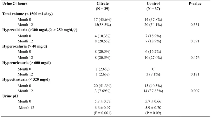
24-hour urine measurement for total urine volume, sodium, potassium, chloride, creatinine, calcium, magnesium, oxalate uric acid, and citrate did not
show signiicant difference between both groups
(Table-2). The average values of citrate were 304.3
± 233.8 mg/d and 259.2 ± 214.7 mg/d in control and in treated group, respectively. Hypocitraturia, with
citrate values lower than 320 mg/day was found in 35 out of the total 76 patients which was 46.05% of total patients, 15 patients in control group (92.9
± 64.96 mg/day) and 20 in treated group (169.5 ±
98.4 mg/day). Mean urine citrate level at month 12
was 305. 3 ± 233.08 mg/day in control group and 405.3 ± 305.44 mg/day in treated group which is statistically signiicant (P = 0.007). Low urine out -put (urine volume lower than 1,500 ml/d) was a
common secondary inding observed in 40.8% of
all patients. Number of patients in both groups who have abnormal 24-hour urine measurement was shown in Table-2.
The change of the stone, assessed at 12 months follow up, is shown in Table-3. In the stone-free group at baseline, 92.3% of treated group and 57.7% of control group were still stone free at 12 months. An increase in stone size was found in 7.7% and 42.3% of treated and control group, respective-ly, which were statistically different. At the same 12-month follow-up, in the group with residual stone
fragments < 4 mm in diameter, 30.8% and 9.1% of
patients were found to be stone-free in the treated and control groups, respectively. 50% of patients in the treated group showed the same stone size, whereas an increase in stone size was found in 7.7% and 54.5% of the treated and control groups, respectively.
DISCUSSION
The most common composition of kidney stones is calcium-based, which is up to 80% of all types of stones (7). The purposes of stone manage-ment are complete stone clearance, prevention of new stone formation and regrowth, preservation of renal function, control of urinary tract infections and, whenever the case, correction of abnormal anatomy and underlying metabolic abnormality. The advancement in minimally invasive surgery, most kidney stones are treated with extracorporeal shock wave lithotripsy (ESWL) and percutaneous
nephro-lithotomy (PCNL). Following these treatments, the
achievement of stone-free condition or of residual fragments with a diameter smaller than 4 mm is de-Table 1 - Proiles of patients at baseline.
Stone free Residual Fragment < 4 mm
Citrate (N = 13)
Control (N = 26)
Citrate (N = 26)
Control (N = 11)
Sex
Male 7 (53.8%) 17 (65.4%) 19 (73.1%) 9 (81.8%)
Female 6 (46.2%) 9 (34.6%) 7 (26.9%) 2 (18.2%)
Age (year) 48.8±8.26
(35 - 64)
54.1± 10.12 (32 - 73)
49.1 ±12.04 (28 - 75)
45.9± 8.93 (31 - 57)
Post treatment
ESWL 8 (61.5%) 16 (61.5%) 17 (65.4%) 9 (81.8%)
ined as a therapeutical success (1,2). The presence
of residual fragments is commonly found following these modern treatments, particularly after ESWL (1,2,8). It is clinically relevant, as a recognized pdisposing factor for new stone formation (1-4), re-current urinary tract infections, pain and, in general, for need of further additional treatments (1).
By analyzing stone-forming activity at two years following ESWL, it was found that the inci-dence of recurrent stones ranged from 8% to 10% in
stone-free patients and from 20% to 22% in patients with residual fragments (8). Another study found re-current calculi, in patients who had stone free status after ESWL, ranging from 7% to 14% per year (9). In
comparing Soygur’s and Fine’s studies at 12 months
after ESWL, the alkaline citrate-treated patients who were stone-free at baseline and remained stone-free
were 100% and 89.5%, respectively. However, there
were found to be only 71.4% and 50% in untreated patients of both studies respectively (2,10). Nine per-Table 2 -24 hours urine metabolic evaluation.
Urine 24 hours Citrate
(N = 39)
Control (N = 37)
P-value
Total volume (< 1500 mL/day)
Month 0 17 (43.6%) 14 (37.8%)
Month 12 15(38.5%) 20 (54.1%) 0.331
Hypercalciuria (>300 mg/d,♂; > 250 mg/d,♀)
Month 0 4 (10.3%) 7 (18.9%)
Month 12 8 (20.5%) 7 (18.9%) 0.391
Hyperoxaluria (> 40 mg/d)
Month 0 8 (20.5%) 6 (16.2%)
Month 12 8 (20.5%) 10 (27.0%) 0.476
Hyperuricouria (> 600 mg/d)
Month 0 1 (2.6%) 0
Month 12 1 (2.6%) 3 (8.1%) 0.171
Hypocitraturia (< 320 mg/d)
Month 0 20 (51.3%) 15 (40.5%)
Month 12 3 (7.69%) 14 (37.83%) 0.007
Urine pH
Month 0 5.8 ± 0.77 5.7 ± 0.66
Month 12 6.6 ± 0.97
(P = 0.001)
5.9 ± 0.70
(P = 0.09)
Table 3 -Stone-forming activity at 12 months (Overall n = 76).
Stone free(%) Residual Fragment < 4 mm (%)
Citrate n = 13
Control n = 26
RR (95% CI)
Citrate n = 26
Control n = 11
RR (95% CI)
Stone free 12(92.3) 15(57.7) 5.33 (0.77 - 36.5)
8(30.8) 1(9.1) 1.38 (0.96 - 1.98)
Stone size unchanged - - 13(50.0) 2(18.2) 1.47
(0.98 - 2.18)
Stone size decreased - - 3(11.5) 2(18.2) 0.83
(0.39 - 1.76)
Stone recurrence / Stone size increased
1(7.7) 11(42.3) 0.19 (0.03 - 1.28)
cent of patients following PCNL had recurrent stones following achievement of stone-free condition, and 63% had either new calculi or continued stone growth after having residual stone fragments (3).
Metabolic abnormalities were also detected
in the majority of patients with recurrent nephroli -thiasis. The wide range of underlying metabolic ab-normalities such as hypercalciuria, hyperoxaluria, hyperuricouria and hypocitraturia can cause the formation of stones in the urinary tract metabolic abnormalities were not affected following ESWL / PCNL therapies. Our common metabolic abnormal-ities are hypocitraturia followed by hypercalciuria and low urine output, which is the same as the pre-vious study from Thailand (11). Effective medical management should therefore be directed primarily towards correction of the underlying abnormalities. As expected, stone-free status and residual frag-ments status following ESWL and PCNL still in-creased the risk for active stone formation (1,2,10).
Such medical treatment signiicantly alleviated
stone-forming activity after ESWL and PCNL in patients who resulted stone-free as well as those with residual fragments (1,2,10). The remission rate
of patients with residual calculi was signiicantly
lower than the stone-free group (1-4).
Citrate is the most potent stone inhibitor (12,13). The mean normal urinary citrate excretion is 640 mg/24 hours of urine. The accepted limit for diagnosing hypocitraturia is 320 mg/24 hours of urine (2). Isolated hypocitraturia has been
iden-tiied in about 13% of patients, and coexists with
other metabolic abnormalities in 15% to 69 % of
calcium stone formers (7,11). Hypocitraturia is
usually found in patients with systemic acidosis, hypokalemia, unbalanced diets, and chronic diar-rhea. The mechanism of action of citrate in the pre-vention of calcium urolithiasis relates to its ability to form a complex with calcium in urine as calcium citrate complex, which is more soluble than calci-um oxalate (7,12,13). The calcicalci-um citrate complex prevents all forms of crystallization by inhibiting spontaneous nucleation of calcium oxalate and/or crystal growth of calcium phosphate and calcium oxalate, by retarding agglomeration of preformed calcium oxalate crystal, and by preventing hetero-geneous nucleation of calcium oxalate;
monosodi-um urate citrate also restores the inhibitory
proper-ties of Tamn-Horsfall protein (7).
Several authors supported the beneit of
alkaline citrates in all patients undergoing
shock-wave lithotripsy for the signiicant improvement in
rates of alleviation the calcium oxalate
stone-form-ing activity and positively inluence the preexiststone-form-ing
stone clearance and dissolution with minor adverse reaction (2,11,14-16). The studies with stone recur-rence as the endpoint demonstrated a reduction in stone-forming rate from 47% to 100%. Of four ran-domized controlled trials, the stone-free rate after at least two years of alkaline citrate treatment was 53.5% and 35% in treatment and placebo groups,
respectively (3,14-16). For three years, in idiopath -ic hypocitratur-ic calcium nephrolithiasis patients who received potassium citrate, the stone
forma-tion per year signiicantly declined from 1.20.6 to
0.1±0.2 stones per patient. This positive effect of potassium citrate also found in calcium stone pa-tients following PCNL (3). Authors concluded that the medical therapy can decrease costs of repeated procedures and recommended it for patients fol-lowing PCNL regardless of their stone-free status. After potassium citrate, minor gastrointestinal side effects such as diarrhea, indigestion, nausea and burning may occur.
Our study demonstrated that hypocitraturia
(urine citrate excretion < 320 mg/24-hours urine)
was the most common metabolic risk factor in our patients after ESWL and PCNL. We have found that in these kinds of patients, the medical therapy with potassium-sodium citrate can prevent and/or reduce the recurrence and stone growth during a 12-month
follow-up. The patients with residual fragment < 4
mm post-treatment were at risk for developing stone growth, as demonstrated in control group compared with the treated group. Our study showed that stone-free status and unchanged stone size status were
sig-niicantly more frequent in the treated group which
is 5.3 fold in stone-free patients and 1.38 fold in
CIRF patients as compared to the untreated group.
Moreover, the oral formulation of
potassium-sodi-um citrate was well-tolerated with signiicantly less
reported side effects. During the follow-up time, only an increase of serum potassium (but within the
-served as related with the therapeutic effects of the medication.
The limitations of this study are the small number size and short follow-up time. Due to the poor compliance with alkaline citrate treatment in stone patients with a long follow-up period in clinical practice, this study can demonstrate the positive effect of potassium-sodium citrate in one year follow-up.
CONCLUSIONS
Hypocitrauria is the most common metabolic
disorder in calcium stone patients. Sodium-potassi-um citrate provides positive effects on stone-forming activities, in a 12- month follow-up in calcium stone patients. We therefore recommend administration of alkali citrates in patients suffering from urolithiasis, following treatment with ESWL and PCNL proce-dures, for effective prevention of stone recurrence and stone growth.
CONFLICT OF INTEREST
None declared.
REFERENCES
1. El-Nahas AR, El-Assmy AM, Madbouly K, Sheir KZ: Predictors of clinical signiicance of residual fragments after extracorporeal shockwave lithotripsy for renal stones. J Endourol. 2006; 20: 870-4.
2. Soygür T, Akbay A, Küpeli S: Effect of potassium citrate therapy on stone recurrence and residual frag-ments after shockwave lithotripsy in lower caliceal calcium oxalate urolithiasis: a randomized controlled trial. J Endourol. 2002; 16: 149-52.
3. Kang DE, Maloney MM, Haleblian GE, Springhart
WP, Honeycutt EF, Eisenstein EL, et al.: Effect of medical management on recurrent stone formation following percutaneous nephrolithotomy. J Urol. 2007; 177: 1785-8; discussion 1788-9.
4. Mattle D, Hess B: Preventive treatment of nephroli -thiasis with alkali citrate--a critical review. Urol Res. 2005; 33: 73-9.
5. Trinchieri A, Ostini F, Nespoli R, Rovera F, Mon -tanari E, Zanetti G: A prospective study of recurrence rate and risk factors for recurrence after a irst renal stone. J Urol. 1999; 162: 27-30.
6. Ljunghall S, Danielson BG: A prospective study of
renal stone recurrences. Br J Urol. 1984; 56: 122-4. 7. Pattaras JG, Moore RG: Citrate in the management of
urolithiasis. J Endourol. 1999; 13: 687-92.
8. Newman DM, Scott JW, Lingeman JE: Two year fol-low- up of patients treated with extracorporeal shock wave lithotripsy. J Endourol. 1988; 2: 163-71. 9. Ljunghall S: Incidence of upper urinary tract stones.
Miner Electrolyte Metab. 1987; 13: 220-7.
10. Fine JK, Pak CY, Preminger GM: Effect of medi -cal management and residual fragments on recurrent stone formation following shock wave lithotripsy. J Urol. 1995; 153: 27-32; discussion 32-3.
11. Stitchantrakul W, Kochakarn W, Ruangraksa C, Dom-rongkitchaiporn S: Urinary risk factors for recurrent calcium stone formation in Thai stone formers. J Med Assoc Thai. 2007; 90: 688-98.
12. Pak CY: Citrate and renal calculi: an update. Miner Electrolyte Metab. 1994; 20: 371-7.
13. Pak CY: Medical management of urinary stone dis-ease. Nephron Clin Pract. 2004; 98: c49-53.
14. Barcelo P, Wuhl O, Servitge E, Rousaud A, Pak CY: Randomized double-blind study of potassium citrate in idiopathic hypocitraturic calcium nephrolithiasis. J Urol. 1993; 150: 1761-4.
15. Hofbauer J, Höbarth K, Szabo N, Marberger M: Al
-kali citrate prophylaxis in idiopathic recurrent cal-cium oxalate urolithiasis--a prospective randomized study. Br J Urol. 1994; 73: 362-5.
16. Ettinger B, Pak CY, Citron JT, Thomas C, Adams--Huet B, Vangessel A: Potassium-magnesium citrate is an effective prophylaxis against recurrent calcium oxalate nephrolithiasis. J Urol. 1997; 158: 2069-73.
_____________________ Submitted for publication: December 01, 2010
______________________ Accepted after revision: May 12, 2011 ___________________
Correspondence address: Dr. Bannakij Lojanapiwat Division of Urology Department of Surgery Chiangmai University Chiangmai, 50200, Thailand Fax: +66 53 945-154