Prostatic carcinomas with neuroendocrine differentiation
ABSTRACT
Purpose: Neuroendocrine carcinomas (NEC) of the prostate are rare, with only a few series hitherto reported. The ob-jective of this study was to assess in a single institution the clinical and morphologic characteristics of neuroendocrine carcinomas diagnosed in needle core biopsies.
Materials and Methods:The current study analyses seven cases diagnosed in needle biopsies at a large tertiary regional cancer center from Northeastern Brazil. Two pathologists reviewed specimens retrospectively, and demographic and morphologic characteristics were compared to 458 acinar tumors diagnosed in the same period.
Results: There were ive small cell carcinomas and two low-grade neuroendocrine carcinomas (carcinoid). NEC were associated with an acinar component in 5/7 cases and the Gleason score of the acinar component was always > 6. The number of cores involved in prostates with NEC was greater (65% compared to 24% of acinar tumors, p < 0.05). The mean PSA at diagnosis was 417.7 (range 5.7-1593, SD 218.3), compared to 100.5 (p = 0.1) of acinar tumors (range 0.3-8545, SD 22.7). Prostates harboring NEC were bigger (p < 0.001, mean volume 240 mL vs. 53 mL of acinar tumors). Treatment of NEC included palliative surgery, chemotherapy, and hormonal therapy.
Conclusions: NEC of the prostate is rare and often associated with a high-grade acinar component. Prostates with NEC tend to be larger and involve a greater number of cores than acinar tumors. PSA at diagnosis does not seem to predict the presence of NE tumors in needle biopsy.
Key words: prostate; neoplasms; carcinoid tumor; neuroendocrine tumor; treatment outcome Int Braz J Urol. 2011; 37: 598-604
Neuroendocrine carcinomas (NEC) of the prostate are rare, representing less than 0.5% of pros-tate carcinomas in the few series reported to date (1-8). The current classiication of neuroendocrine carcino -mas is based on the World Health Organization 2004 lung tumor classiication, and it divides those tumors into well-differentiated neuroendocrine carcinomas (carcinoid tumors), moderately-differentiated neu-roendocrine carcinomas (atypical carcinoid tumors), and poorly-differentiated neuroendocrine carcino-mas, which include two morphologic distinct entities (small cell carcinomas and large cell neuroendocrine
carcinomas) (9). The classiication is based solely on histomorphology and relies on both light microscopy and immunohistochemical studies.
Albeit rare, the most common neuroendo-crine carcinoma of the prostate is by far small cell car-cinoma. Furthermore, it is estimated that up to 10% of prostate cancer in patients with androgen-resistant disease after long-term androgen deprivation therapy are high grade NEC, most with associated acinar ad-enocarcinoma (10).
Recognition of this entity via needle biopsies is critical, as its therapy differs signiicantly from that of usual acinar high-grade prostatic adenocarcinoma.
Clinical Urology
International Braz J Urol Vol. 37 (5): 598-604 September - October, 2011
Prostatic carcinomas with neuroendocrine differentiation
diagnosed in needle biopsies, a morphologic study of 7 cases
among 465 sequential biopsies in a tertiary cancer center
MVA Lima, C Nogueira, JAA Oliveira, FJ Muniz Neto, M Franco, F Tavora
Hospital do Câncer, Instituto do Câncer do Ceara (MVAL, CN, JAAO, FJMN, FT), Fortaleza,
Cea-ra and Escola Paulista de Medicina (MF, FT), São Paulo, BCea-razil
MATERIALS AND METHODS
Seven cases of neuroendocrine carcinomas primary to the prostate were collected over 4 years (2006-2010) from the Department of Pathology ar-chives of the Cancer Hospital of the Ceara Cancer Institute among 465 sequential needle biopsies from the in-house Urology Department Service. Patients with neuroendocrine tumors primary to other sites were excluded from the study.
The morphologic data independently collect-ed and reviewcollect-ed by two pathologists (FT and CDM) were basic morphology to include small or non-small cell pattern, percentage of cores involved, associa-tion with convenassocia-tional acinar adenocarcinoma and the Gleason grading, mitotic rate, presence of tumor necrosis and any other morphologic indings.
Immunohistochemical studies were per-formed on the available parafin blocks in all seven cases. Immunohistochemistry was performed in our laboratory using the standard streptavidin-biotin-peroxidase procedure. Primary monoclonal antibod-ies to PSA (dilution 1:100), chromogranin (1:2000), synaptophysin (1:50), ki67 (1:100) and p63 (1:300) (Dako Inc., Carpinteria, USA) were applied to 5-mm thick 10% formalin-ixed, parafin-embedded tissue sections. The sections underwent a process of de-parafinization, rehydration, and washing in xylene, graded alcohols, and distilled water. Blockage of en-dogenous peroxide activity was performed by incu-bation with 3% H2O2. The sections were placed in 10 mM citrate buffer at pH 6 with subsequent antigen re -trieval procedure. The antigen-antibody reaction was visualized using the avidin-biotin peroxidase com-plex and diaminobenzidine as the chromogen. Slides were counterstained with hematoxylin. Positive re-sults consisted of dark brown nuclear (p63, ki67) and cytoplasmic (chromogranin, synaptophysin) staining and cytoplasmic and luminal granular staining of se-cretory epithelial cells by PSA. Appropriate positive and negative controls were included. Only staining that was moderate or strong was considered positive.
Clinical follow-up was possible in all but one case by retrospective clinical chart review by one of the authors (MVL).
Clinical and histopathologic variables were compared among categorized groups using the χ2
test or Student t- test. A p value less than 0.05 was considered signiicant. The software SPSS 5.0 (Chi -cago, IL) was used for statistical analyses.
RESULTS
The clinical and pathological characteristics of the seven cases are summarized in Tables 1 and 2. Those cases were retrieved spanning four years and included all needle biopsies performed at the Urolo-gy Department at our institution among 465 sequen-tial biopsies (1.5%). The mean age at diagnosis was 69.8 years. Metastatic NEC from any other site or direct extension from the bladder or gastrointestinal tract were excluded clinically in all cases. The se-rum PSA values at the time of initial diagnosis were available in 5 patients and ranged from 7.3 to 1449.0 ng/mL (mean 461.1 ng/mL, median 194.15 ng/mL).
Prostatic carcinomas with neuroendocrine differentiation
Figure 1 - Low power of small cell carcinoma (left) associated with Gleason 8 acinar adenocarcinoma (right). Hematoxilin-eosin, 40x.
Figure 2 - Small cell carcinoma associated with acinar adenocarcinoma. On the upper right corner, small cell carcinoma predominates, whereas acinar Gleason pattern 3 can be seen in the left lower corner. Hematoxilin-eosin, 200x.
Figure 3 - Small cell carcinoma in close association with Gleason 4+3 acinar adenocarcinoma (same case as Figure-4). Hematoxilin-eosin, 100x.
Figure 4 -Medium-power view of small cell carcinoma. The tumor tends to fragment on processing. Hematoxilin-eosin, 100x.
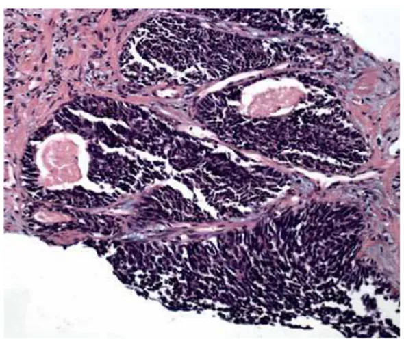
Figure 5 - Small cell carcinoma. Punctuate tumor necrosis is a common inding. Also note the desmoplastic stroma surrounding tumor nests. Hematoxilin-eosin, 200x.
601
Prostatic carcinomas with neuroendocrine dif
ferentiation
acinar adenocarcinoma
of associated adenocarcinoma
of cores involved
by NEC
percent of
involved cores
score in NEC
component
67 (%)
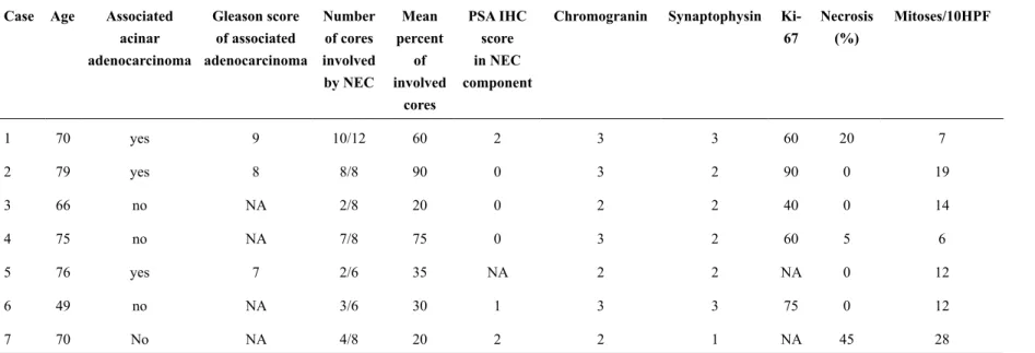
1 70 yes 9 10/12 60 2 3 3 60 20 7
2 79 yes 8 8/8 90 0 3 2 90 0 19
3 66 no NA 2/8 20 0 2 2 40 0 14
4 75 no NA 7/8 75 0 3 2 60 5 6
5 76 yes 7 2/6 35 NA 2 2 NA 0 12
6 49 no NA 3/6 30 1 3 3 75 0 12
7 70 No NA 4/8 20 2 2 1 NA 45 28
Table 2 - Clinical and follow-up characteristics of neuroendocrine carcinomas.
Case Surgery QT HT Status at follow-up PSA at diagnosis Clinical stage Follow-up time
(month)
1 No Yes No DOD 7.33 T4N0M1c 8.1
2 TUR/orchiectomy Yes Orchiectomy LWD 285.0 T4N0M0 21.3
3 no No No LFU NA NA NA
4 no Yes No LWD 103.3 T4N1M1b 26.6
5 no No Yes LWD 1449.0 T4N0M1b 22.2
6 Palliative colostomy No No DOD NA T4N1Mx 2.1
7
Prostatic carcinomas with neuroendocrine differentiation
ive available cases. PSA immunohistochemistry was only weakly positive in NEC in 2 cases (Fig -ures 7-10).
During the same period, data on 458 con-ventional type acinar adenocarcinomas were re-viewed and compared with the seven cases on this study. Of the 458 cases, there were 191 Gleason 6, 131 Gleason 7, 72 Gleason 8, 33 Gleason 9 and 7 Gleason 10. The mean age at diagnosis correspon-dent to Gleason 6-10 were 69.1, 70.3, 74.2, 73.6 and 76.2 (p < 0.001). Within the acinar tumors, high grade tumors (Gleason > 7) also correlated with a high PSA at diagnosis (p < 0.001), but not with prostate volume (p = 0.3). The ratio of PSA/
volume signiicantly correlated with a high Glea -son score (p < 0.05).
The number of cores involved in prostates with NEC was greater (65% compared to 24% of acinar tumors, p < 0.05). The mean PSA at diag -nosis was 417.7 (range 5.7-1593, SD 218.3), com-pared to 100.5 (p = 0.1) of acinar tumors (range 0.3-8545, SD 22.7). Prostates harboring NEC were bigger (p < 0.001, mean volume 240 mL vs 53 mL of acinar tumors).
Follow-up was available in six patients. Treatment of our patients included chemotherapy and hormonal therapy, as well as palliative sur-gery. Most patients were diagnosed in an advanced
Figure 7 - High grade neuroendocrine carcinoma with strong positivity for chromogranin immunostain.
Figure 8 -PSA immunostain showing strong positivity in benign prostate glands, whereas the neuroendocrine tumor is faint to absent.
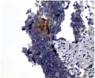
Figure 9 - Occasional neuroendocrine carcinoma tumor cells were positive for PSA immunostain.
stage, precluding the possibility of radical prosta-tectomy. Three patients underwent palliative sur-gery: one was treated with transurethral resection only for obstruction, a second with transurethral resection followed by surgical castration (orchiec-tomy) and a third with colostomy for intestinal ob-struction by metastatic tumor. Three patients were treated with chemotherapy in association with eto-poside phosphate (VP-16) and cisplatin (CDDP). In one of these patients, chemotherapy was sus-pended after one cycle due to obstructive renal failure, whereas in another patient the regimen was modiied to taxol after the fourth cycle, but with no measurable response. Mean follow-up was 13.7 months, with a range of 2.1 months in two patients with distant metastatic disease at diagnosis (bone in one, and bone and liver in the second), to 26.6 months in a patients who is alive with disease at last follow-up (Table-2).
DISCUSSION
The morphologic features of NEC of the prostate are similar to those of other sites, including the common pulmonary small cell carcinomas (9). In prostate neuroendocrine carcinoma series, however, a common inding is the association with conven -tional acinar tumors, suggesting a common pathway of tumor differentiation, or a neuroendocrine trans-formation from the better-differentiated carcinoma to neuroendocrine tumor (7,11). In the current study, only of the cases showed associated conventional type cancer; however, it is noteworthy that these were all diagnosed on needle biopsies, and one can not exclude another tumor components if the tumors were resected and examined throughout.
The diagnosis of high-grade neuroendo-crine carcinomas in a needle biopsy may be chal-lenging to the pathologists, especially because of the important clinical implication, which is exclu-sion from tentative surgical treatment with curative intent. None of the patients in the series were taken to radical prostatectomy, with two patients under-going transurethral resection for obstructive dis-ease palliative management. This data underscores the need for new therapeutic strategies to treat these tumors, which may include the use of protocols that
have been effective against neuroendocrine carci-nomas arising in other organ systems (6,12-14).
In cases where the diagnosis of small cell carcinoma is dificult, either due to the limited ma -terials available, or due to lack of clear neuroen-docrine differentiation, where the main differential is always with high-grade Gleason 9 or 10 acinar prostate adenocarcinomas, and poorly differenti-ated urothelial tumors invading the prostate, immu-nohistochemistry can be helpful. The vast majority of these tumors express at least one neuroendo-crine marker. Wang et al. reported a rate of 94%, being CD56 the most sensitive (7). One caveat is that conventional adenocarcinomas, up to 100% in some studies, may focally express these same markers, reinforcing the need for careful morpho-logic evaluation by the pathologist (5,15,16). More recent discoveries have suggested that prostatic speciic membrane antigen (PSMA), CD44 and protein (P501S) may help with indentifying neuro-endocrine expression in tumors (7,17).
High-grade neuroendocrine carcinomas have been reported in association with obstructive symptoms in the setting of androgen-independent disease. In this scenario, serum PSA levels tend to be low to undetectable. In the current series, all cases were diagnosed de novo with a high mean PSA, indicating that those tumors or the associated acinar tumor are able to express high quantities of PSA. Interestingly, we have found no association of serum PSA levels and PSA detection on the tis-sue by immunohistochemistry (Tables 1 and 2).
CONCLUSIONS
Prostates harboring neuroendocrine carci-noma tend to be larger and involve a greater num-ber of cores than acinar tumors. Association with conventional acinar tumors is common. Serum PSA levels vary greatly and its value at diagnosis does not seem to predict the presence of NE tumors in needle biopsy.
CONFLICT OF INTEREST
Prostatic carcinomas with neuroendocrine differentiation
REFERENCES
1. Tanaka M, Suzuki Y, Takaoka K, Suzuki N, Muraka-mi S, Matsuzaki O, et al.: Progression of prostate can-cer to neuroendocrine cell tumor. Int J Urol. 2001; 8: 431-6; discussion 437.
2. Sarma DP, Weilbaecher TG: Small-cell carcinoma of prostate. Urology. 1989; 33: 332-5.
3. Oesterling JE, Hauzeur CG, Farrow GM: Small cell anaplastic carcinoma of the prostate: a clinical, patho-logical and immunohistopatho-logical study of 27 patients. J Urol. 1992; 147: 804-7.
4. Manson AL, Terhune D, MacDonald G: Small cell carcinoma of prostate. Urology. 1989; 33: 78-9. 5. di Sant’Agnese PA, Cockett AT: Neuroendocrine
dif-ferentiation in prostatic malignancy. Cancer. 1996; 78: 357-61.
6. di Sant’Agnese PA: Neuroendocrine differentiation in carcinoma of the prostate. Diagnostic, prognos-tic, and therapeutic implications. Cancer. 1992; 70(1 Suppl): 254-68.
7. Wang W, Epstein JI: Small cell carcinoma of the pros-tate. A morphologic and immunohistochemical study of 95 cases. Am J Surg Pathol. 2008; 32: 65-71. 8. Helpap B, Köllermann J, Oehler U: Neuroendocrine
differentiation in prostatic carcinomas: histogenesis, biology, clinical relevance, and future therapeutical perspectives. Urol Int. 1999; 62: 133-8.
9. Brambilla E, Travis WD, Colby TV, Corrin B, Shi-mosato Y: The new World Health Organization clas -siication of lung tumours. Eur Respir J. 2001; 18: 1059-68.
10. Shah RB, Mehra R, Chinnaiyan AM, Shen R, Ghosh D, Zhou M, et al.: Androgen-independent prostate cancer is a heterogeneous group of diseases: lessons from a rapid autopsy program. Cancer Res. 2004; 64: 9209-16.
11. Evans AJ, Humphrey PA, Belani J, van der Kwast TH, Srigley JR: Large cell neuroendocrine carcinoma of prostate: a clinicopathologic summary of 7 cases of a rare manifestation of advanced prostate cancer. Am J Surg Pathol. 2006; 30: 684-93.
12. Stein ME, Bernstein Z, Abacioglu U, Sengoz M, Miller RC, Meirovitz A, et al.: Small cell (neuroendo-crine) carcinoma of the prostate: etiology, diagnosis, prognosis, and therapeutic implications--a retrospec-tive study of 30 patients from the rare cancer network. Am J Med Sci. 2008; 336: 478-88.
13. Segawa N, Inamoto T, Ibuki N, Mizutani Y, Azuma H, Tsuji M, et al.: Neuroendocrine differentiation in adenocarcinoma of the prostate during hormonal treat-ment : a case report. Hinyokika Kiyo. 2010; 56: 49-54. 14. Köllermann J, Helpap B: Neuroendocrine differentia -tion and short-term neoadjuvant hormonal treatment of prostatic carcinoma with special regard to tumor regression. Eur Urol. 2001; 40: 313-7.
15. Ordóñez NG: Value of thyroid transcription factor-1 immunostaining in distinguishing small cell lung car-cinomas from other small cell carcar-cinomas. Am J Surg Pathol. 2000; 24: 1217-23.
16. Yao JL, Madeb R, Bourne P, Lei J, Yang X, Tickoo S, et al.: Small cell carcinoma of the prostate: an immu-nohistochemical study. Am J Surg Pathol. 2006; 30: 705-12.
17. Simon RA, di Sant’Agnese PA, Huang LS, Xu H, Yao JL, Yang Q, et al.: CD44 expression is a feature of prostatic small cell carcinoma and distinguishes it from its mimickers. Hum Pathol. 2009; 40: 252-8.
_____________________
Submitted for publication: January 12, 2011
___________________ Accepted after revision: April 15, 2011
______________________
Correspondence address:
Dr. Marcos Venicio Lima
Hosp. do Câncer do Inst do Câncer do Ceará Rua Papi Júnior, 1222