ABSTRACT
Objectives: To search for relationships between phasic (P) and terminal (T) DO with age, urodynamic indings and sphincter behavior during involuntary detrusor contraction in woman.
Materials and Methods: Urodynamic studies (triple lumen catheter 7F, seated position) of 164 successive women re -ferred for LUTS with diagnosis of DO were reviewed. Patients were stratiied in 4 sub-groups: pre- (18-44 y), peri- (45-54 y), post-menopause (55-74 y) and oldest old (≥ 75 y). The urethral sensor was positioned at the level of the maximum urethral closure pressure for sphincter behavior analysis. A variation of at least 5 cm H2O in pressure (detrusor or urethra) was chosen to assert DO or sphincter response. Sphincter response was classiied as relaxation (re) before or during DO, or steady (st).
Results: Occurrence of P and TDO was similar: 77 P and 87 T. The PDO group was signiicantly younger (p = 0.0003). TDO was more frequent in patients with a history of neurological disease.
The percentage of PDO remained almost constant in age groups, while that of TDO increased with age from 6.7% to 23.2% (p = 0.0013).
Uninhibited contraction occurred at a smaller bladder volume in the P group: 149 ± 95 vs. 221 ± 113 mL (p < 0.0001). Steady sphincter predominated in the TDO subgroup: 45.9% vs. 32.1% and increased signiicantly in each DO sub-group of ≥ 75 y.
Conclusion: Steady sphincter during both P and TDO, and occurrence of TDO appear as speciic of aging. The last result could be related to structural changes in the detrusor muscle with aging.
Key words: Urinary bladder, overactive; urinary incontinence; Women; Urodynamics Int Braz J Urol. 2011; 37: 773-780
INTRODUCTION
Detrusor overactivity (DO) is a frequent urodynamic diagnosis in women with urge syn-drome with or without incontinence. DO is charac-terized by involuntary detrusor contraction (IDC) during the illing phase and can be idiopathic when there is no deined cause (IDO) or neurogenic when there is a relevant neurological condition (NDO) (1). According to the ICS recommendations, it is usual to distinguish phasic (P) (wave(s) with or without leakage) from terminal (T) DO (single in-voluntary detrusor contraction occurring at
cysto-metric capacity, which cannot be suppressed, and results in incontinence, usually resulting in blad-der emptying) (1). To make a distinction between P and TDO is sometimes dificult: high vesical pres -sure or/and pain during cystometry might lead to stop the illing and to under-estimate DO. Distinc -tion of TDO from decreased bladder compliance needs to repeat the cystometry at lower illing rate to assert or to invalidate DO.
As DO is believed to underlie overactive bladder (OAB) symptoms, most studies focus on relationships between OAB and DO (2-6). If, the impact of DO on bladder function in women has
Phasic or terminal detrusor overactivity in women: age,
urodynamic indings and sphincter behavior relationships
Françoise A. Valentini, Brigitte G. Marti, Gilberte Robain, Pierre P. Nelson
been widely studied (7-11), few studies have been conducted to analyze the relationships between each type of DO with age, history of neurological disease, urodynamic indings including sphincter behavior during IDC (2, 12-14).
Our purpose was to look for relationships between P or TDO and these conditions or indings.
MATERIALS AND METHODS
Between June 2005 and December 2007, 684 women were referred in our outpatient clinic for investigation of lower urinary tract (LUT) dysfunc -tion. Nineteen were excluded (complete spinal cord injury, severe dementia (mini mental state < 20) and grade ≥ 2 prolapse). Among the 665 eligible women, the urodynamic diagnosis of DO was made in 164; of these, 72 had a history of neurological disease (Table-1). The DO population was then stratiied in 4 age groups: pre- (18-44y), peri- (45-54y), post-menopause (55-74y) and oldest old (≥ 75y).
All patients had evaluation including medi -cal history and usual medication, bladder diary for
at least 48 hours including voiding times and voided volumes during day- and night-time, physical ex-amination and dipstick urinalysis.
Cystometry was performed with a triple lu -men catheter 7F at a illing rate of 50 mL/min in seated position using a Dorado® unit from Laborie.
Pressures were zeroed to atmosphere with the transducers placed at the level of the upper edge
Table 1 - Description of the DO population with a history of neurological disease. In each age-group, the number of patients is presented as total (a) and in parentheses bP and cT DO: a(bP+cT).
18-44 y 45-54 y 55-74 y ≥ 75 y ∑
Encephalic lesion (stroke, meningioma)
1 (P) 5(1P+4T) 9(3P+6T) 7(1P+6T) 22(6P+16T)
Parkinson’s disease 0 1(T) 1(P) 4(1P+3T) 6(2P+4T) Multiple sclerosis 8(4P+4T) 7(4P+3T) 6(3P+3T) 1(T) 22(11P+11T) Supra-sacral spinal cord lesion 4(2P+2T) 4(1P+3T) 8(3P+5T) 4(1P+3T) 20(7P+13T) Sub-sacral and Peripheral 1(P) 1 (P) 0 0 2(2P)
∑ 14(8P+6T) 18(7P+11T) 24(10P+14T) 16(3P+13T) 72
of the symphisis pubis. For urethral pressure re-cording, the catheter eye was positioned at the level of the maximum urethral closure pressure (located from a urethral proilometry bladder empty). Ab -dominal pressure was recorded using a punctured intra-rectal balloon catheter.
The urethral sensor was positioned at the level of the maximum urethral closure pressure (MUCP) for sphincter behavior analysis.
To avoid instrumental errors, we consid-ered that there was an episode of DO when the detrusor pressure increased of at least 5 cmH2O in pressure; in the same way, a variation of ± 5 cm -H2O in urethral pressure was chosen to evaluate the sphincter response. In case of rhythmic rectal tractions, the change in vesical pressure was con-sidered. Shifting of the urethral transducer during illing and non rhythmic rectal activity during IDC were criteria for exclusion.
One voiding cycle was recorded during each cystometry. For 18 patients a second cystometry at a lower illing rate of 20 mL/min was performed to con -clude between decreased bladder compliance and DO.
Sphincter response was classiied as re -laxation (re) before or during IDC, or remaining steady (st).
This study was conducted in accordance with the declaration of Helsinki. According to the local practice of our Ethics Committee, there is no formal Institutional Review Board approval required for retrospective studies.
STATISTICAL ANALYSIS
Data are presented as mean ± SD and range. The Wilcoxon signed rank test was used for compari -son of related samples, analysis of variance (ANOVA), the t-test and the Chi 2 test to compare unrelated sam -ples. Statistical analysis was performed using SAS, version 5.0 (SAS Institute, Inc., Cary, NC). All statisti -cal results were considered signiicant at p < 0.05.
RESULTS
In the whole DO population, there were 77 (46.9%) PDO and 87 (53.1%) TDO, with 28 and 44 women respectively who had a history of neurologi-cal disease (Table-1). A history of neurologineurologi-cal dis -ease was not signiicantly different (n.s.) between the DO sub-groups.
Main LUT symptoms were urgency with or without incontinence (118 patients) and frequency
(35 patients). Other symptoms were 5 stress urinary incontinence, 3 bedwetting and 3 incomplete reten -tion. Incontinence was the main complaint for 123 (75%) of the patients.
Table-2 shows the distribution in the differ-ent of both populations with DO (PDO or TDO) and without DO.
Age incidence (Table-2):
Comparing the four age groups, there was no signiicant incidence of age on occurrence of DO. TDO was more frequent than PDO with aging (p = 0.006).
In the DO population, PDO was predomi-nant in the 18-44 y age group (p = 0.008), and TDO in the ≥ 75 y one (p < 0.0001) with a ratio 3/1, while in the 45-54 y and 55-74 y age groups the ratio was close to 1.
In the entire population, increase of TDO was signiicant with aging between age (p = 0.0036) while the decrease of PDO was not signiicant.
The PDO group was signiicantly younger (52 ± 19) than the TDO group (63 ± 16) (p = 0.0003).
There was no signiicant difference in age between women with or without neurological dis-ease in DO subgroups.
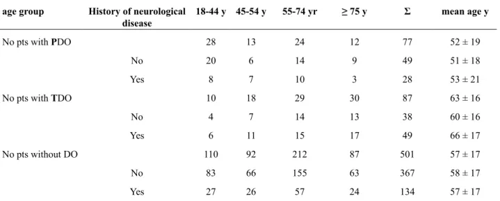
Table 2: Number of women in each age group. No difference in age between the 3 groups: PDO, TDO and no DO whatever the neurological history.
age group History of neurological disease
18-44 y 45-54 y 55-74 yr ≥ 75 y Σ mean age y
No pts with PDO 28 13 24 12 77 52 ± 19
No 20 6 14 9 49 51 ± 18
Yes 8 7 10 3 28 53 ± 21
No pts with TDO 10 18 29 30 87 63 ± 16
No 4 7 14 13 38 60 ± 16
Yes 6 11 15 17 49 66 ± 17
No pts without DO 110 92 212 87 501 57 ± 17
No 83 66 155 63 367 58 ± 17
Yes 27 26 57 24 134 57 ± 17
History of neurological disease (HND) (Table-3)
For women without HND, occurrence of each kind of DO was signiicantly different with age (p = 0.0114) with a decrease of PDO and an increase of TDO; for women with HND, no difference was observed.
In the PDO sub-group, HND was predomi -nant in the peri- and post-menopausal age groups (53.8 and 41.6%), while in the pre-menopause and the oldest groups the percentage of HND was close to that observed in the eligible population (28.5 and 25% vs. a mean value of 26.7%). The frequency of HND was very high (60.0 and 61.1%) in the pand peri-menopausal age groups with TDO pand
re-mained at a signiicant level in the other age groups (48.3 and 43.3%).
In the NDO population, neurological disor-der secondary to supra-pontine (SP) lesions (ence-phalic lesion and Parkinson’s disease) was observed in 8/28 (28%) of the PDO group and 20/44 (45%) of the TDO group. The distribution of SP lesions showed an increase with ageing in the TDO group.
Urodynamic indings
The bladder volume of occurrence of the irst IDC in P group was smaller than that at the IDC
Table 3 - Ratio of women with neurological disease (N) in each sub group of DO and in each age group.
age group 18-44 y 45-54 y 55-74 y ≥ 75 y
% PNDO/ (PDO) 28.5 53.8 41.6 25.0
% TNDO/ (TDO) 60.0 61.1 48.3 43.3
% N/entire population 24.3 28.4 26.8 27.3
Table 4: Comparison of the maximal urethral closure pressure (MUCP) with the expected value for age in each DO group (women with or without a history of neurological disease (HND)).
P T
MUCP with HND without HND Σ with HND without HND Σ
Low 4 6 10(12.9%) 1 7 8 (9.2%)
Normal 7 17 24(31.2%) 20 19 39(44.8%)
High 17 26 43(55.8%) 23 16 39(44.8%)
in the T group: 144 ± 95 vs. 219 ± 114 mL (p < 0.0001); the functional bladder capacity in group P (296 ± 105 mL) was signiicantly higher than that in the T group (246 ± 121 mL) (p = 0.0045). The functional capacity in the population without DO was 391 ± 149 mL (p < 0.0001).
The detrusor pressure (pdet) was 37 ± 25 cm H2O at the onset of low (when intubated low was obtained) in the PDO population and 35 ± 19 cmH2O at the onset of leakage in the T DO population (n.s.). In each DO sub-group, there was no signiicant dif -ference between patients without (39 ± 29 cm H2O) and those with HND (31 ± 14 cm H2O); the same result was observed in each age group. There was no signiicant effect of aging on pdet.
The maximum urethral closure pressure (MUCP) decreased with aging but was most often found normal or higher (Table-4) compared with the value expected for age [(110 - age) ± 20% in cm H2O] (15). Mean expected value was 62.8 ± 22.3 cm H2O when the mean observed value was 69.9 ± 37.4
cm H2O (p = 0.0021).
IDC characteristics in the PDO popula-tion (Table-5)
the NDO sub-group and 16 in the IDO sub-group. Characteristics of the IDC are given in the Table-3. There was no signiicant variation in the du -ration, time to maximum and amplitude whatever the neurological status.
Recorded sensation (desire to void) showed mainly an occurrence of the irst IDC before or at the irst desire to void (FDV) (p = 0.033) (Table-6). That behavior was independent of a HND.
Sphincter behavior (Table-7)
Steady sphincter was predominant in the TDO subgroup: 45.9% vs. 32.1% (n.s.). In each DO sub-group, the ratio of steady sphincter increased in the ≥ 75 y sub-group (P: 50.0% vs. 32-33-36%; T: 65% vs. 40-35-34%); in the same way, the ratio of sphincter relaxation before IDC decreased in the ≥ 75 y sub-group (P: 0% vs. 29-42-23%; T: 17% vs. 33-29-34%).
DISCUSSION
The distinction between IDO and NDO has been frequently studied (16,17), but scarce data ex -ist regarding the conditions underlying occurrence and expression of P or TDO. Occurrence of TDO has been related to aging and particularly to elderly patients with neurological lesions such as
cerebro-vascular accident (3,12,18). In his study, Tong (14) reported a high incidence of TDO with abnormal bladder sensation and unfelt PDO in men with be-nign prostatic hyperplasia.
Our purpose was to search for a condition which could be an indicator for occurrence of P or TDO. The irst idea was to test aging because it is well known that LUT function changes in elderly patients. Because about half of the population (44%) had DO and a history of neurological disease, that last condition was also analyzed. Finally, because DO is a urodynamic diagnosis, some urodynamic param-eters (pdet at the onset of low for PDO, or at the onset of leakage for TDO, MUCP and sphincter behaviour during IDC) have been investigated. In their paper, Romanzi et al. (19) compared patients (men and women) according to the main presenting symptom and the neurological status and concluded that the characteristics of the cystometric tracing during IDC were not distinct to aid in differential diagnosis but might have prognostic and therapeutic signiicance.
Occurrence of TDO is signiicantly associ -ated with aging; an inversion of the ratio PDO/TDO from 3:1 to is also observed. That result is consis -tent with an association between TDO and changes in LUT function due to aging. TDO would more probably due to alteration in detrusor function or ab-normal micturition relex than to sphincter incompe
-Table 5 - Spatio-temporal characteristics of the successive IDC in the PDO population; comparison between women with or without history of neurological disease (HND).
Duration (s) Time to maximum (s) Amplitude (cm H2O)
Pts without HND 14.9 ± 1.7 6.6 ± 0.5 11.1 ± 3.3
Pts with HND 16.1 ± 1.8 7.5 ± 1.1 14.3 ± 1.9
p n.s. n.s. n.s.
Table 6 - Recorded sensation on UDS at occurrence of the irst IDC in the PDO group (FDV: irst desire to void; NDV: normal desire to void; SDV: strong desire to void). The irst IDC occurs signiicantly before or at the irst desire to void (FDV) (p = 0.033).
Age group 1rst IDC 18-44 y 45-54 y 55-74 y ≥ 75 y Σ
Up to FDV 14 (50%) 4 (31%) 11(46%) 7 (58%) 36
> FDV to NDV 7 2 5 1 15
tence because, in the oldest group, pdet at leakage is signiicantly lower than MUCP (p < 0.0001). With regard to aging-related DO, a characteristic structur-al pattern (20) and age-related changes in muscarinic receptor functions have been reported (21) but not related to each kind of DO.
The percentage of women with PDO is sig-niicantly higher than that of TDO in the 18-44 y sub -group (p = 0.008). A hypothesis to explain that result could be a high incidence of spinal lesions (spina biida, multiple sclerosis) in younger. Unfortunately, in this age group the number of spinal lesion is the same in P and TDO groups.
Among the whole population, occurrence of DO was higher in the group with history of neurologi-cal disease (44% vs. 18%) as it has been previously reported (17-22). It has been veriied that there was no bias of recruitment, as the percentage of patients with a neurological disease was similar in the different age groups: 26.8 ± 1.3%. Aging and history of neuro -logical disease appear to be conditions for occurrence of TDO; consequently, the role of encephalic lesions (stroke, meningioma and Parkinson’s disease) which frequency increases with ageing is a question. Unfor -tunately, that sub-group comprises only nine women, which is too small a group to conclude.
As expected, the functional bladder capacity (FBC) is lower in the DO population for which main motive for urodynamics is urge or mixed inconti-nence compared to the population without DO.
Comparing P and TDO patients, FBC is sig -niicantly higher in the PDO group. That result is consistent with the ability of PDO patients to coun-terbalance the increase in detrusor pressure and to prevent voiding. pdet does not differ at the onset of low for PDO women and at the onset of leakage for
TDO women. That result conirms the inability to abort IDC for TDO women. For our female popu -lation, the values of pdet at IDC are lower than the values reported by Romanzi et al. (19) whose study included men with urethral obstruction leading to detrusor hypertrophy.
MUCP decreases with aging but its value re -mains normal and higher the value expected for the age which can be the consequence of long-lasting DO and unconscious reinforcement of the striated sphincter.
An intuitive proposal that the larger the am -plitude of the IDC, the more severe DO has been invalidated by Miller et al. (23), as it is severely confounded by the urethral sphincter function. That result is consistent with our indings; we do not ob -serve differences between duration, time to maxi-mum, and amplitude in successive IDC whatever the neurological status, and in addition, MUCP is nor -mal or higher than the expected value for age. An unexplained result is the value of detrusor pressure generated during IDC which is much lower than the values reported by Romanzi et al. (19). In his paper (2), P Abrams suggests that PDO tends to be char -acterized by contractions of increasing amplitude as the bladder volume increases and that this pattern is seen in most IDO of middle age men and women. We don’t observe any increase of the IDC amplitude with bladder illing. The number of IDC in the PDO group is lower for the NDO group (only 28% had 3 IDC vs. 43% in the IDO group); that result could be a limitation of the comparison between characteristics (duration, time to maximum, and amplitude) of the IDC between NDO and IDO but in this study there is no signiicant difference between these parameters during the irst and second IDC.
Table 7 - Relationship between sphincter behavior and a history of neurological disease (HND) at IDC in each DO group. Sphincter relaxation before or during IDC is signiicantly observed in PDO women without HND (p = 0.0045).
PDO TDO
Sphincter behavior With HND Without HND Σ With HND Without HND Σ
Relaxation before IDC 4 14 18 (24.33%) 12 11 23 (27.05%)
Relaxation during IDC 8 21 29 (34.52%) 13 10 23 (27.05%)
In the PDO group, the irst IDC is mainly observed up to irst desire to void, which could imply abnormal intrinsic bladder relexes (24) to bladder illing.
Incidence of steady sphincter in the oldest old group might be the consequence of aging on ure -thral function, as the threshold of ure-thral sensation increased in the elderly (3). The same conclusion can be proposed for the decrease of sphincter relaxation before IDC with aging.
In the population with a history of neurolog-ical disease, one observes a higher occurrence of a steady sphincter during IDC in the PDO group and a lower occurrence of sphincter relaxation before IDC when the neurological condition is an incomplete spinal cord injury.
Our study has several limitations, including its retrospective nature as well as the limited num-ber of patients in each type of neurological lesion; its merit is that it is to be the irst report assessing to ind characteristics for each kind of DO.
CONCLUSIONS
Steady sphincter during non-inhibited detru-sor contraction for both P and TDO, and occurrence of TDO appear as speciic of aging. In the elderly, occurrence of a steady sphincter may be associated with loss of sensory nerve function in the urethra and occurrence of TDO could be related to the structural changes in the detrusor muscle with aging. Looking at a history of neurological disease, there is a trend of increase for both P and TDO with aging, except in the oldest group where TDO predominates.
ABBREVIATIONS: DO = Detrusor Overactivity
FBC = Functional Bladder Capacity FDV = First Desire to Void
HND = History of Neurological Disease IDC = Involuntary Detrusor Contraction IDO = Idiopathic Detrusor Overactivity LUT = Lower Urinary Tract
MUCP = Maximum Urethral Closure pressure NDO = Neurogenic Detrusor Overactivity NDV = Normal Desire to Void
OAB = Overactive Bladder pdet= Detrusor pressure
PDO = Phasic Detrusor Overactivity STV = Strong Desire to Void
TDO = Terminal Detrusor Overactivity
CONFLICT OF INTEREST
None declared.
REFERENCES
1. Abrams P, Cardozo L, Fall M, Grifiths D, Rosier P, Ulmsten U, et al.: The standardisation of terminology of lower urinary tract function: report from the Stan -dardisation Sub-committee of the International Con -tinence Society. Neurourol Urodyn. 2002; 21: 167-78. 2. Abrams P: Describing bladder storage function: over -active bladder syndrome and detrusor overactivity. Urology. 2003; 62(5 Suppl 2): 28-37; discussion 40-2. 3. Guralnick ML, Grimsby G, Liss M, Szabo A,
O’Connor RC: Objective differences between over -active bladder patients with and without urodynami-cally proven detrusor overactivity. Int Urogynecol J Pelvic Floor Dysfunct. 2010; 21: 325-9.
4. Hashim H, Abrams P: Overactive bladder: an update. Curr Opin Urol. 2007; 17: 231-6.
5. Schaefer W: Re: Is the bladder a reliable witness for predicting detrusor overactivity? H. Hashim and P. Abrams. J Urol, 175: 191-195, 2006. J Urol. 2006; 176: 1255-7; author reply 1256-7.
6. Andersson KE: Mechanisms of Disease: central ner -vous system involvement in overactive bladder syn-drome. Nat Clin Pract Urol. 2004; 1: 103-8.
7. Pisterer MH, Grifiths DJ, Schaefer W, Resnick NM: The effect of age on lower urinary tract function: a study in women. J Am Geriatr Soc. 2006; 54: 405-12. 8. Kayigil O, Metin A, Atmaca AF: Obstructive urody -namic indings in idiopathic detrusor overactivity. Int Urol Nephrol. 2007; 39: 445-8.
9. Garnett S, Swithinbank L, Ellis-Jones J, Abrams P: The long-term natural history of overactive bladder symptoms due to idiopathic detrusor overactivity in women. BJU Int. 2009; 104: 948-53.
11. Lee SR, Kim HJ, Kim A, Kim JH: Overactive bladder is not only overactive but also hypersensitive. Urol-ogy. 2010; 75: 1053-9.
12. Kessler TM, Madersbacher H: Urodynamic phenom -ena in the aging bladder. Urologe A. 2004; 43: 542-6. 13. Schaefer W, Tadic S, Grifiths DJ, Resnick NM:
Overactive bladder and detrusor overactivity: but what does the sphincter do? J Urol 2007;177(suppl 4):556. #abstract 1677.
14. Tong YC: Comparisons of urodynamic indings and voiding habits in patients with concomitant benign prostatic hyperplasia and detrusor overactivity pre-senting with or without the symptom of urgency. Urol Int. 2007; 78: 219-25.
15. Constantinou CE: Urethrometry: considerations of static, dynamic, and stability characteristics of the female urethra. Neurourol Urodyn. 1988; 7: 521-39. 16. Defreitas GA, Lemack GE, Zimmern PE, Dewey RB,
Roehrborn CG, O’Suilleabhain PE: Distinguishing neurogenic from non-neurogenic detrusor overactiv-ity: a urodynamic assessment of lower urinary tract symptoms in patients with and without Parkinson’s disease. Urology. 2003; 62: 651-5.
17. Lemack GE, Frohman EM, Zimmern PE, Hawker K, Ramnarayan P: Urodynamic distinctions between idiopathic detrusor overactivity and detrusor overac-tivity secondary to multiple sclerosis. Urology. 2006; 67: 960-4.
18. Geirsson G, Fall M, Lindström S: Subtypes of overac -tive bladder in old age. Age Ageing. 1993; 22: 125-31. 19. Romanzi LJ, Groutz A, Heritz DM, Blaivas JG: In -voluntary detrusor contractions: correlation of urody -namic data to clinical categories. Neurourol Urodyn. 2001; 20: 249-57.
20. Elbadawi A, Yalla SV, Resnick NM: Structural basis of geriatric voiding dysfunction. II. Aging detrusor: normal versus impaired contractility. J Urol. 1993; 150: 1657-67.
21. Andersson KE: Antimuscarinic mechanisms and the overactive detrusor: an update. Eur Urol. 2011; 59: 377-86.
22. Ashok K, Wang A: Detrusor overactivity: an over -view. Arch Gynecol Obstet. 2010; 282: 33-41. 23. Miller KL, DuBeau CE, Bergmann M, Grifiths DJ,
Resnick NM: Quest for a detrusor overactivity index. J Urol. 2002; 167: 578-84; discussion 584-5.
24. Fowler CJ: Integrated control of lower urinary tract--clinical perspective. Br J Pharmacol. 2006; 147(Sup -pl 2): S14-24.
_____________________
Submitted for publication:
January 04, 2011
___________________
Accepted after revision: April 05, 2011
______________________ Correspondence address: Dr. Françoise A. Valentini
Université Pierre et Marie Curie (Paris 6) 4 Place Jussieu, 75005 Paris, France Fax: + 33 1 4959-4697