ABSTRACT
In this study, we aimed to state the relationship between testis, epididymis and vas deference, in adult cases with non-palpable testis.
Between January 1996 and December 2009, we evaluated 154 adult cases with nonpalpable testes. Mean age was 23 years (20-27 years). Explorations were performed by open inguinal incision, laparoscopy, and by inguinal incision and laparoscopy together on 22, 131 and 1 patient, respectively.
Of all the unilateral cases, 32 were accepted as vanishing testis. In ive of these cases, vas deference was ending inside the abdomen, and in the others, it was ending inside the scrotum. In the remaining 99 unilateral and 22 bilateral cases, 143 testes were found in total.
Testes were found in the inguinal canal as atrophic in one case, at the right renal pedicle level with dysmorphic testis in one case, and anterior to the internal ring between the bladder and the common iliac vessels at a smaller than normal size in 119 cases. One (0.69%) case did not have epididymis. While epididymis was attached to the testis only at the head and tail locations in 88 (61.53%) cases, it was totally attached to the testis in 54 (37.76%) cases.
There is an obviously high incidence rate of testis and vas deference anomalies, where epididymis is the most frequent one. In cases with abdominal testes, this rate is highest for high localised abdominal testes.
Key words: testis; surgery; abnormalities; Vas Deferens Int Braz J Urol. 2011; 37: 727-732
INTRODUCTION
Testes are the predominant endocrine or-gans, which have been localised posterior to the abdominal cavity in the early fetal period. Although they are in the abdomen in most (90.54%) of the cas-es until the 23rd week, they begin to migrate down
to the scrotum around 28th week (1). During this
mi-gration, testes can occasionally be localised at the higher or lower abdomen, inner opening of the in-guinal canal and between the opening of the external inguinal canal and the scrotum (2). It is hypothesized that the epididymis has a major role in the localisa-tion of the testis in the scrotum. In fact, the situalocalisa-tion which states that, epididymal anomalies are present
in more than 50% of the higher abdominal testes, supports this theory (3,4).
In this study, we report our results of testis, epididymis and vas deference abnormalities in pa-tients with nonpalpable testes by open surgical ex-ploration and laparoscopic interventions.
MATERIALS AND METHODS
Explorations were performed by open ingui-nal and laparoscopic intervention.
During laparoscopic interventions, 3 port ac-cesses (sub-umbilical 1 port, and pararectal 2 ports) were used. In 9 of bilateral nonpalpable testis cas-es, for who single session laparoscopic orchiopexy
Findings concerning testis, vas deference, and epididymis in adult
cases with nonpalpable testes
Coskun Sahin, Mehmet Kalkan, Soner Yalcinkaya
was performed; a 4th port was placed trans-scrotally,
whereas in 13 of these cases, double session orchio-pexy was performed. Of the 32 cases with vanish-ing testis, vas deference excision was performed by inguinal exploration in 5 cases, and by laparoscopic intervention in the remaining 27 cases. Testis di-mensions were calculated according to the graphi-cal method. This method includes graphics of testis
igures and predeined testicular volumes on paper
(Figure-1). Abdominal testicular dimensions were compared with normal scrotal testicular values.
Table 1 - Findings concerning the vas deference.
Finding no
Absence of vas deference 1
Independent of the testis 1
Blunt ending at the abdomen 2
Blunt ending at the scrotum 20
Together with the testis 113
Total 137
Figure 1 -Scheme for graphic measurement of testicular volume.
Epididymal conigurations were evaluated with a 1 to 5 classiication system which deines
the relation between epididymis and testis. Normal epididymis is completely attached to the testis at the upper pole of it. However, it is attached to the
testis by a ibrous ilm at the lower pole. Such a coniguration is known as type I, and the conigura -tion where the epididymis is completely attached to the testis is known as type II. In type III and IV, head and tail portions are attached respectively to the testis and in type V the epididymis is complete-ly unattached to the testis.
Laparoscopic orchiectomy was performed on unilateral nonpalpable testis cases. Excised testicular tissues and nubbins were evaluated pathologically.
RESULTS
re-gion in 22 patients with bilateral nonpalpable testes. In 99 of the 131 cases with unilateral nonpalpable testis, testes were found at either anterior to the in-ternal ring, or over the common iliac vessels, or be-tween the bladder and the common iliac vessels.
In all of the cases with intra-abdominal tes-tis, testis volumes were determined as 12.5-22.5 cm³ (average 17.5 cm³). Mean testicular volume was 22.5 cm³ in the control group. While the testes were tense (Tight) in 3 cases, they were less dense than the normal density value in the remaining cases (Table-2). One case did not have epididymis (Type V
epididymal coniguration, Figure-2). While the epi -didymis was attached to the testis only at the head
and tail locations (Type I epididymal coniguration,
Figure-3) in 88 of the remaining 143 testes, it was totally attached to the testis (Type II epididymal
con-iguration, Figure-4) in the other 54 testes (Table-3).
Carcinoma in-situ was not determined in any of the orchiectomy applied cases. No spermato-genetic activity was detected in testis specimens.
Sertoli cells were present in only 4 testicular tissues (Sertoli cell only syndrome). All other testicles had
leydig cells.
DISCUSSION
Cryptorchidism is a congenital anomaly seen in 3 % of term born children. Cases with non-palpable testes comprise 20-34% of such anomalies (5,6). It is known that, the frequency of testis-epi-didymis anomalies is higher in high localised testes (7). In the literature, the frequency of epididymal anomalies is reported to be between 36% and 75% and such anomalies are divided into 5 groups (8). However, some investigators claim that there is not a correlation between the testis localisation, and epididymal anomaly (3). It had also been reported that, the frequency of epididymal anomalies among healthy children with undescended testis was 40% (8). Normal epididymis is completely attached to the testis at the upper pole. However, it is attached to
the testis by a ibrous ilm at the lower pole. In this
study of fetal testes, Favorito had determined that as high as 89 % of the cases were in type I, 7.53 % were in type II, 2.05 % were in type III, and 0.68 %
were in type IV coniguration. He did not detect any type V coniguration (3). In their studies about chil
-Table 2 - Testis anomalies.
Normal size
(n)
Small size*
(n)
Dysmorphic testis
(n)
Atrophic size**
(n)
Total
(n)
High abdominal testis - - 1 - 1
Low abdominal testis 17 94 - - 111
Inguinal testis - - - 1 1
Total (n) 17 94 1 1 113
*: Testicular Volume = 12.5 - 22.5 mL (17.5 mL) **: Testicular Volume = 1 mL
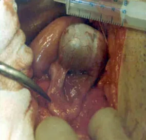
Figure 2 -Right testes is localised in the right renal pedicle
dren aged between 1 month and 18 years, Turek et al. reported that the frequency of type II cases, where the epididymis was completely attached to the testis, was 12% (9). In our study we determined that 88 (61.53 %) cases were type I and 54 (37.76 %) cases
were type II coniguration. While type V conigu -ration was determined in only 1 (0.69 %) case, we
did not coincide with type III and IV conigurations.
The case with type V epididymal anomaly had also testicular anomaly located at the higher abdomen. The testis in this case was at the renal pedicle level with the dimensions of 1x1.5x5 cm. When hold by a grasper, it appeared as a tubular organ, and it did not look like a testis. In fact, decisive diagnosis was performed pathologically on this case. It is obvious that, in patients with nonpalpable testes, such high abdominal testes will not be found during inguinal exploration, and will be evaluated as agenetic.
Figure 3 -Head and tail portions of epididymis attached
to the testis (type I coniguration).
Figure 4 -Epididymis is completely attached to the testis (type II coniguration).
Table 3 - Epididymal conigurations.
Type I Type II Type III Type IV Type V
High abdominal testis - - - - 1 ( 0.9% )
Low abdominal testis 72 ( 64% ) 39 ( 34.2% ) - -
-Inguinal testis - 1 ( 0.9% ) - -
There are different theories about testicular descent in embryogenic life. Epididymal factor is
one of them (8). In our Type V epididymal conigu -ration subject who didn’t contain an epididymis, testis remained in its embryogenic location.
Adult testis dimensions are usually 4x3x2.5 cm. Testis volume is usually measured by Prader or-chidometer or the graphic method (10-12). We used the graphical method to measure the testis volumes (Figure-1). Except the higher abdominal located testis, all testes were in ellipsoidal structure. In one patient, the testis was inside the inguinal canal at an atrophic size (1x1 cm). Lower abdominal testes are usually located either just proximal to the inter-nal ring, over the common iliac vessels, or between the bladder and the common iliac vessels. In these patients, the testis volumes were measured between 12.5 and 22.5 cm³ (average 17.5 cm³). Average tes-tis volume in the control group of 20 adults who did not have any scrotal pathology was measured as 22.5 cm³. While normal testes were tense and tight when they were palpated, abdominal testes were softer. Higher abdominal testes however, were even softer than the abdominal testes, and the testicular walls were so elastic that they could contact each other upon squeezing. Although the testicular vol-ume of the abdominal testes in our cases seemed
to be high at irst glance, this situation can be ex -plained by the age group of our cases, which is be-tween 20 and 27 years.
Testes that are not detected by laparosco-py and inguinal exploration are known as vanish-ing testes. The etiological reason underlyvanish-ing this phenomenon on is thought to be intrauterine testes torsions (10). We did not detect any testes in 32 of our patients. In 8 cases, which had open explo-ration, vas deference was observed to be blunted at the scrotum. In one of these patients, testis was found inside the abdomen, and vas deference was ending at the scrotum independent of the testis. As seen in this case, observing blunt ended vas defer-ence during exploration does not mean that testis is
absent. Testicular vessels should deinitely be seen.
In 2 patients however, vas deference was observed to extend higher from the small pelvis and end over the common iliac vessels with the testicular ves-sels. In patients with vanishing testes, viable
tes-ticular tissue ratio was reported to be 0% to 16%
(13). Some authors are recommending inguinal ex -ploration for laparoscopically undetected testes in cases with nonpalpable testes (14). Only 4 testicu-lar nubbin were contained viable testicutesticu-lar tissues. It is reported that laparoscopic intervention is un-necessary in palpable scrotal testicular nubbins as there was no testis in the abdomen (15).
Inguinal exploration was performed on the
irst 4 of the vanishing testis cases, whose vas def -erence and testicular vessels were observed to enter the internal inguinal ring during laparoscopy. While atrophic testis was detected in 1 of these 4 patients, vas deference was ending blunted in the remaining three. After we gained more experience on laparos-copy, vas deference was laparoscopically excised instead of applying inguinal exploration, in these cases. Technically, following the circular incision of peritoneum at the level of internal inguinal ring, testicular vessels and vas deference were excised as being tied with end clip, after they were taken inside the abdomen. In 28 of the 32 patients, who were evaluated as having vanishing testes, no tes-ticular tissue was detected pathologically, whereas in 4 cases viable testicular tissue was found. Vi-able testicular tissue detection rate is low as seen in these cases. Nearly all of these testicles are located in the scrotal cavity as these testicular nubbins are palpable position in the event of cancer, we don’t recommend prophylactic excision.
Abdominal testes were found to be smaller and softer in comparison with the normal ones. We determined that, the incidence of higher abdominal testes was extremely low, but the testis-epididymis anomaly was obvious in such testes. If laparoscopic exploration is not performed in higher abdominal
testicles, it may not be always possible to ind them
during open abdominal surgery. However we think that laparoscopy is not necessary in every nonpal-pable case; also it is not needed to perform any in-tervention including laparoscopy in palpable scro-tal nubbin cases.
CONFLICT OF INTEREST
9. Turek PJ, Ewalt DH, Snyder HM 3rd, Duckett JW: Normal epididymal anatomy in boys. J Urol. 1994; 151: 726-7.
10. Tanagho EA, McAninch JW: Smith’s General Urol -ogy. 15th (ed.), New York. McGraw-Hill Company. 2000.
11. Takihara H, Sakatoku J, Fujii M, Nasu T, Cosentino MJ, Cockett AT: Signiicance of testicular size mea -surement in andrology. I. A new orchiometer and its clinical application. Fertil Steril. 1983; 39: 836-40. 12. Chipkevitch E, Nishimura RT, Tu DG, Galea-Rojas
M: Clinical measurement of testicular volume in ado -lescents: comparison of the reliability of 5 methods. J Urol. 1996; 156: 2050-3.
13. Nishizawa S, Suzuki K, Tachikawa N, Nukui A, Ku -mamaru T, Shioji Y, et al.: The vanishing testis: di -agnosis and histological indings. Nihon Hinyokika Gakkai Zasshi. 2000; 91: 537-41.
14. Merry C, Sweeney B, Puri P: The vanishing testis: anatomical and histological indings. Eur Urol. 1997; 31: 65-7.
15. Sahin C, Yigit T, Ozbey I: Adult nonpalpable testis: is laparoscopy always required? J Laparoendosc Adv Surg Tech A. 2002; 12: 431-4.
_____________________
Submitted for publication: January 24, 2011
___________________
Accepted after revision: May 05, 2011 ______________________
Correspondence address: Dr. Soner Yalcinkaya
Antalya Education and Research Hospital Varlık Mah. Kazım Karabekir Cad. Soğuksu, 07070, Antalya, Turkey Fax: + 90 242 249 4462
E-mail: [email protected] REFERENCES
1. Moore KL, Persaud TVN: The Developing Human (Clinically Oriented Embryology). 5th ed. Philadel-phia. WB Saunders Company; 1993. pp. 265-301. 2. Sampaio FJ, Favorito LA: Analysis of testicular mi
-gration during the fetal period in humans. J Urol. 1998; 159: 540-2.
3. Favorito LA, Sampaio FJ: Anatomical relationships between testis and epididymis during the fetal period in humans (10-36 weeks postconception). Eur Urol. 1998; 33: 121-3.
4. Kirsch AJ, Escala J, Duckett JW, Smith GH, Zderic SA, Canning DA, et al.: Surgical management of the nonpalpable testis: the Children’s Hospital of Phila -delphia experience. J Urol. 1998; 159: 1340-3. 5. Husmann DA, Levy JB: Current concepts in the
pathophysiology of testicular undescent. Urology. 1995; 46: 267-76.
6. Sampaio FJ, Favorito LA, Freitas MA, Damião R, Gouveia E: Arterial supply of the human fetal testis during its migration. J Urol. 1999; 161: 1603-5. 7. Kiely EA: Scientiic basis of testicular descent and
management implications for cryptorchidism. Br J Clin Pract. 1994; 48: 37-41.