Licenciado sob uma Licença Creative Commons
[T]
Intervening factors in the walking of children presenting
myelomeningocele
[)]
Fatores intervenientes na marcha de crianças com mielomeningocele
[A]
Dirce Shizuko Fujisawa[a], Marcia Larissa Cavallari da Costa Gois[b], Josilainne Marcelino Dias[c], Egle de Oliveira Netto Moreira Alves[d], Marcelo de Souza Tavares[e], Jefferson Rosa Cardoso[f]
[a] PT, PhD, professora do Programa de Mestrado em Ciências da Reabilitação UEL - UNOPAR e do Departamento de
Fisioterapia da Universidade Estadual de Londrina UEL , Londrina, PR - Brasil, e-mail: [email protected]
[b] PT, Residente em Pediatria Fisioterapia, Universidade Estadual de Londrina UEL , Londrina, PR - Brasil, e-mail:
[c] PT, Mestranda em Ciências da Reabilitação UEL - UNOPAR , Universidade Estadual de Londrina UEL , Londrina, PR -
Brasil, e-mail: [email protected]
[d] PT, MSc, professor do Departamento de Fisioterapia da Universidade Estadual de Londrina UEL , Londrina, PR - Brasil,
e-mail: [email protected]
[e] MD, PhD, professor do Departamento de Pediatria da Universidade Federal de Minas Gerais, Minas Gerais, MG - Brasil,
e-mail: [email protected]
[f] PT, PhD, professor do Programa de Mestrado em Ciências da Reabilitação UEL - UNOPAR e do Departamento de
Fisioterapia da Universidade Estadual de Londrina UEL , Londrina, PR - Brasil, e-mail: [email protected]
[R]
Abstract
Introduction: All children presenting myelomeningocele are capable of walking. Certain interventions
can influence the walking prognosis of these children: physical therapy, medication, and nutritional orien-tation. Objectives: The aim of this study was to verify the association between ability to walk in children
with myelomeningocele and clinical, socioeconomic and therapeutic factors. Method: This
cross-section-al study was conducted at the University (ospitcross-section-al. The participants were children aged two years-old or more, diagnosed with myelomeningocele. Data collection was conducted by physical therapy assessment and medical records. The dependent variable was walking and the independent variables were clinical, socioeconomic and therapeutic factors. Results: Forty-one children were evaluated, with a median age of
276
myelomeningocele are capable of walking, given that they present normal functioning of the upper limbs, adequate spinal stability, capacity to raise the pelvis, and present hip and trunk mobility . Walkingand the requirement for orthotics and as-sistive devices for locomotion are not the same in children presenting the same neurological dysfunc-tion .
The clinical manifestations and complications could demand clinical intervention and multipro-fessional follow-up . Certain interventions can influence the walking ability of children with myelo-meningocele: physical therapy, which can promote motor development and gait training; medication, required to treat urinary infection; and nutritional orientation as related to alimentary control in order to avoid obesity. Moreover, orthopedic corrections are fundamental to promote the alignment of the lower limbs and should be conducted prior to the onset of walking. Thus, the aim of this study was to verify the association between the ability to walk in children with myelomeningocele with clinical, so-cioeconomic and therapeutic factors.
Introduction
Myelomeningocele is one of the most serious and frequent congenital anomalies. Studies dem-onstrate that the occurrence of myelomeningocele involves genetic, environmental and dietary factors - . The incidence is variable, ranging from to per . births, according to geography and the ethnic origin of the parents . Clinical mani-festations are flaccid paralysis; diminished muscu-lar force; muscumuscu-lar atrophy; diminished or loss of sensitivity and tendon reflexes; and alterations in vesicle, intestinal function and hydrocephalus .
The main concern for the health care team and parents after myelomeningocele diagnosis is the survival of the children, since this requires fetal correction or surgical intervention in the first to hours of life and antibiotic therapy. Later, the therapeutic focus shifts to the child’s functional in-dependence, especially, in relation to the possibil-ity of walking.
The ability to walk is directly related to the neurological segments . All children presenting
, and fractures p = , . Socioeconomic factors showed not to be significant in relation to ability to walk. Surgery p = , and the use of assistive devices p = , were also associated with the ability to walk. Conclusion: The determinant clinical factor for walking prognosis was the neurological area. The use
of assistive devices and surgical intervention were shown to be necessary for promoting walking activity. [#] [P]
Keywords: Gait. Children. Myelomeningocele. [#] [B]
Resumo
Introdução: Todas as crianças com mielomeningocele são capazes de andar. Certas intervenções podem
influenciar no prognóstico de marcha destas crianças: fisioterapia, medicação e orientação nutricional.
Objetivo: Foi verificar a associação entre a capacidade de andar em crianças com mielomeningocele e fatores
clínicos, socioeconômicos e terapêuticos. Método: Estudo transversal realizado no Hospital Universitário. Os
participantes foram crianças de dois ou mais anos de idade com diagnóstico de mielomeningocele. A coleta
de dados foi realizada por avaliação de um fisioterapeuta e em prontuários médicos. A variável dependente
foi o prognóstico de marcha e as variáveis independentes foram os fatores clínicos, socioeconômicos e ter-apêuticos. Resultados: Foram avaliadas 41 crianças com idade média de cinco anos (2-9). Os fatores clínicos que apresentaram associação com a marcha foram o segmento neurológico (p < 0,0001) e as fraturas (p = 0,022). Fatores socioeconômicos não se mostraram estatisticamente significantes em relação à capacidade de andar. Cirurgia (p = 0,017) e o uso de dispositivos de apoio (p = 0,023) também revelaram associação com a marcha. Conclusão: O fator clínico determinante para a capacidade de desenvolver marcha foi o segmento neurológico. A intervenção cirúrgica e o uso de dispositivos auxiliares demonstraram ser necessários na
promoção da marcha.[#]
[K]
fractures, tethered cord and nutritional status. Nutri-tional status was based on the Z score for weight-for-stature index, with evaluation conducted by the nutritionist on the multidisciplinary team.
Descriptive analysis of the sample was presented as mean ± standard deviation or median - % , depending on normality assumptions. Categorical variables were presented in the form of absolute and relative frequencies. For association between the pri-mary outcome walking and the explicative variables, chi-square rxc tests were used. To measure the ef-fect of exposure to the independent variables on the dependent variable ability to walk , multiple logis-tic regression test Stepwise method was used. )n this procedure, the most significant results F test of these associations were selected for the initial model. Statistical significance α was determined as % p ≤ , . The Statistical Package for Social Science SPSS® software, version . , was used for all analysis.
Results
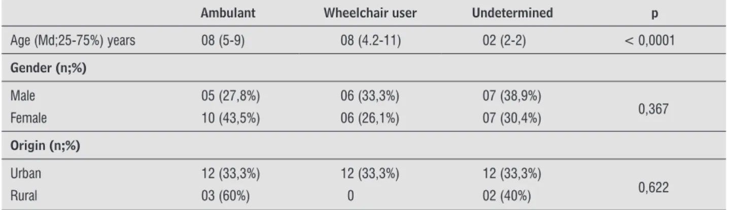
Forty-one children with myelomeningocele were evaluated, with a median age of - years-old, including boys , % and girls , % . Considering the locomotion mode, , % were ambulant and , % were wheelchair users. Fourteen participants , % were considered un-determined. Ambulant children and those who used a wheelchair were older than those who were yet to determine their mode of locomotion p< , . Only five children , % came from families resi-dent in the rural area. The demographic data, accord-ing to locomotion mode, are presented in Table .
The most observed neurological lesions was in the upper n = , , % and lower lumbar spine n = , , % . The most frequent complications were deformities and urinary infection. (ydrocephalus was observed in children , % , all of whom had been submitted to ventricular peritoneal deri-vation. Trophic ulceration occurred in children
, % , mainly at ischiatic, sacral, malleolar and calcaneus regions. The occurrence of fractures and tethered cord was , %; i.e., children presented these complications. Concerning nutritional status, children , % were malnourished. Moreover, an association was observed between the ability to walk and clinical factors, neurological area and frac-ture occurrence Table .
Method
This cross-sectional study was conducted at the University (ospital. Only participants with a diag-nosis of myelomeningocele, aged two years or older, were selected, since this period is usually sufficient to define the mode of locomotion, and the sample size was considered by convenience. The subjects parents provided a voluntary written informed consent, which received the approval of the Ethics Committee on (uman Experimentation of the )nstitution involved / .
Data were collected by physical therapy as-sessment and additional data was determined by consultation of the patient’s medical records. The protocol consisted of four parts: identification data, socioeconomic conditions, therapeutic aspects clinical and surgical interventions and physi-cal therapy evaluation. The identification data re-quired information regarding age, gender, area of origin urban or rural and education status. The therapeutic aspects investigated were: therapies performed onset, frequency and interruption , in-cluding physical therapy and surgical intervention; myelomeningocele closure, ventricular peritoneal derivation, orthopedic correction and others. The physical therapy evaluation aimed to identifying the motor development phase, motor condition co-ordination, movement amplitude, tonus, muscular retraction and shortening , neurological segment, presence and type of deformity, use of orthotics and assistive devices and mode of locomotion.
The neurological area was determined by evalu-ation of motor and sensory function, in accordance with the American Spinal )njury Association’s AS)A )nternational Standards for Neurological Classification of Spinal Cord )njury . Classification into thoracic, upper lumbar L /L , lower lumbar L /L and sacral lesions was done after deter-mining the neurological area .
The locomotion mode was considered as walker, wheelchair user and undetermined. Walker children presented gait as their principal mode of locomotion, whereas wheelchair users mostly or exclusively use a wheelchair for locomotion. The undetermined condi-tion represented those children who had yet to de-fine their principal locomotion mode.
278
Table 2 - Association between locomotion and clinical factors
Ambulant Wheelchair user Undetermined p
Neurological area (n;%)
Thoracic 01 (9,1%) 07 (63,6%) 03 (27,3%)
< 0,001 Upper lumbar 01 (8,3%) 05 (41,7%) 06 (50%)
Lower lumbar 07 (58,3%) 0 05 (41,7%)
Sacral 06 (100%) 0 0
Scars (n;%)
Yes 07 (41,2%) 07 (41,2%) 03 (17,6%)
0,131
No 08 (33,3%) 05 (20,8%) 11 (45,8%)
Deformities (n;%)
Yes 10 (34,5%) 10 (34,5%) 09 (31%)
0,493
No 05 (41,7%) 02 (16,7%) 05 (41,7%)
Fractures (n;%)
Yes 0 04 (57,1%) 03 (42,9%)
0,022
No 15 (44,1%) 08 (23,5%) 11 (32,4%)
Hydrocephalus (n;%)
Yes 07 (33,3%) 06 (28,6%) 08 (38,1%)
0,848
No 08 (40%) 06 (30%) 06 (30%)
Urinary infection (n;%)
Yes 10 (34,5%) 10 (34,5%) 09 (31%)
0,493
No 05 (41,7%) 02 (16,7%) 05 (41,7%)
Tethered cord (n;%)
Yes 05 (71,4%) 01 (14,3%) 01 (14,3%)
0,117
No 10 (29,4%) 11 (32,4%) 13 (38,2%)
Nutritional status
Malnourished 08 (36,4%) 06 (27,3%) 08 (36,4%)
0,874
Eutrophic 04 (30,8%) 04 (30,8%) 05 (38,5%)
Overweight 03 (50%) 02 (33,3%) 01 (16,7%)
Table 1- Demographic data
Ambulant Wheelchair user Undetermined p
Age (Md;25-75%) years 08 (5-9) 08 (4.2-11) 02 (2-2) < 0,0001
Gender (n;%)
Male 05 (27,8%) 06 (33,3%) 07 (38,9%)
0,367 Female 10 (43,5%) 06 (26,1%) 07 (30,4%)
Origin (n;%)
Urban 12 (33,3%) 12 (33,3%) 12 (33,3%)
0,622
Rural 03 (60%) 0 02 (40%)
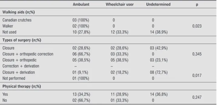
Table 3 - Association between locomotion mode and therapeutic factors
Ambulant Wheelchair user Undetermined p
Walking aids (n;%)
Canadian crutches 03 (100%) 0 0
0,023
Walker 02 (100%) 0 0
Not used 10 (27,8%) 12 (33,3%) 14 (38,9%)
Types of surgery (n;%)
Closure 02 (28,6%) 02 (28,6%) 03 (42,9%)
0,345 Closure + orthopedic correction 06 (66,7%) 03 (33,3%) 0
Closure + orthopedic 05 (38,5%) 05 (38,5%) 03 (23,1%)
Correction + derivation – – –
Closure + derivation 01 (9,1%) 02 (18,2%) 08 (72,7%)
0,017
Not performed 01 (100%) 0 0
Physical therapy (n;%)
Yes 13 (34,2%) 11 (28,9%) 14 (36,8%)
0,247
No 02 (66,7%) 01 (33,3%) 0
submitted to any surgical intervention. All wheel-chair users and those children who had yet to de-fine their mode of locomotion had undergone surgi-cal intervention. Among the therapeutic factors, the use of assistive devices p = , and surgical in-terventions p= , showed an association with walking ability Table .
The statistically significant results of associations between explicative variables on the primary out-come ambulant activity were selected for the mod-el used in multivariate analysis. The following vari-ables were included in this model: age, neurological area, fracture occurrence, type of surgery and assis-tive devices. The final logistic regression model was unable to identify variables that could predict walk-ing ability in children presentwalk-ing myelomenwalk-ingocele.
Discussion
The predominance of female gender girls: boys was relatively low in comparison to the find-ings of other studies . Children presenting my-elomeningocele acquire the ability to walk around
months-old; thus, at years it is not possible to determine whether the child will be ambulant or a wheelchair user . A study conducted in South Africa showed that ambulant activity was more fre-quent in patients from Cape Town urban area than Children were members of families consisting of
at least three persons and of low income. Unemploy-ment was observed in families , % , at the time of the evaluation. Of the families that presented a monthly income, , % earned one to three times the minimum wage and , % earned over three times the minimum wage. Only families presented no description of monthly income in the social worker’s evaluation.
Difficulties referred to the proposed treatment were reported by participants , % . These problems referred to difficulties attaining access to transport, purchase of materials, medication, or-thotics and scheduling of consultations, exams and surgeries. Socioeconomic conditions presented no association with walking ability.
Of children evaluated, only , % did not receive physical therapy regularly, two ambulant and one wheelchair user. Orthotics were used by children , % , ankle-foot orthosis AFO , hip-knee-ankle-foot orthosis (KAFO and in-sole. Assistive devices were not used by children
, % . Of the children who used such assistive devices, , % required Canadian crutches and
, % required walkers.
280
Children presenting neurogenic bladder can de-velop clinical complications resulting from urinary incontinence. Urinary infection is a frequent com-plication in children presenting neurogenic bladder, as verified in this study. Moreover, girls that present urinary incontinence show greater risk of develop-ing low self-esteem .
(ydrocephalus frequency , % was lower than rates reported in previous studies - , and showed no significance in relation to walking prog-nosis in myelomeningocele children. (owever, func-tional independence in adolescents presenting my-elomeningocele, evaluated by means of the Functional )ndependence Measure F)M , suffered modifica-tions according to the presence or absence of hydro-cephalus, locomotion dominion, personal hygiene and transferences .
Decubitus ulcers are a common complication, occurring in % of myelomeningocele children as result from diminished or loss of sensitivity . Ulcers identified in children , % in the pres-ent study, demand increased attpres-ention from the child, family members and professionals involved in relation to preventive care.
Seven children had a history of fractures in the lower extremities, four were wheelchair users and three had yet to define their mode of locomotion. The fractures occurred in the femur, the proximal and distal thirds, and the tibia; they were painless and not related to trauma. Wheelchair users suf-fered fractures at high, thoracic and lower lumbar spine, which, according to Quan et al. present low mineral density . The occurrence of fractures is probably result of osteoporosis , osteopenia , lack of ambulant activity and less frequent move-ment in the lower extremities , as well as other aspects that might be involved. Thus, fractures oc-cur as a result of the influence of several factors, in-cluding the local of neurological lesion and mode of locomotion adopted, rather than as an intervenient factor in walking ability.
)dentification of , % patient with tethered cord is in agreement with that reported by Didelot . Parents and professionals involved in accompa-nying myelomeningocele children should maintain constant supervision of signs and symptoms of teth-ered cord, since this can lead to a worsening gait pattern in certain cases .
The nutritional status of the myelomeningocele children differed from investigations conducted in those residents in the rural area . )n the present
study, it was not possible to evaluate the interference of this factor on walking prognosis, since only five myelomeningocele patient lived in the rural area.
The lumbar spine was most affected, confirmed by other studies , , though when considered in isolation, no predominance between upper and lower lumbar segments was found. )n our study, no child presented lesion at cervical spine.
Neurological area interfered in walking ability, given that wheelchair users presented compromise of the thoracic and upper lumbar; whereas ambu-lant children presented lower lumbar and sacral impairments. Functional ability and gait pattern of myelomeningocele patients are variable, and mainly dependent on the local of neurological lesion . Therefore, neurological lesion is a significant clini-cal factor in walking ability in children presenting myelomeningocele. )n their study, )borra, Pagés and Cuxart also identified neurological area as the main factor determining walking prognosis in children presenting myelomeningocele .
)t is worth pointing out that of the wheel-chair users, only four presented orthostatism and/ or nonfunctional ambulant activity. The multidisci-plinary team should be aware of the importance of performing orthostatism and nonfunctional ambu-lant activity, due to its physiological benefits. During the evaluation, children’s guardians revealed they did not possess the orthotics and other equipment required for performing such activities.
lesions, since it diminishes the degree of dorsiflexion and prevents tibia advancement , . The use of orthoses with mechanisms that favor walking in children presenting higher neurological lesions, such as RGO Reciprocating Gait Orthoses and (GO (ip Guidance Orthosis was not found .
The use of Canadian crutches and walkers only occurred among ambulant children; thus, although this was statistically significant, it referred to the need for assistive devices among those who pre-sented the possibility of walking. Accordingly, the use of assistive devices is not a determining factor in walking ability of children presenting myelome-ningocele, but a necessary resource for walking.
)t should be highlighted that even wheelchair us-ers should make use of assistive devices when per-forming nonfunctional ambulant activity, since sev-eral physiological benefits result from orthostatism and ambulant activity.
Myelomeningocele closure was performed within the first two days of life, in most cases; only one child was not submitted to surgical intervention and pre-sented a reduced diameter non-opened. The chil-dren , % who underwent ventricular peritoneal derivation were submitted to surgery during the first week of life. Some studies report surgical interven-tion indices greater than % in the treatment of hydrocephalus . No child was submitted to fetal surgery, though some authors have reported that my-elomeningocele closure prior to birth leads to dimin-ished occurrence of hydrocephalus, consequently avoiding ventricular peritoneal derivation - .
)n the present study, verification showed that am-bulant children and wheelchair users were submit-ted to surgical interventions with similar frequencies in some cases, with greater differences among chil-dren who had yet to determine their mode of loco-motion. Myelomeningocele closure was performed in seven children ambulant, wheelchair users and undetermined . Myelomeningocele closure fol-lowed by ventricular peritoneal derivation occurred in children ambulant, wheelchair users and undetermined . Myelomeningocele closure, later followed by ventricular peritoneal derivation and or-thopedic corrections was identified in children ambulant, wheelchair users and undetermined .
Myelomeningocele closure, later followed by orthopedic corrections, occurred in nine children ambulant and wheelchair users . Orthopedic corrections were more frequent among ambulant other countries, which report the risk of overweight
and obesity - . The frequency of malnourished children is probably due to the socioeconomic con-ditions of their respective families, suggesting that more in-depth studies regarding nutritional status and the aspects involved should be developed.
The socioeconomic conditions of the majority of children’s families proved to be precarious. The children who were members of families in which the principal earner was unemployed survived on the monthly benefit provided by the government to assist in the treatment of persons with disabili-ties. The value of this benefit is R$ , , approxi-mately US$ , , which is insufficient for the survival of the family and was not used to subsidize the child’s care and treatment requirements. The benefit provided by the government aimed at subsi-dizing the treatment of persons with disabilities, is only offered to children of families whose monthly income is less than % of the minimum wage. The minimum wage in Brazil is currently R$ , ap-prox. US$ , . Reiterating that all the families included at least three members and that monthly income covered housing, food and hygiene and the child’s treatment costs, even those families earning over three times the minimum wage presented dif-ficulties in conducting the treatment indicated.
According to their mothers, the three children who did not receive physical therapy interrupted this treatment due to difficulties regarding transport to the service; i.e., they did not have the financial re-sources or access to public transport. Children with undetermined mode of locomotion received physi-cal therapy regularly, since this is required to de-velop the physical and motor conditions necessary for walking or wheelchair use. (owever, the need for physical therapy persists even after determining the locomotion mode, since complications, such as deformities and contractures, can evolve with wors-ening of the gait pattern. The majority of children required assistance to complete daily life activities; thus, maximization of their functionality is likely to be developed in therapeutic programs .
282
. Au KS, Ashley-Koch A, Northrup (. Epidemiologic and genetic aspects of spina bifida and other neural tube defects. Dev Disabil Res Rev. ; : - . . Sandler AD. Children with spina bifida: key clinical
issues. Pediatr Clin North Am. ; : - . . Bartonek A. Motor development toward ambulation
in preschool children with myelomeningocele: a pro-spective study. Pediatr Phys Ther. ; : - . . Norrlin S, Strinnholm M, Carlsson M, Dahl M. Factors
of significance for mobility in children with myelo-meningocele. Acta Paediatr. ; : - . . Didelot WP. Currents concepts in
myelomeningo-cele. Curr Opin Orthop. ; : - . . Maynard FM JR, Bracken MB, Creasey G, Dittuno JF,
Donovan W(, Ducker TB, et al. )nternational Stan-dards for Neurological and Functional Classification of Spinal Cord )njury. American Spinal )njury Asso-ciation. Spinal Cord. ; : - .
. Verhoef M, Barf (A, Post MWM, van Asbeck FWA, Gooskens R(JM, Prevo AJ(. Functional indepen-dence among young adults with spina bifida, in rela-tion to hydrocephalus and level of lesion. Dev Med Child Neurol. ; : - .
. )borra J, Pagés E, Cuxart A. Neurological anormali-ties, major orthopaedic deformities and ambula-tion analysis in a myelomeningocele populaambula-tion in Catalonia Spain . Spinal Cord. ; : - . . Buccimazza S, Molteno C, Dunne T. Pre-school fol-low-up of a cohort of children with myelomeningo-cele in Cape Town, South Africa. Ann Trop Paediatr.
; : - .
. Valtonen KM, Goksör LA, Jonsson O, Mellström D, Alaranta (T, Viikari-Juntura ER. Osteoporosis in adults with meningomyelocele: an unrecognized problem at rehabilitation clinics. Arch Phys Med Rehabil. ;
: - .
. Gabrieli APT, Vankoski S, Dias LS, Milani C, Lourenço A, Filho LJ. Laboratorial analysis of the myelomenin-gocele gait of lower lumbar level and unilateral hip instability. Acta Ortop Bras. ; : - .
. Gutierrez EM, Bartonek A, (aglund-Akerlind Y, Saraste (. Characteristic gait kinematics in persons with lumbosacral mielomeningocele. Gait Post. ;
: - .
children than wheelchair users and had not been performed on children yet to define their locomo-tion mode. Orthopedic correclocomo-tions were performed on children, of which were ambulant, indicat-ing that they are required to promote walkindicat-ing capac-ity. Alignment of the lower extremities in children presenting myelomeningocele can be performed by means of orthopedic corrections. Thus, given the re-sults obtained, it is possible to affirm that orthopedic corrections are required to promote walking capacity in myelomeningocele children, in agreement with fact that the surgical factor was statistically identified as a significant factor in walking capacity .
Conclusion
The local of neurological lesion was a determinant clinical factor for walking prognosis in children pre-senting myelomeningocele. Participants prepre-senting upper lumbar and thoracic lesions were wheelchair users, whereas ambulant children presented lower lumbar and sacral neurological deficits. Fractures were also inversely associated with walking ability, though they are result of many aspects, including lack of physical and ambulant activity. Unfavorable socioeconomic conditions make conduction of treat-ment more difficult, but apparently do not interfere in walking prognosis in children presenting myelo-meningocele. Among therapeutic factors, the use of assistive devices and surgical interventions were as-sociated with walking prognosis in children present-ing myelomenpresent-ingocele, demonstratpresent-ing that these are required to promote ambulant activity. )n this study it was not possible to conclude that the intervenient factors were predictive of walking ability of children presenting myelomeningocele.
References
. Piatt J(. Treatment of myelomeningocele: a review of outcomes and continuing neurosurgical consider-ations among adults. J Neurosurg Pediatr. ;
- .
. Wolf S), Alimusaj M, Rettig O, Döderlein L. Dynamic assist by carbon fiber spring AFOs for patients with myelomeningocele. Gait Post. ; : - . . Campbell J(. Outcome study: the progression of
spinal deformity in paraplegic children fifted with reciprocating gait orthoses. J Prosthet Orthot. ;
: - .
. Manning SM, Jennings R, Madsen JR. Pathophysiology, prevention, and potential treatment of neural tube de-fects. Ment Retard Dev Disabil Res Rev. ; : - . . Farmer D. Fetal surgery. BMJ. ; : - . . Danielsson AJ, Bartonek A, Levey E, Mc(ale K,
Sponseller P, Saraste (. Associations between ortho-paedic findings, ambulation and health-related qual-ity of life in children with myelomeningocele. J Child Orthop. ; : - .
Received: / /
Recebido: / /
Approved: / /
Aprovado: / /
. Moore C, Kogan BA, Parekh A. )mpact of urinary incon-tinence on self-concept in children with spina bifida. J Urol. ; : - .
. Quan A, Adams R, Ekmark E, Baum M. Bonemineral density in children with myelomeningocele. Pediatrics.
; :E .
. Chapman D. Context effects on the spontaneous leg movements of infants with spina bifida. Pediatr Phys Ther. ; : - .
. . Schoenmakers MAGC, Gooskens R(JM, Gulmans VAM, (anlo PW, Vandertop WP, Uiterwaal CSPM, et al. Long-term outcome of neurosurgical untethering on neurosegmental motor and ambulation levels. Dev Med Child Neurol. ; : - .
. Van den Berg-Emons (JG, Bussmann JBJ, Meyerink (J, Roebroeck ME, Stam (J. Body fat, fitness and level of everyday physical activity in adolescents and young adults with meningomyelocele. J Rehab Med. ;
: - .