ORIGINAL
RES
EAR
CH
Correspondence to: Roberta Munhoz Manzano – FIB – Rua José Santiago, Q.15 – Jardim Ferraz – CEP: 17100-000 – Bauru (SP), Brazil – E-mail: [email protected] Presentation: Dec. 2013 – Accepted for publication: Oct. 2014 – Financing source: none – Conflict of interests: nothing to declare – Presentation at a scientific event: V Symposium of Rehabilitation of Hospital Estadual de Bauru (2011) and VII Jornada Científica das FIB (2012) – Approval at the Ethics Committee n. 0068/11.
ABSTRACT | Diottix® was calibrated at 25 Hz to achieve
the frequency indicated in literature as being effective to mobilize the airways secretions. However, the amplitude and frequency of the waves generated by the equipment in different regions of the chest still need to be investigated. The objective of this study was to analyze the frequency and amplitude of waves generated by Diottix® in chests of healthy subjects. Diottix® was used in the anterior and pos-terior regions of the chest. The mechanical waves were cap-tured using stethoscopes connected to electret microphones, which were connected to a digital oscilloscope. Frequency and amplitude data were recorded by the stethoscope, posi-tioned in six points in the anterior region and six in the pos-terior region of the chest, following the positions commonly used in pulmonary auscultation. Signals were recorded and transferred to a computer with software for their analysis. The frequency of waves did not present a significant change (from 24.9 to 26.4 Hz). The wave amplitude in the anterior
versus the posterior region in each area of the lung, the upper, middle and lower, had differences. Diottix® produces frequencies in the chest according to the calibrated; thus, it can be a complementary resource to bronchial hygiene maneuvers. The amplitudes of waves seem to be affected by other structures like bone parts and heart.
Keywords | Chest Wall Oscillation; Respiratory Therapy; Vibration.
Analysis of acoustic frequency and wave amplitude
generated by the Oscillatory Thoracic Thixotropic
Device (Diottix®) in human chest
Análise da frequência acústica e amplitude das ondas sonoras geradas pelo Dispositivo
Oscilatório Torácico Tixotrópico (Diottix®) no tórax humano
Análisis de la frecuencia acústica y amplitud de las ondas sonoras generadas por el
Dispositivo Oscilatorio Torácico Tixotrópico (Diottix®) en el tórax humano
Roberta Munhoz Manzano1,2, Rodrigo Leonel Dos Santos1,3, José Roberto de Alcântara1, Daniel Donaire Albino4, Audrey Borghi-Silva5, Alexandre Ricardo Pepe Ambrozin2
Study conducted at the Physical Therapy Clinic of Faculdades Integradas de Bauru (FIB) – Bauru (SP), Brazil.
1Physical Therapy Course at FIB – Bauru (SP), Brazil.
2Department of Physical Therapy at Universidade Estadual Paulista “Júlio de Mesquita Filho” (UNESP) – Marília (SP), Brazil. 3Department of Physical Therapy of Hospital Sírio-Libanês – São Paulo (SP), Brazil.
4Department of Electric Engineering of Universidade Paulista (UNIP) – Bauru (SP), Brazil.
5Laboratory of Cardiopulmonary Physical Therapy, Universidade Federal de São Carlos (UFSCar) – São Carlos (SP), Brazil.
RESUMO | O Diottix® foi calibrado a 25 Hz para atingir a fre-quência indicada na literatura como eficaz a fim de mobili-zar secreções de vias aéreas. A amplitude e a frequência das ondas geradas pelo equipamento nas diferentes regiões do tórax ainda precisam ser investigadas. O objetivo de estudo foi analisar a frequência e a amplitude das ondas geradas pelo Diottix® no tórax de indivíduos saudáveis. A aplicação do Diottix® foi realizada nas regiões anterior e posterior do tórax. As ondas mecânicas foram captadas utilizando este-toscópios conectados a microfones de eletreto, os quais esta-vam ligados a um osciloscópio digital. Os dados de frequência e amplitude foram captados pelo estetoscópio, posicionado em seis pontos na região anterior e seis na posterior do tórax, seguindo as posições comumente utilizadas na ausculta pul-monar. Os sinais foram registrados e transferidos para um computador por meio de um software para análise deles. A frequência das ondas não apresentou variação significa-tiva (24,9 a 26,4 Hz). A amplitude de onda na região anterior
versus posterior em cada segmento do pulmão, terço supe-rior, médio e infesupe-rior, apresentou diferença. O Diottix® produz frequências no tórax de acordo com o calibrado; desta forma, pode ser um recurso complementar às manobras de higiene brônquica. As amplitudes de ondas parecem ser afetadas por outras estruturas, que incluem as partes ósseas e o coração.
INTRODUCTION
One of the basic objectives of Respiratory Physical herapy is to facilitate the mucociliary clearance by bron-chial hygiene maneuvers aiming at maintaining airway permeability, preventing the accumulation of secretions and facilitating gas exchange1. he constant exposure of
respiratory mucosa to diferent types of harmful agents, like micro-organisms and air pollutants, can increase the production of mucus in the respiratory tract, activating the mechanism of mucociliary depuration2. he mucus
binds to products of cellular degradation and inhaled sub-stances, and is transported toward the cephalic direction, by the ciliary movement of the respiratory epithelium, with frequency ranging between 8 and 15 Hz3.
In some respiratory conditions, like chronic bronchi-tis, bronchiectasis and cystic ibrosis, there is the hyper-production of mucus, and mucociliary depuration can be reduced, which favors the accumulation of secretions and contributes with the onset of infections. In this context, the development and the use of methods favor muco-ciliary transport3.
King et al. found a frequency that reduces the viscosity of the mucus oscillating between 3 and 16 Hz4. In 1990,
the same authors concluded that the high-frequency oscil-lation in the thoracic wall of dogs doubled the tracheal clearance in 13 Hz; however, the one in the airway did not increase5. Tomkiewicz et al. observed that viscosity is
reduced after 30 minutes, with an oscillation of 22 Hz6.
Recently, Mueller et al. reported that the ideal frequency to mobilize secretions is around 13 Hz7.
Respiratory Physical herapy disposes of resources that try to mobilize secretions in the airways and increase expectoration, thus contributing to improve ventilation,
oxygenation and, therefore, pulmonary function. Among the used procedures are postural drainage techniques, controlled breathing, vibrocompression, tracheobronchial aspiration, assisted cough, high-frequency percussion and oral oscillation, which can be used alone or combined8-11.
he eicacy of these Respiratory Physical herapy maneu-vers is still discussed in literature11-15.
Chatburn11 described some equipment that generates
high frequency in the thorax, and each of these frequen-cies was very diverging. he intrapulmonary percussive ventilation device operates between 1.7 and 5 Hz; the Percussionator (Breas IMP2) works between 1 and 6 Hz; the Percussive Neb, between 11 and 30 Hz, and he Vest, between 2 and 25 Hz11.
he modalities of manual therapy and auxiliary devices for bronchial hygiene present some limitations in terms of clinical practice, like, for instance, the presence of osteo-porosis, which makes the performance of manual thoracic percussions impossible. On the other hand, the reduced level of consciousness of the patients can limit the use of high-frequency oral oscillation devices16.
A device that tries to complement the bronchial hygiene resources was proposed, called oscillatory tho-racic thixotropic device (Diottix®- patent for approval with registration number (Utility Model) UM 91016975). Diottix® was calibrated at 25 Hz17, however, it is not
known which frequency this device generates in human thorax and what the behavior of mechanical waves in the diferent regions of this organ is.
In this context, the oscilloscope, which is used to assess the frequency in Hz as well as the generated wave amplitudes, can be used to analyze these properties of the Diottix® in a healthy human thorax, and therefore detect the diferences between its regions. hese results
RESUMEN | Diottix® fue calibrado en 25 Hz para alcanzar la
frecuencia indicada en la literatura como eficaz para movilizar secreciones de las vías aéreas. La amplitud y frecuencia de ondas generadas por el equipamiento en las diferentes regiones del tórax aun necesitan de más investigaciones. El objetivo de eso estudio fue analizar la frecuencia y amplitud de ondas genera-das por el Diottix® en el tórax de sujetos saludables. La aplica-ción del Diottix® fue realizada en las regiones anterior y poste-rior del tórax. Las ondas mecánicas fueron captadas utilizándose estetoscopios ligados a micrófonos de electret, los cuales esta-ban ligados a uno osciloscopio digital. Los datos de frecuencia y amplitud fueron captados por lo estetoscopio posicionado en seis puntos en la región anterior y seis en la posterior del tórax,
siguiendo las posiciones comúnmente utilizadas en la ausculta pulmonar. Los sígnales fueron registrados y transferidos para una computadora a través de un programa para su análisis de datos. La frecuencia de ondas no presentó variación significativa (del 24,9 al 26,4 Hz). La amplitud de onda en la región anterior versus
posterior en cada segmento del pulmón, tercio superior, medio e inferior, presentó diferencia. Lo Diottix® produce frecuencias en el tórax según el calibrado. Por lo tanto, puede ser uno recurso complementar a las manobras de higiene de los bronquios. Las amplitudes de ondas parecen ser afectadas por otras estructu-ras, las cuales incluyen partes óseas y el corazón.
can be used for comparison with other evaluations in pathological processes, like the increasing secretion in airways, caused by several chronic pulmonary diseases. herefore, this study aimed at analyzing the frequency and the amplitude of the waves generated by Diottix® in the thorax of healthy humans.
METHODOLOGY
his is a prospective and cross-sectional study. Male and healthy individuals, aged more than 18 years old, with-out previous report of chronic pulmonary diseases, par-ticipated in the study. Participants did not present any changes in the chest wall and had not been submitted to surgical procedures in the chest wall. All of the par-ticipants signed the informed consent form. he project was approved by the Research Ethics Committee, pro-tocol 0068/11.
Individuals presenting respiratory diseases, smokers or those who refused to participate in the study were excluded.
To characterize the sample, body mass and height were measured, and body mass index was calculated (BMI) by dividing the weight for the square of the height.
Participants were submitted to the pulmonary func-tion test by spirometry, which was conducted according to the criteria by the American horacic Society (ATS, in 1999) and the Guidelines for Pulmonary Function Tests, in 2002, in a portable spirometer18,19 (IQ TeQ Spirometer,
version 4.891, South Africa). After a ive-minute rest, three forced, reproductive and acceptable vital capacity tests were performed. he analyzed variables were forced
vital capacity (FVC), forced expiratory volume in the irst second (FEV1) and the ratio VEF1/CVF.
Preparation to capture waves
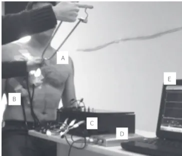
Mechanical waves were obtained by using three stetho-scopes connected to electret microphones, which were attached to a digital oscilloscope (Model DSO 2090 40 mHz, two USB channels, QinkDao, China). he data obtained by the oscilloscope were transmitted to a com-puter and registered by the software DSO-2090 USB, version 7.0.0.0. With the oscilloscope, the frequency was obtained in Hertz (Hz), and the amplitude, in millivolts (Mv), in two independent channels. herefore, to cap-ture waves in the upper, middle and lower thorax, it was necessary to use two oscilloscopes. Figure 1 shows one with both channels, and Figure 2 presents the experi-mental design.
Six points were determined in the anterior region (three in each hemithorax) and six in the posterior part of the thorax (three in each hemithorax), according to the posi-tions that were usually used in pulmonary auscultation20.
In the anterior region, the irst point was located 10 cm away from the nipple, in apical direction. he second one was 2 cm medially away from the nipple, and the third one, 10 cm below the nipple, approximately 10 cm to the side. he procedure was repeated in the contralateral hemithorax. In the posterior region, six points were also determined, three in each hemithorax. he irst one was located 5 cm away from the spinal process of C7, in the
Figure 1. DSO 2090 with two channels
Figure 2. Illustrative image of the experimental design. (A) Application of Diottix®; (B) stethoscopes placed in the anterior region of the right hemi-thorax; (C) connected to the electret microphone; (D) connected to the oscilloscope and (E) interfaced with the software to analyze the amplitude and frequency of signals
A
E
B
C
lateral direction; the second one was 1 cm below the cen-ter of the medial border of the scapula toward the medial direction, and the third one was 2 cm below the inferior angle of the scapula in the caudal direction21.
To ixate the electret microphone, one ear tip was removed from each stethoscope, which was installed inside the metallic arch that connects the conduction tube of the sound to the diaphragm. he other metallic arch was occluded to address the sound to the microphone.
Data were collected in a silent place, therefore look-ing to eliminate the efects of external noise and enable the qualiied capture of sounds.
Application of Diottix®
With the stereotypes attached to the anterior region of the thorax, Diottix®was applied in the same region, and this application was called anterior auscultation with anterior application (AAAA). With the stethoscope still in the anterior region of the thorax, the device was used in the posterior area, which was called ante-rior auscultation with posteante-rior application (AAPA).
When the stethoscopes were attached to the posterior region of the thorax and the Diottix® was applied in the
posterior region, it was called posterior auscultation with posterior application (PAPA), and when stethoscopes were in the posterior region and Diottix®was applied in the anterior region, it was called posterior auscultation with anterior application (PAAA).
he application of Diottix® was performed by the approximation of sinuous prolongations, by a tweez-ing movement and the posterior rough removal from the ingers17.
Statistical analysis
Measures of frequency and amplitude of mechanical waves generated by Diottix®were compared between the points captured in the right and left hemithorax, and anterior and posterior of the thorax, by using a paired sample t-test if distribution was normal, and the Wilcoxon test if it was not normal. For the relationship between the apex, the middle third and the base, the one-way analy-sis of variance (Anova) was used, with the Holm-Sidak Method as a discriminatory post-test. When data did not present the pre-requirements of normal distribution and the same variance, the Kruskal-Wallis test was used. Statistical analysis was conducted with the software Sigma Stat for Windows (version 3.5), with a signiicance level (p<0.05) and a 95% conidence interval.
RESULTS
Fifteen male subjects were assessed. Data regarding age, weight, height, BMI, spirometry of the studied sample are presented in Table 1.
Table 1. Characterization of the sample and pulmonar function
Characteristic Mean±SD
Age (years) 23.8±4.9
Weight (kg) 72.1±13.4
Height (cm) 172±9.2
BMI (kg/cm2) 24.3±1.6
FVC (%) 95.2±16.9
FEV1 (%) 92.4±18.4
FEV/FVC(%) 87.2±0.7
SD: standard deviation; BMI: body mass index; FVC: forced vital capacity; FEV1: forced expiratory volume in the first second
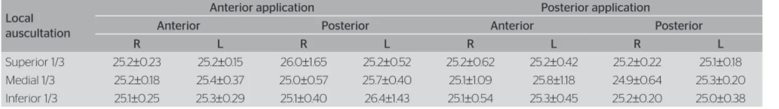
Table 2. Wave frequency in sites for the application of Diottix® and pulmonary regions
Local auscultation
Anterior application Posterior application
Anterior Posterior Anterior Posterior
R L R L R L R L
Superior 1/3 25.2±0.23 25.2±0.15 26.0±1.65 25.2±0.52 25.2±0.62 25.2±0.42 25.2±0.22 25.1±0.18 Medial 1/3 25.2±0.18 25.4±0.37 25.0±0.57 25.7±0.40 25.1±1.09 25.8±1.18 24.9±0.64 25.3±0.20 Inferior 1/3 25.1±0.25 25.3±0.29 25.1±0.40 26.4±1.43 25.1±0.54 25.3±0.45 25.2±0.20 25.0±0.38
Data in mean±standard deviation. There were no diferences between applications and regions of auscultation
Table 3. Amplitude of wave in places of Diottix® application and in pulmonary regions
Local auscultation
Anterior application Posterior application
Anterior Posterior Anterior Posterior
R L R L R L R L
Superior 1/3 48.0±17.3 66.3±15.8 20.5±9.7 17.6±14.5 19.7±4.0 20.5±8.3 75.3±9.5# 45.3
±16.9&
Medial 1/3 49.7±16.7 45.7±18.7* 16.6±8.5 17.8±13.1 17.9±5.3 17.0±4.3 73.6±9.9# 49.2
±19.3 Inferior 1/3 58.2±17.1 41.2±16.5* 25.6±4.8 19.7±16.3 18.0±5.4 20.1±3.8 61.2±17.1 59.3±17.3&
he wave frequency did not have signiicant variation (24.9 to 26,4 Hz), regardless of the application and aus-cultation point which are presented in Table 2.
he amplitude of the wave, comparing the right and left hemothorax, in each thoracic segment, did not pres-ent any diferences in all forms of application (Table 3). he comparison of wave amplitude in AAAA versus AAPA in each lung segment, right upper third versus left, right middle third versus left and right lower third versus left, showed signiicant diference (Table 3).
he amplitude of the wave presented diferences in AAAA in the left thorax, between the upper and middle thirds, and the upper thirds with the inferior left thorax, presenting more amplitude in the upper third (Table 3).
DISCUSSION
he main indings in this study are that the frequency of Diottix® remained unaltered in the diferent regions of the thorax in comparison to the superior of the left tho-rax, and in the posterior of the right thorax by relating to the anterior region of the same hemithorax when the application is posterior and the amplitude is lower in the let posterior region in comparison to the anterior region; however, it was higher in the inferior region.
he sound wave assessed in this study is periodical, that is, it happens in intervals with the same period. his wave system is deined as a harmonic oscillator, once it always presents a constant period. In a harmonic oscil-lator, the equation that describes mass dislocation, or the behavior of its velocity with time, is always expressed in a sinusoidal function, which provides the following vari-ables: amplitude, meaning the higher dislocation of mol-ecules from the middle in relation to the mean point of vibration, and frequency, which is the number of cycles per unit of time22.
Amplitude is related to the length of vibration, which is higher when auscultation is close to the place Diottix® was applied (AAAA and PAPA). herefore, when aus-cultation is away from the place of application (AAPA and PAAA), the wave amplitude is lower, that is, the waves generated by the diapason create higher disloca-tion of the molecules close to the point of applicadisloca-tion and lose intensity while getting more distant from this point. It also relates to the density of organic tissues, in case of pulmonary parenchyma, and amplitude and peak of amplitude are higher22. On the other hand, mediastinal
organs, such as heart and large vessels, can be responsible
for the lower values found in the left hemithorax during posterior auscultation with posterior application, even when Diottix® was used close to the auscultation site.
he amplitude and the frequency of the wave gener-ated by the Flutter® device VRP1 were assessed by the photoacoustic technique using electret microphones and showed frequency variation of 1 to 35 Hz20. he
ampli-tude of wave generated by the Flutter® VRP1 was uni-form in the right hemithorax, so that, in the left side, it was larger in the base when compared to the apical and medial regions. his fact was explained by the authors due to the attenuation of the propagation of sound waves generated by the Flutter® VRP1 device in the heart20.
In this study, the amplitude in the base of the left hemi-thorax was higher when Diottix® was applied in the pos-terior region with pospos-terior auscultation, however, this diference was not signiicant.
Flutter® is a device that matches positive expiratory pressure (PEP) and high-frequency oscillations, thus generating waves by the pressure variation. hey reach the basal regions, which present higher quantities of pul-monary tissue and that, consequently, would promote higher pressure variation. Diottix®, on the other hand, is applied directly on the thoracic region; therefore, it does not depend on the pressure oscillation in the air-ways, but only on the tissues that feel this vibration. So, the higher the amount of tissue, the stronger the reduc-tion of amplitude of the wave.
Silveira compared the use of Flutter® VRP1 with conventional physical therapy among people with cys-tic ibrosis, and reported that the device can generate frequencies of approximately 15 Hz16. he frequency
manual respiratory physical therapy can reach during the maneuvers is controversial in literature, as well as its eicacy8. It is possible to observe that, the one found
in literature about oral oscillation devices is diverging; therefore, Diottix® has a diferential, according to which the frequency ranges little, between 24.9 and 26.4 Hz. his instrument was capable of reaching the proposed frequency, with the advantage that the frequency varia-tion is minor, unlike Flutter® VRP1. In this context, it is still necessary to conduct new studies with the objec-tive of comparing its efects with Flutter® VRP1 or even with conventional respiratory physical therapy.
low lows24. Diottix® was built to maintain the frequency
ixed, however, our study showed that the amplitude ranges considerably when applied in the thorax, since this vari-able depends on biological tissues that sufer the action from the equipment, thus justifying the wide variation. Besides, the comparison with previous studies becomes hard, since the variables in this study were obtained directly from the thorax, while, in others, variables were obtained directly from Flutter® VRP1 or from Acapella24.
Another group compared the Acapella, the Flutter® VPR1 and the Shaker. Frequencies were similar in all of the devices, and the amplitude of the wave was diferent between them10. he Acapella® Choice, Acappela®Blue,
Acapella® Green were also compared with the water bottle. All of the tested devices reached 12 to 15 Hz7 in
the airway frequency.
In this study, despite the amplitude variation, the fre-quency remained constant, which is a great advantage for Diottix®, since the frequency of oral oscillators and bronchial hygiene maneuvers can range considerably.
Study limitations
Even though this study has important information about the frequency and amplitude of the waves provoked by Diottix®, it is still necessary to conduct validation and reproducibility studies about this equipment.
he frequency evaluation was made with cutaneous sensors, which can change the frequency of vibration due to the inluence of other tissues, like skin. herefore, studies assessing the pressure generated in the tho-rax when Diottix® is performed need to be produced. he investigation was led in the thorax of healthy sub-jects, in the absence of hypersecretion, so it was neces-sary to understand the behavior of these variables in the hypersecretion patient However, in this analysis, it was important to investigate the behavior of the amplitude and frequency of the wave at the absence of pulmonary disease. In this context, further studies must be focused on the investigation about the use of the device to pro-mote bronchial hygiene.
CONCLUSION
Diottix® produces frequencies in the thorax according to the device calibration, so it can be a complementary resource to bronchial hygiene maneuvers for respira-tory physical therapists. However, the wave amplitudes
seem to be afected by other structures, which include bone parts and the heart.
ACKNOWLEDGEMENTS
We would like to thank Luiz Vicente Franco de Oliveira and Camila Gimenes for suggesting and performing spirometry.
REFERENCES
1. Jones AP, Rowe BH. Withdrawn: bronchopulmonary hygiene physical therapy for chronic obstructive pulmonary disease and bronchiectasis. Cochrane Database of Systematic Reviews. The Cochrane Library. 2013(11):CD000045.
2. Trindade SH, Mello Junior JF, Mion OG. Métodos de estudo do transporte mucociliar. Rev Bras Otorrinolaringol. 2007;73(5):704-12.
3. Van Der Schans CP. Bronquial Mucus Transport.Respir Care 2007;52(9):1150-6.
4. King M, Phillips DM, Gross D, Vartian V, Chang HK, Zidulka A. Enhanced tracheal mucus clearance with high frequency chest wall compression. Am Rev Respir Dis. 1983;128(3):511-5.
5. King M, Zidulka A, Phillips D, Wright D, Gross D, Chang HK. Tracheal mucus clearance in high-frequency oscillation: efect of peak flow rate bias. Eur Respir J. 1990;3(1):6-13.
6. Tomkiewicz RP, Biviji AA, King M. Efects of oscillating air flow on the rheological properties and clerability of mucous gel simulants. Bioreheology. 1994;31(5):511-20.
7. Mueller G, Bersch-Porada I, Koch-Borner S, Raab AS, Jonker M, Baumberger M, et al. Laboratory evaluation of four diferent devices for secretion mobilization: acapella choice, green and blue versus water bottle. Respir Care. 2014;59(5):673-7.
8. Bott J, Blumenthal S, Buxton M, Ellum S, Falconer C, Garrod R, et al. Guidelines for the physiotherapy management of the adult, medical, spontaneously breathing patient. Thorax. 2009;64(Suppl 1):i1-i51.
9. França EE, Ferrari P, Fernandes P, Cavalcanti R, Duarte A, Martinez BP, et al. Fisioterapia em pacientes críticos adultos: recomendações do Departamento de Fisioterapia da Associação de Medicina Intensiva Brasileira. Rev Bras Ter Intensiva. 2012;24(1):6-22.
10. Santos AP, Guimarães RC, Carvalho EM, Gastaldi AC. Mechanical Behaviors of Flutter VRP1, Shaker, and Acapella Devices. Respir Care. 2013;58(2):298-304.
11. Chatburn RL. High-frequency assisted airway clearance. Respir Care. 2007;52(9):1224-37.
12. Nellessen A, Hernandes NA, Pitta F. Physiotherapy and rehabilitative interventions in patients with chronic respiratory diseases: exercise and non-exercise treatment. Panminerva Med. 2013;55(2):197-209.
14. Castro AM, Calil SR, Freitas SA, Oliveira AB, Porto EF. Chest physiotherapy efectiveness to reduce hospitalization and mechanical ventilation length of stay, pulmonary infection rate and mortality in ICU patients. Respir Med. 2013;107:68-74.
15. Stiller K. Physiotherapy in Intensive Care. An Updated Systematic Review. Chest. 2013;144(3):825-47.
16. Silveira ACT, Cunha CS, Pacheco DB, Silva NMA. Uso da oscilação oral de alta frequência em pacientes ventilados mecanicamente, um estudo prospectivo e revisão de literatura. Cad UniFOA. 2007;2(4):105-10.
17. Alcântara JR, Santos RL, Albino DD, Manzano RM. Desenvolvimento de aparelho de diapasão como uma ferramenta auxiliar nas manobras de higiene brônquica para fisioterapeutas. ConScientae Saúde. 2012;11(4):529-34.
18. Hankinson JL, Odencrantz JR, Fedan KB. Spirometric reference values from a sample of the general U.S. population. Am J Respir Crit Care Med. 1999;159:179-87.
19. Sociedade Brasileira de Pneumologia e Tisiologia. Diretrizes para Testes de Função Pulmonar. J Pneumol. 2002;28(3 Suppl):74-9.
20. Oliveira LH, Sá PF, Moraes ER, Barja PR, Avalos DA, Oliveira LV. Análise das frequências acústicas geradas pelo Flutter VRP1 no tórax humano. IX Encontro Latino Americano de Iniciação Científica e V Encontro Latino Americano de Pós-graduação, Universidade do Vale do Paraíba. Vale do Paraíba: Univap; 2006. p. 1615-8.
21. Carvalho VO, Souza GE. O estetoscópio e os sons pulmonares: uma revisão da literatura. Rev Med (São Paulo). 2007;86(4):224-31.
22. Garcia EAC. Biofísica. São Paulo: Sarvier; 2002.
23. Mcllwaine PM, Wong LT, Peacock D, Davison AG. Long-term comparative trial of positive expiratory pressure versus oscillating positive expiratory pressure (flutter) physiotherapy in the treatment of cystic fibrosis. J Pediatr. 2001;138(6):845-50.