Impact of ultrasound-guided polidocanol foam sclerotherapy in
patients with venous ulcers
Impacto da escleroterapia com espuma de polidocanol guiada por ultrassom em
pacientes com úlcera venosa
Melissa Andreia de Moraes Silva1
*, Álefy Zanelato Pereira Araujo
1, Jéssica Funchal do Amaral1,
Seleno Glauber de Jesus-Silva1, Rodolfo Souza Cardoso1, Fausto Miranda Júnior2
Abstract
Background: Ultrasound-guided polidocanol foam sclerotherapy is used to treat patients with venous ulcers. It is a minimally invasive procedure and is simple to perform, but it has high relapse rates. Objectives: To report short to medium term results in patients with venous ulcers treated using ultrasound-guided polidocanol foam sclerotherapy. Methods: A sample of 19 patients who had been treated with ultrasound-guided polidocanol foam sclerotherapy between January 2013 and December 2014 were followed-up. Time taken for ulcers to heal, improvement of clinical symptoms, recanalization of treated veins, and relapse of symptoms and of venous ulcers were analyzed. Results: Fifteen of the patients analyzed were female (78.9%) and four were male (21.1%). Overall mean age was 53 years. Follow-up times ranged from 448 days to 1,276 days (mean of 791 days). he mean duration of active ulcers was 53 months. At postoperative follow-up assessments, total recanalization was observed in 15.7%, partial recanalization in 21%, and occlusion in 47.3% of the veins that had been treated. here was only one case of ulcer relapse. Analysis of mean Venous Clinical Severity Scores (VCSS) revealed a signiicant diference from before to after the procedure, with a variation of 11.2 (p < 0.01). Conclusions: Ultrasound-guided foam sclerotherapy has high rates of therapeutic success and achieves high rates of venous ulcer healing.
Keywords: sclerotherapy; varicose ulcer; healing; relapse.
Resumo
Contexto: A escleroterapia com espuma de polidocanol guiada por ultrassom tem sido utilizada no tratamento de pacientes com úlceras venosas. É um procedimento minimamente invasivo e de fácil execução, porém apresenta taxas de recidiva elevadas. Objetivos: Relatar a evolução a curto e médio prazo de pacientes com úlcera venosa tratados com escleroterapia com espuma de polidocanol guiada por ultrassom. Métodos: Foram reavaliados 19 pacientes submetidos ao tratamento de escleroterapia com espuma de polidocanol guiada por ultrassom no período de janeiro de 2013 a dezembro de 2014. Foram analisados tempo de cicatrização da úlcera, melhora de sintomas clínicos, recanalização das veias tratadas, recidiva dos sintomas e da úlcera venosa. Resultados: Foram analisados 15 pacientes do sexo feminino (78,9%) e quatro do sexo masculino (21,1%). A média geral de idade foi de 53 anos. O tempo de seguimento dos pacientes variou de 448 dias a 1.276 dias (média de 791 dias). O tempo médio de presença das úlceras foi de 53 meses. Na avaliação pós-procedimento, foram observadas recanalização total em 15,7%, recanalização parcial em 21% e oclusão em 47,3% das veias tratadas. Apenas em um caso foi observada recidiva da úlcera. Pela avaliação das médias do Venous Clinical Severity Score (VCSS), houve diferença signiicativa antes e após o procedimento, com variação entre os grupos de 11,2 (p < 0,01). Conclusões: A escleroterapia por espuma guiada por ultrassom apresenta altas taxas de sucesso terapêutico, com índices de cicatrização de úlceras venosas elevados.
Palavras-chave: escleroterapia; úlcera varicosa; cicatrização; recidiva.
1 Faculdade de Medicina de Itajubá – FMIt, Cirurgia Vascular, Itajubá, MG, Brazil.
2 Universidade Federal de São Paulo – UNIFESP, Escola Paulista de Medicina, Cirurgia Vascular, São Paulo, SP, Brazil. Financial support: None.
Conlicts of interest: No conlicts of interest declared concerning the publication of this article. Submitted: April 21, 2017. Accepted: July 24, 2017.
INTRODUCTION
Varicose veins are manifestations of chronic venous
insuficiency (CVI) resulting from long duration venous hypertension.1,2 They affect 25% of women
and 15% of men of working age,1 have a major
socioeconomic impact, and are highly detrimental to patients’ quality of life.
Brazilian studies have reported a CVI prevalence of 35.5% of the population,3 while estimates of the
prevalence of venous ulcers vary from 0.2 to 1.0%.4
In turn, other studies published in the country have investigated the prevalence of advanced signs related to varicose veins, inding mean rates of 19.7% for edema, 5.7% for hyperpigmentation, 1.4% for eczema, and 0.6% for dermatoibrosis.5
Since 1994, a classiication of venous diseases based on clinical data (C), etiology (E), anatomic distribution (A) and pathophysiology (P), known as the CEAP classiication,6 has been adopted worldwide,
although certain modiications were introduced in 2004 to improve it.7 Clinical assessment alone does
not determine the anatomic levels involved, for which additional tests and examinations are needed.8 The most
widely used noninvasive method is color Doppler ultrasonography, which is a painless method that can be employed as often as necessary, offering the advantage of conirming diagnosis both by evaluation of the diameter of veins and by identiication of the presence of relux or occlusions. It is one of the available methods for accurate determination of the distribution and extent of venous disease.9
Sclerotherapy with polidocanol foam, one of the methods used to treat CVI, offers the advantages of being a minimally invasive and easily executed procedure that can be provided in outpatients settings and which enables patients to return home and resume daily activities early. There are no limitations to using the technique after relapses, since in cases of recanalization the same patients can be treated again with the same method.10 Polidocanol, in the form
of foam, is the agent generally used in this type of treatment and it is painless and has a low incidence of allergic reactions. Treatment success rates have been reported by Gonzalez-Zeh et al.,11 who showed that
after 1 year the procedure had achieved successful ablation in 77% of those treated.
The current technique is based on methods described by Tessari et al.,12 in which a sclerosing agent and air
are mixed together to form foam using two syringes and a three-way stopcock. Foam injection can be facilitated using Doppler ultrasound to guide the
puncture and to observe the foam progressing through the venous segment as the procedure is performed.13,14
The objective of the present study is to describe the short and medium term outcomes of patients with lower limb CVI with ulcers who were treated with ultrasound-guided polidocanol foam sclerotherapy.
METHODS
This is a descriptive study of patients with CEAP C6 grade CVI who were treated with ultrasound-guided polidocanol foam by the vascular surgery service of a teaching hospital between January 2013 and December 2014. Data from the initial treatments were acquired from a database maintained by the service. The patients were reassessed at consultations held from September 2015 to December 2015, using structured interviews. The study enrolled all patients within the age range of 18 to 80 years of age who had had venous ulcers and had been treated with ultrasound-guided polidocanol foam sclerotherapy. Patients were excluded if they were pregnant, under the age of 18 or over the age of 80, had a recent or early history of deep venous thrombosis that had not been recanalized, or had chronic or acute arterial disease.
The database was searched for information such as: patient name, age, sex, medical record, affected limb, comorbidities, vein treated, and date of procedure. Additionally, information was acquired from the Doppler examination conducted prior to treatment, covering relux in the deep and supericial vein systems.
At the reassessment consultation, patients underwent another physical examination, supplementary information related to ulcer healing and the procedures conducted was collected, and observations were made on whether or not ulcer relapse had occurred. At the same consultation, a trained professional used Doppler ultrasonography to assess veins that had been treated. The indings were recorded as maintenance of occlusion, total recanalization, or partial recanalization.
Parameters needed to calculate the international Venous Clinical Severity Score (VCSS) were included in the before and after sclerotherapy analysis. This score evaluates the severity of CVI in terms of numerical parameters for pain, varicose veins, venous edema, inlammation, hardening, number of ulcers, duration of ulcer, size of largest ulcer, compression therapy, and skin pigmentation.15
parametric t test for paired samples was used to
analyze VCSS results for before and after treatment. This study was approved by the institutional Research Ethics Committee and the Plataforma Brasil, under ruling number 1.111.919.
RESULTS
A total of 73 patients with venous ulcers were treated during the period analyzed. Of these, 19 patients (26%) attended for clinical reassessment, after being invited by telephone, with follow-up times ranging from 448 days to 1,276 days (mean of 791 days). The major dificulty with recruiting patients originally treated was caused by the fact that the majority (52 patients) live in towns that are a long way from the treating service, with the result that the sample reassessed was smaller than the total cohort of patients treated.
Of these 19 patients who presented for reassessment, 15 were female (78.9%) and 4 were male (21.1%). The overall mean age was 63.6 years. Nine patients had associated comorbidities (47.3%). The most prevalent of these was systemic arterial hypertension, in six of the 19 patients (31.5%), followed by diabetes mellitus, in two of the 19 patients (10.5%), and cirrhosis of the liver, in one patient (5.3%).
The limb most frequently affected by venous ulcers was the left lower limb, in 10 of the 19 patients (52.6%). The mean duration of ulcer activity was 53 months.
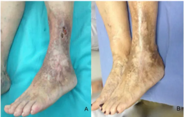
Thirteen of the 19 patients (68.4%) were treated in a single session, while six of the 19 (31.5%) patients were given more than one administration of foam, the proportion of ulcer healing was 89% (Figures 1A and 1B), and mean time taken for healing was 31.94 days (Figure 2).
The pattern of venous relux observed prior to treatment in the limbs to be treated was irregularly distributed between deep, superficial, and perforating-communicating venous systems. Just one patient had relux in the deep vein system (5.2% of limbs). The greater proportion of cases (12 patients) exhibited relux along the entire extension of the great saphenous vein. One patient had segmental relux in the great saphenous vein in one leg only. The small saphenous vein was considered incompetent in four limbs, in one of which there was also relux in the great saphenous vein. Two patients (10% of cases) only had relux in the perforating-communicating system.
The reassessment after the procedure detected total recanalization in 21%; partial recanalization in 26%, and maintenance of occlusion in 53%. However, ulcer relapse only occurred in one case (5%), 728 days after
the initial treatment. This case had total recanalization of the great saphenous vein that had been treated.
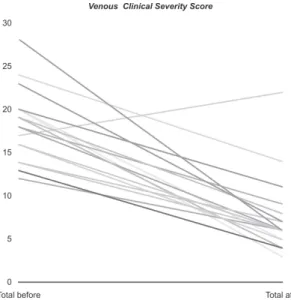
Before treatment, the VCSS scores ranged from 12 to 28 (mean of 18.7). After treatment, the scores varied from 3 to 22 (mean of 7.5). Clinical deterioration was only observed in one case, with the VCSS score increasing from 17 to 22. There was a signiicant change in VCSS scores from before to after the procedure (p < 0.01), with a mean difference of 11.2. Using a 95% conidence interval, the absolute values ranged from 8.2 to 14.1 (Figure 3).
DISCUSSION
Sclerotherapy is an effective and attractive option for treatment of chronic venous insuficiency with reflux observed in both superficial and perforating-communicating systems. It can be easily performed in outpatients settings, and this is particularly true of foam sclerotherapy.
The follow-up time reported in the present study is greater than in many published studies. With follow-up
Figure 1. (A) Active venous ulcer on lateral surface of left leg; (B) After treatment with ultrasound-guided polidocanol foam sclerotherapy, with healing time of 30 days.
times ranging from 448 days to 1,276 days (mean of 791 days), it was possible to analyze the long-term effects of sclerosing treatment in a speciic group of patients with ulcers, focusing on relapses and recanalization of the veins treated.
The patients analyzed in this study have a heterogeneous epidemiological proile. It is a small sample, but it conforms to the pattern reported in previously published work, encompassing patients of varying ages and with multiple comorbidities.10,15
In a study conducted to evaluate immediate results in order to assess the eficacy of the procedure, it was shown that a single session was suficient to treat 58% of patients with CVI. Total elimination of varicose veins was achieved in 87% of the sample,16 indicating
a high therapeutic success rate. However, that sample cannot be compared to the patients in the present study, since the outcome analyzed was elimination of varicose veins, whereas in the present study it was occlusion of the vein or healing of the ulcer.
With relation to patients with severe CVI, Silva et al.17
showed that ultrasound-guided foam sclerotherapy had a signiicant effect on patients. Ulcers healed in 84.2% of cases, with a mean healing time of 37 days. Recanalization was observed in 31.5%, but the rate of recanalization with symptoms and recurrence of ulcers was 11.8%.
The present study observed rates of ulcer healing similar to those reported in the literature. The rate of 89% (at 791 days) is similar to that seen in a study by Howard et al., who observed an ulcer healing rate of 86% at 12 months, showing the long-term eficacy
of foam sclerotherapy at grade C6 of the CEAP CVI classiication.18 The venous ulcer recurrence rate in the
present study was 5.2%. According to Grover et al., in a sample of 50 limbs with a median follow-up time of 15.2 months, there was recurrence in 4/50 (8%) at 12 months.19
The partial and total recanalization rates observed in the present study were 26% and 21%, respectively. In some aspects these results are similar to those reported by Howard et al.,18 who observed complete
recanalization in 27% and partial recanalization in 58%. One year after treatment, Wiliamsson et al.20
observed that 86% (25/29) of veins examined with Doppler ultrasound were occluded, one was partially occluded, and three were recanalized. These values are different to the indings of the current study. Several different factors inluence recanalization rates, such as the diameters of the veins treated, for example. However, this information was not analyzed in this study.
The VCSS score was another criterion analyzed before and after the procedure. Masuda et al.21 noted
signiicant improvements in the scores for 37 patients with active ulcers after treatment with polidocanol foam. Just one of the patients in the present study had a higher score after the procedure, indicating deterioration. This was a patient whose ulcer remained active, despite wearing elastic compression after treatment.
Despite the small sample size, it was possible to analyze certain variables that are important to a real understanding of this type of treatment for CVI. The plan is to continue following these patients, and include additional cases, to enable another analysis in the future.
CONCLUSIONS
Ultrasound-guided foam sclerotherapy achieved high treatment success rates and high and sustained rates of venous ulcer healing over the short and medium term. There was a considerable proportion of recanalization of treated veins; but in the majority of cases this did not cause disease severity to exacerbate.
REFERENCES
1. Abreu J, Pitta G, Miranda F Jr. Doppler ultrasonography of the femoral popliteal segment in patients with venous ulcer. J Vasc Bras. 2012;11(4):277-85. http://dx.doi.org/10.1590/ S1677-54492012000400005.
2. Figueiredo M, Filho AD. Avaliação do efeito da meia elástica na hemodinâmica venosa dos membros inferiores de pacientes com insuficiência venosa crônica. J Vasc Bras. 2004;3(3):231-7.
3. Moura RMF, Gonçalves GS, Navarro TP, Britto RR, Dias RC. Correlação entre classificação clínica CEAP e qualidade de vida na doença venosa crônica. Rev Bras Fisioter. 2010;14(2):99-105. PMid:20464164. http://dx.doi.org/10.1590/S1413-35552010005000007.
4. Cornwall JV, Doré CJ, Lewis JD. Leg ulcers: epidemiology and aetiology. Br J Surg. 1986;73(9):693-6. PMid:3756430. http://dx.doi. org/10.1002/bjs.1800730905.
5. Sandri GA. Tratamento endovascular das obstruções venosas crônicas do segmento iliocaval. J Vasc Bras. 2011;10(2):137-44. http://dx.doi.org/10.1590/S1677-54492011000200008.
6. França LH, Tavares V. Insuficiência venosa crônica: uma atualização. J Vasc Bras. 2003;2(4):18-28.
7. Eklöf B, Rutherford RB, Bergan JJ, et al. Revision of the CEAP classification for chronic venous disorders: consensus statement. J Vasc Surg. 2004;40(6):1248-52. PMid:15622385. http://dx.doi. org/10.1016/j.jvs.2004.09.027.
8. Saliba OA Jr, Giannini M, Rollo HA. Métodos de diagnóstico não-invasivos para avaliação da insuficiência venosa dos membros inferiores. J Vasc Bras. 2007;6(3):266-75. http://dx.doi.org/10.1590/ S1677-54492007000300010.
9. Santos RF, Porfírio GJ, Pitta GB. A diferença na qualidade de vida de pacientes com doença venosa crônica leve e grave. J Vasc Bras. 2009;8(2):143-7. http://dx.doi.org/10.1590/S1677-54492009000200008.
10. Shadid N, Ceulen R, Nelemans P, et al. Randomized clinical trial of ultrasound-guided foam sclerotherapy versus surgery for the incompetent great saphenous vein. Br J Surg. 2012;99(8):1062-70. PMid:22627969. http://dx.doi.org/10.1002/bjs.8781.
11. Gonzalez-Zeh R, Armisen R, Barahona S. Endovenous laser and echo-guided foam ablation in great saphenous vein reflux: one-year follow-up results. J Vasc Surg. 2008;48(4):940-6. PMid:18639418. http://dx.doi.org/10.1016/j.jvs.2008.05.062.
12. Tessari L, Cavezzi A, Frullini A. Preliminary experience with a new sclerosing foam in the treatment of varicose veins. Dermatol Surg. 2001;27(1):58-60. PMid:11231246.
13. Campos V Jr. Estudo comparativo entre escleroterapia com espuma de polidocanol e cirurgia convencional no tratamento de varizes primárias dos membros inferiores em portadores de úlcera venosa [tese]. São Paulo: Faculdade de Medicina, Universidade de São Paulo; 2014.
14. Rabe E, Otto J, Schliephake D, Pannier F. Efficacy and safety of great saphenous vein sclerotherapy using standardised polidocanol foam (ESAF): a randomised controlled multicentre clinical trial. Eur J Vasc Endovasc Surg. 2008;35(2):238-45. PMid:17988905. http:// dx.doi.org/10.1016/j.ejvs.2007.09.006.
15. Rutherford RB, Padberg FT Jr, Comerota AJ, Kistner RL, Meissner MH, Moneta GL. Venous severity scoring: an adjunct to venous outcome assessment. J Vasc Surg. 2000;31(6):1307-12. PMid:10842165. http://dx.doi.org/10.1067/mva.2000.107094.
16. Kakkos SK, Bountouroglou DG, Azzam M, Kalodiki E, Daskalopoulos M, Geroulakos G. Effectiveness and safety of ultrasound-guided foam sclerotherapy for recurrent varicose veins: immediate results. J Endovasc Ther. 2006;13(3):357-64. PMid:16784324. http://dx.doi. org/10.1583/05-1781.1.
17. Silva MAM, Burihan MC, Barros OC, Nasser F, Ingrund JC, Neser A. Resultados do tratamento da Insuficiência Venosa Crônica grave com espuma de polidocanol guiada por ultrassom. J Vasc Bras. 2012;11(3):206-11. http://dx.doi.org/10.1590/ S1677-54492012000300007.
18. Howard JK, Slim FJ, Wakely MC, et al. Recanalisation and ulcer recurrence rates following ultrasound-guided foam sclerotherapy. Phlebology. 2015;31(7):506-13. PMid:26224059. http://dx.doi. org/10.1177/0268355515598450.
19. Grover G, Tanase A, Elstone A, Ashley S. Chronic venous leg ulcers: effects of foam sclerotherapy on healing and recurrence. Phlebology. 2016;31(1):34-41. PMid:25351907. http://dx.doi. org/10.1177/0268355514557854.
20. Williamsson C, Danielsson P, Smith L. Catheter-directed foam sclerotherapy for chronic venous leg ulcers. Phlebology. 2014;29(10):688-93. PMid:24072751. http://dx.doi.org/10.1177/0268355513505506.
21. Masuda EM, Kessler DM, Lurie F, Puggioni A, Kistner RL, Eklöf B. The effect of ultrasound-guided sclerotherapy of incompetent perforator veins on venous clinical severity and disability scores. J Vasc Surg. 2006;43(3):551-6, discussion 556-7. PMid:16520171. http://dx.doi.org/10.1016/j.jvs.2005.11.038.
*
Correspondence
Melissa Andreia de Moraes Silva Avenida BPS, 492/1001 CEP 37500-177 - Itajubá (MG), Brazil Phone: +55 (35) 99904-7400 E-mail: [email protected]
Author information
MAMS - MD, Board-certiied in Vascular Surgery and Vascular Ultrasonography; Professor, Disciplina de Cirurgia Vascular, Faculdade de Medicina de Itajubá (FMIt), Itajubá, MG, Brazil. AZPA, JFA - Medical students (6th year), Faculdade de Medicina de Itajubá (FMit). SGJS, RSC - MDs, Boards-certiied in Vascular Surgery and Interventional Radiology; Professors, Disciplina de Introdução aos Procedimentos Minimamente Invasivos, Faculdade de Medicina de Itajubá (FMIt). FMJ - MD, Professor, Disciplina de Cirurgia Vascular, Escola Paulista de Medicina, Universidade Federal de São Paulo (UNIFESP).
Author contributions
Conception and design: MAMS Analysis and interpretation: MAMS, AZPA, JFA Data collection: AZPA, JFA Writing the article: MAMS, AZPA, JFA Critical revision of the article: MAMS, AZPA, JFA, SGJS, RSC, FMJ Final approval of the article*: MAMS, AZPA, JFA, SGJS, RSC, FMJ Statistical analysis: N/A. Overall responsibility: MAMS