Ischemia and reperfusion by retrograde flow: experimental
comparative study
Isquemia e reperfusão por circulação retrógrada: estudo comparativo experimental
Cesar Roberto Busato1
*
, Carlos Alberto Lima Utrabo1, Leandro Cavalcante Lipinski1, Keizi Dayane de Lima1, Márcio Dias Guilherme Filho1, Nicolas Brandalize Medeiros1, Samela Basi Fagundes1, Willman Josviak1
Abstract
Background: here are few options for treating critical ischemia in limbs with no distal patency. Diverting low through the venous circulation is an option supported by evidence from numerous published studies. Objectives: To compare the behavior of clinical and laboratory variables between the hind limbs of pigs subjected to ischemia and to ischemia with reperfusion by retrograde circulation and between these intervention groups and a control group. Methods: Ten pigs were divided into 2 groups. In group 1 (n=5), controls, patterns of physiological variables such as low according to Doppler ultrasound, temperature, blood gas analysis results, lactate, creatine kinase, and blood pressure were evaluated. In group 2 (n=5), after an initial ischemia period with mean duration of 27 minutes and 30 seconds, provoked by interrupting low through the femoral arteries, the animals were subjected to venous arterialization of the left hind limb while the right hind limb was maintained in ischemia. Variables were analyzed separately for each hind leg at 0, 2, 3, 4, and 6 hours after reperfusion and compared against each other and the control group. Results: Analysis of variables from both procedures showed decreases in BE and PO2 and signiicant increases in lactate and creatine kinase, in relation to the control group. In arterialized ischemic limbs, we observed low on Doppler ultrasound, and arterial pressures and temperatures were higher than in the ischemic limbs. Conclusions: Comparative analysis of the extremities in ischemia and arterialized ischemia showed, in relation to the control group, metabolic acidosis with signiicant increases in lactate and creatine kinase, suggesting cellular damage, and there were signs of retrograde reperfusion in arterialized extremities.
Keywords: venous arterialization; ischemia; reperfusion.
Resumo
Contexto: Isquemia crítica de membro inferior sem leito distal tem opções restritas para tratamento. Desviar o luxo de maneira retrógrada através da circulação venosa é alternativa amparada em evidências de inúmeros trabalhos publicados. Objetivos: Comparar o comportamento de variáveis clínicas e laboratoriais em extremidades de suínos submetidas a isquemia e a isquemia com reperfusão por circulação retrógrada entre si e em relação e a um grupo controle. Métodos: Dez suínos foram separados em dois grupos. No grupo 1 (n=5), controle, avaliaram-se padrões isiológicos de variáveis como luxo ao Doppler, temperatura, gasometria, lactato, creatinoquinase (CK) e pressão arterial. No grupo 2 (n=5), após um período médio de isquemia de 27 minutos e 30 segundos, consequente à interrupção do luxo nas artérias femorais, os animais foram submetidos a arterialização venosa no membro posterior esquerdo e a manutenção da isquemia no direito. As variáveis foram analisadas separadamente durante momentos 0, 2, 3, 4 e 6 horas após a reperfusão para efeito de comparação entre si e com o grupo controle. Resultados: A análise das variáveis mostrou, em ambos os procedimentos, queda de BE e pO2, com elevação signiicativa de lactato e CK em relação ao grupo controle. Nos membros isquêmicos arterializados, encontramos luxo ao Doppler e maiores pressões arteriais e temperaturas quando comparadas ao membro em isquemia. Conclusões: A análise comparativa das extremidades em isquemia e isquemia arterializada mostrou, em relação ao grupo controle, um quadro de acidose metabólica, com signiicativo aumento de lactato e CK, que sugerem dano celular e sinais de reperfusão retrógrada nas extremidades arterializadas.
Palavras-chave: arterialização venosa; isquemia; reperfusão.
1Universidade Estadual de Ponta Grossa – UEPG, Departamento de Medicina, Ponta Grossa, PR, Brazil. Financial support: None.
Conlicts of interest: No conlicts of interest declared concerning the publication of this article. Submitted: November 07, 2016. Accepted: May 11, 2017.
INTRODUCTION
In cases of critical ischemia in which distal arterial bed patency is lacking, it is not possible to shunt blood to an arterial extremity distal of the obstruction.
Diverting low in a retrograde direction through the
venous circulation is a feasible alternative that is supported by evidence from countless published studies.1-13 The concept is based on the theory that in the absence of primary arterial blood pressure in arterioles, blood supplied by arterialization of the distal venous system is capable of supplying the peripheral tissues and providing adequate oxygenation.3-5,13
Many different variables have been used to evaluate tissue hypoxia in animal models, such as assessment
of arterial low using Doppler ultrasound (US),14,15 temperature measurements,16 blood gas analysis,17 lactate18 and creatine phosphokinase (CP) assays,19 and measurements of arterial blood pressure at extremities.20 The objective of this study is to compare the behavior of these clinical and laboratory variables in pigs’ hind limbs after induction of ischemia, with and without reperfusion via retrograde circulation, against each other and in relation to a control group.
METHODS
This project was approved by the Animal Research
Ethics Committee (CEUA 009/2013) and conducted in the Operating Techniques and Experimental Surgery Laboratory at the Universidade Estadual de Ponta Grossa (UEPG) Medical Faculty, Brazil. Ten Large
White-Landrace cross pigs were acquired and divided
into two groups. In group 1 (n=5), the control group,
analyses were conducted of physiological indicators
of the quality of arterial low using Doppler US, temperature, arterial blood gas analysis (pH, base
excess, bicarbonate, partial oxygen pressure and
partial carbon dioxide pressure), lactate, CP, and
arterial blood pressure in the hind limbs, by femoral artery dissection. These analyses were all performed on animals that had been selected for use in practical Operating Technique classes before surgical procedures were conducted.
In group 2 (n=5), the intervention group, the same
variables measured in group 1 were determined for the right hind limb after induction of ischemia and the left hind limb after induction of ischemia and arterialization.
The animals in both groups were premedicated
with ketamine (14 mg/kg), xylazine (0.2 mg/kg), and acepromazine (0.4 mg/kg). Anesthesia was induced with propofol (5 mg/kg) and maintained with inhaled
isolurane at a minimum alveolar concentration of
1.2 to 1.7%.
In all hind limbs studied, common femoral arteries and veins were dissected. In limbs in the control
group (group 1) these vessels were only used for
data collection. In the hind limbs of animals in group 2, veins were used to draw blood samples by direct puncture, while arteriotomies were performed at the distal and proximal extremities of the common femoral arteries and cannulated with number 14 intravascular
catheters, (ESCALPE intravenous catheter without entry tip, with Telon wall sterilized by ethylene oxide; SOLIDOR®), ligated, and occluded, when ischemia
time measurement was initiated.
In limbs designated for arterialization after ischemia,
the external (small) saphenous vein was dissected. Systemic anticoagulation was administered with 5,000 UI of heparin, followed by proximal ligature,
venotomy, and rupture of downstream valves using a Lengua valvulotome, and then distal dilatation with heparinized saline via the nº 4 probe, before cannulation
and ixation with the number 14 intravascular catheter (SOLIDOR®). The proximal extremities of the common
femoral arteries were connected to the saphenous
veins using a silicone catheter (EXTENSOR for 20 cm Luer Lock Reversible Catheter, 2 10F male connectors–; HARTMANN®), which was deined
as the start of arterialization, T0.
Once the surgical procedures had been completed, at 0, 2, 4, and 6 hours of reperfusion, respectively
times T0, T1, T3 and T4 (Figure 1), tests were performed to detect blood low at the extremities with a portable Doppler US unit (DV 600 Vascular Doppler; Martec®), temperature was measured with an infrared thermometer (FR1DZ1 non-contact infrared thermometer; G-TECH®). Blood low was tested
at the saphenous artery in each limb. Temperature measurements were performed at three different times,
at a ixed point in the interdigital space of the plantar
region and, on the dorsal surface at three points around 3 cm apart from each other along the extremity of each limb. The distance between the thermometer and the skin was approximately 3 to 5 cm.
At 0, 3, and 6 hours of reperfusion, times T0, T2
and T4 respectively (Figure 1), heparinized syringes
were used to collect 3 mL blood samples for blood gas analysis, via the catheters at the distal extremities
of the arteriotomies (supericial femoral artery); and
venous blood was drawn by direct puncture of the
femoral vein for lactate and CP assays; and, a ruler
and a saline column were used to determine blood
Throughout the study, 5,000 UI of heparin were
administrated every 3 hours.
After all procedures had been completed, animals in
group 2 were euthanized as directed by Federal Veterinary Medical Council resolution 1000/2012 (Conselho Federal de Medicina Veterinária).
Statistical analysis
The results were subjected to analysis of normality of distribution using the Anderson Darling test. The chi-square test with Yates’ correction was used
to compare qualitative variables, and Student’s t test
was used for quantitative variables, with statistical
signiicance set at p < 0.05.
RESULTS
The variables measured in limbs arterialized after a mean ischemia time of 27 minutes and 30 seconds were compared with variables measured in the ischemic limbs and control limbs.
Arterial flow
Doppler examination of arterial flow in the arterialized ischemic limbs at T0 showed absence of
low in one limb, pulsating venous pattern in three, and pulsating arterial pattern in one; at T1 four limbs with blood low had pulsating venous low patterns; at T3, pulsating arterial low was observed in one and pulsating venous low in three; at T4, three limbs exhibited pulsating venous low and two limbs had
no blood low. All ischemic limbs exhibited absence of low throughout the study.
For the purposes of analysis, only data from arterialized ischemic limbs that had low detected by Doppler US were considered: four animals up to
T3 and three animals at T4.
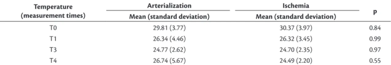
Temperature
Although there was no environmental control, the mean variations in temperature, at the extremities in the study group, exhibited similar curves, with means
signiicantly lower to those measured for the control group over time (Table 1). When compared against each other, the mean temperatures for the arterialized
ischemic limbs exhibited a non-signiicant (p= 0.55) difference; temperatures were 2.25 °C higher than the ischemic limbs at T4 (Table 2).
Blood gas analysis
The pH of arterialized ischemic limbs and ischemic limbs began at slightly higher levels than the pH of
the control group and followed similar curves, with
a non-signiicant fall at T2, a little more accentuated than at T4 (Tables 3, 4, and 5). When compared
against each other, they did not exhibit signiicance differences (Tables 6, 7, and 8).
Base excess
Both arterialized ischemic limbs and limbs
maintained in ischemia exhibited a progressive fall
Table 1. Comparison of temperatures in arterialized limbs and limbs in ischemia in relation to the control group, at four measurement times.
Temperature (measurement
times)
Arterialization Control
p
Ischemia Control
p Mean
(standard deviation)
Mean (standard deviation)
Mean (standard deviation)
Mean (standard deviation)
T0 29.81 (3.77) 35.65 (0.55) 0.01 30.37 (3.97) 35.65 (0.55) 0.02
T1 26.34 (4.46) 35.65 (0.55) 0.002 26.32 (3.45) 35.65 (0.55) 0.0005
T3 24.77 (2.62) 35.65 (0.55) 0.0001 24.70 (2.35) 35.65 (0.55) 0.0001
T4 26.74 (5.67) 35.65 (0.55) 0.010 24.49 (2.20) 35.65 (0.55) 0.0001
Student’s t test.
Table 2. Comparison of temperatures in arterialized ischemic limbs and limbs in ischemia at diferent measurement times. Temperature
(measurement times)
Arterialization Ischemia
p Mean (standard deviation) Mean (standard deviation)
T0 29.81 (3.77) 30.37 (3.97) 0.84
T1 26.34 (4.46) 26.32 (3.45) 0.99
T3 24.77 (2.62) 24.70 (2.35) 0.97
T4 26.74 (5.67) 24.49 (2.20) 0.55
Student’s t test.
Table 3. Comparison of biochemical variables and arterial blood pressure levels in arterialized limbs and limbs in ischemia in relation to the control group at measurement time T0.
Variables
Arterialization Control
p
Ischemia Control
p Mean
(standard deviation)
Mean (standard deviation)
Mean (standard deviation)
Mean (standard deviation)
pH 7.32 (0.11) 7.30 (0.15) 0.83 7.31 (0.12) 7.30 (0.15) 0.92
BE 2.68 (1.02) 2.38 (2.47) 0.83 2.18 (1.38) 2.38 (2.47) 0.89
HCO3- 29.45 (1.77) 30.62 (4.17) 0.62 29.15 (2.29) 30.62 (4.17) 0.55
pO2 166.33 (61.91) 298.10 (128.32) 0.10 158.93 (58.34) 298.10 (128.32) 0.09
pCO2 60.75 (16.19) 68.82 (34.49) 0.68 60.60 (17.63) 68.82 (34.49) 0.68
Lactate 18.55 (2.99) 17.14 (6.96) 0.72 21.20 (10.28) 17.14 (6.96) 0.50
CP 1,647.50 (590.89) 940.00 (364.18) 0.06 1,612.50 (563.68) 940.00 (364.18) 0.07
Arterial blood
pressure 55.75 (15.76) 35.86 (22.86) 0.18 47.50 (22.49) 35.86 (22.86) 0.47
Table 4. Comparison of biochemical variables and arterial blood pressure levels in arterialized limbs and limbs in ischemia in relation to the control group at measurement time T2.
Variables
Arterialization Control
p
Ischemia Control
p Mean
(standard deviation)
Mean (standard deviation)
Mean (standard deviation)
Mean (standard deviation)
pH 7.26 (0.08) 7.30 (0.15) 0.65 7.25 (0.06) 7.30 (0.15) 0.55
BE 1.13 (1.97) 2.38 (2.47) 0.44 0.68 (1.41) 2.38 (2.47) 0.26
HCO3- 29.30 (1.47) 30.62 (4.17) 0.57 29.08 (1.28) 30.62 (4.17) 0.50
pO2 143.65 (89.98) 298.10 (128.32) 0.08 167.53 (143.26) 298.10 (128.32) 0.19
pCO2 57.80 (23.85) 68.82 (34.49) 0.61 68.40 (11.02) 68.82 (34.49) 0.98
Lactate 40.30 (19.38) 17.14 (6.96) 0.04 36.10 (21.17) 17.14 (6.96) 0.10
CP 1,747.00 (556.34) 940.00 (364.18) 0.03 1,704.25 (554.79) 940.00 (364.18) 0.04
Arterial blood
pressure 42.63 (19.61) 35.86 (22.86) 0.65 37.75 (15.76) 35.86 (22.86) 0.89
Table 5. Comparison of biochemical variables and arterial blood pressure levels in arterialized limbs and limbs in ischemia in relation to the control group at measurement time T4.
Variables
Arterialization Control
p
Ischemia Control
p
Mean (standard deviation)
Mean (standard deviation)
Mean (standard deviation)
Mean (standard deviation)
pH 7.21 (0.12) 7.30 (0.15) 0.41 7.20 (0.09) 7.30 (0.15) 0.34
BE -0.50 (3.58) 2.38 (2.47) 0.41 1.07 (2.72) 2.38 (2.47) 0.11
HCO3- 29.07 (1.61) 30.62 (4.17) 0.57 27.87 (2.48) 30.62 (4.17) 0.35
pO2 95.83 (13.82) 298.10 (128.32) 0.04 94.67 (3.89) 298.10 (128.32) 0.04
pCO2 76.23 (21.75) 68.82 (34.49) 0.75 71.87 (15.09) 68.82 (34.49) 0.89
Lactate 61.93 (19.70) 17.14 (6.96) 0.003 55.33 (19.15) 17.14 (6.96) 0.006
CP 2,020.67 (621.81) 940.00 (364.18) 0.02 1,879.33 (425.96) 940.00 (364.18) 0.02
Arterial blood
pressure 33.17 (19.20) 35.86 (22.86) 0.87 36.67 (13.58) 35.86 (22.86) 0.96
Student’s t test.
Table 6. Comparison of biochemical variables and arterial blood pressure levels in arterialized limbs and limbs in ischemia at measurement time T0.
Variables Arterialization Ischemia p
Mean (standard deviation) Mean (standard deviation)
pH 7.32 (0.11) 7.31 (0.12) 0.91
BE 2.68 (1.02) 2.18 (1.38) 0.58
HCO3- 29.45 (1.77) 29.15 (2.29) 0.84
pO2 166.33 (61.91) 158.93 (58.34) 0.87
pCO2 60.75 (16.19) 60.60 (17.63) 0.99
Lactate 18.55 (2.99) 21.20 (10.28) 0.64
CP 1,647.50 (590.89) 1,612.50 (563.68) 0.93
Arterial blood pressure 55.75 (15.76) 47.50 (22.49) 0.57
Student’s t test.
Table 7. Comparison of biochemical variables and arterial blood pressure levels in arterialized limbs and limbs in ischemia at measurement time T2.
Variables Arterialization Ischemia p
Mean (standard deviation) Mean (standard deviation)
pH 7.26 (0.08) 7.25 (0.06) 0.85
BE 1.13 (1.97) 0.68 (1.41) 0.72
HCO3- 29.30 (1.47) 29.08 (1.28) 0.83
pO2 143.65 (89.98) 167.53 (143.26) 0.79
pCO2 57.80 (23.85) 68.40 (11.02) 0.45
Lactate 40.30 (19.38) 36.10 (21.17) 0.78
CP 1,747.00 (556.34) 1,704.25 (554.79) 0.92
Arterial blood pressure 42.63 (19.61) 37.75 (15.76) 0.71
Student’s t test.
Table 8. Comparison of biochemical variables and arterial blood pressure levels in arterialized limbs and limbs in ischemia at measurement time T4.
Variables Arterialization Ischemia p
Mean (standard deviation) Mean (standard deviation)
pH 7.21 (0.12) 7.20 (0.09) 0.91
BE 0.50 (3.58) 1.07 (2.72) 0.58
HCO3- 29.07 (1.61) 27.87 (2.48) 0.52
pO2 95.83 (13.82) 94.67 (3.89) 0.89
pCO2 76.23 (21.75) 71.87 (15.09) 0.79
Lactate 61.93 (19.70) 55.33 (19.15) 0.70
CP 2,020.67 (621.81) 1,879.33 (425.96) 0.76
Arterial blood pressure 33.17 (19.20) 36.67 (13.58) 0.81
which was more accentuated in limbs maintained in
ischemia (Tables 3, 4, and 5). When arterialized limbs were compared with those maintained in ischemia,
there was a non-signiicant (p=0.58) difference of 1.57 at T4 (Tables 6, 7, and 8).
Bicarbonate concentration
Both arterialized ischemic limbs and limbs
maintained in ischemia exhibited a progressive fall in
the concentration of bicarbonate (HCO3
-), which was
more accentuated in the ischemic limbs, especially at
T4 (Tables 3, 4, and 5), but without signiicance either in relation to the control group or to the arterialized
limbs (Tables 6, 7, and 8).
Partial oxygen pressure
Partial oxygen pressures (pO2) in the arterialized ischemic limbs and the ischemic limbs started from lower levels than the control limbs and followed similar
curves, with signiicant drops at T4 (Tables 3, 4, and 5)
Comparisons between them did not reveal signiicance (Tables 6, 7, and 8).
Partial carbon dioxide pressure
Partial carbon dioxide pressure (pCO2) values were similar and lower than in the control group at
T0, followed by a progressive increase in mean pCO2 in limbs maintained in ischemia and a mild fall in
mean pCO2 in the arterialized ischemic limbs at T2. The means for both groups exhibited increases at T4, although the increase was greater in the ischemia with
arterialization group (Tables 3, 4, and 5). Comparisons
between means were not signiicant (Tables 6, 7, and 8).
Lactate
Mean lactate values started from a baseline level similar to that in the control group and exhibited similar curves, with a progressive increase at T2, which was
signiicant for arterialized ischemic limbs, and a more accentuated increase at T4 that was signiicant both for arterialization and ischemia (Tables 3, 4, and 5).
Comparisons between means were not signiicant (Tables 6, 7, and 8).
Creatine phosphokinase
Mean CP values started from higher baseline levels than the control group, exhibiting signiicant
increases both in arterialized ischemic limbs and in
those in ischemia at T2 and T4 (Tables 3, 4, and 5).
However, means did not exhibit signiicant differences (Tables 6, 7, and 8).
Arterial blood pressure
The ratios of distal arterial pressures to proximal arterial pressures were used for the purposes of calculations. In arterialized ischemic limbs, the mean of this ratio started from a higher baseline than in the control group and than in limbs in ischemia. The means in both intervention groups exhibited a drop at T2, although the ratio for arterialized ischemic limbs remained higher. At T4, the means approached
those in the control group (Tables 3, 4, and 5) and
were not signiicantly different from each other (Tables 6, 7, and 8).
The curves for absolute distal pressures exhibited
similar patterns to the ratios, also without signiicance.
DISCUSSION
There are no experimental studies of ischemia
and reperfusion (I/R) by arterialization of veins
that have tested the variables of interest in animals. Our model induced acute ischemia, which is different from the chronic form, in which a period of ischemia without necrosis can lead to production of stimuli for arteriogenesis.
Although both procedures were conducted on the same animal, one on each hind limb, the variables
studied were determined separately. Use of the
contralateral limb is common in studies of unilateral ischemia,21,22 because it means that the measurements can be taken over the same substrate, although it is not possible to entirely rule out interference from one limb with the other. The experimental model used in this study reproduces the conditions of ischemia and reperfusion by venous arterialization, with retrograde
transfer of the higher pressure arterialized venous low (proximal arteries) to the lower pressure arterial bed (distal arteries).13
When Doppler US did not detect flow at the
extremity, the data collected from the arterialized ischemic limb were disregarded.
Doppler examination showed pulsating arterial
and pulsating venous low patterns, as are seen in arteriovenous istulas.23
Sasajima et al. studied deep venous arterialization
in mice, showing that rupture of the valves at the level of the femoral vein was accompanied by an increase in skin temperature in the region of the hip and thigh and at the knee joint. They also showed that hyperthermia of the distal extremity only occurred when the valves in the popliteal vein were ruptured.10
The increased mean temperature of arterialized ischemic limbs, observed from T3 onwards, suggests
that the system is patent and conirms the utility of
Tissue edema occurs during reperfusion, with potential exacerbation of tissue damage and the
systemic response. In the context of I/R, local and systemic changes involve many different systems:
endothelial, circulatory, metabolic, acid-base, etc.20
Szokoly et al. demonstrated that in rats subjected to I/R in the hind limbs there was a continuous and signiicant drop in venous pH compared to baseline in the irst hour. This was accompanied by changes in pCO2 and pO2, which exhibited moderate signs of respiratory compensation.20
Mondek et al. reported a pilot study in which the maximum level of acidosis occurred 2 hours after the start of reperfusion to a limb that had been ischemic due to vascular clamping. The blood sample was drawn from the ipsilateral femoral vein.24
Findings from this study showed metabolic acidosis, with a drop in the BE value and little change to HCO3
- The fall in BE suggests buffering, which
allows a certain stability in pH levels. The variations in pCO2 and pO2 were nonspeciic, with a signiicant drop in pO2 in both limbs, combined with an increase
in pCO2 at the end of the experiment. These indings
are compatible with other studies of I/R.20,25
Szokoly et al. and Mondek et al. conducted studies in which pH was measured in the collector vein of the
limb, because reperfusion was via arterial pathways.20,24 In our study, we conducted blood gas analysis of samples from the distal extremity of the femoral artery, since reperfusion was via venous vessels, and
our intention was to evaluate retrograde low.
During surgery for abdominal aortic aneurysms
that involved I/R of extremities, Sako et al. observed transitory increases in lactate and a reduction in pH
in iliac veins after reperfusion.26
Theodoraki et al. studied the transhepatic lactate
gradient during I/R in hepatectomies and observed
increased hepatic lactate production 50 minutes after reperfusion. They also demonstrated a positive correlation between intraoperative systemic lactate levels and the transhepatic lactate gradient, suggesting
that hepatic reperfusion made a signiicant contribution
to the systemic hyperlactatemic state.27
The mean lactate values observed in the intervention groups in this study started from a similar baseline level to the control group and exhibited similar curves,
with a progressive and signiicant increase that was
more accentuated in arterialized ischemic limbs, in
which reperfusion was retrograde (low present on Doppler US).
Woodruff et al. conducted a study to evaluate the capacity of a drug to counter injury provoked
by ischemia and subsequent reperfusion (I/R), demonstrating increases in CP in the group subjected to I/R, which were not observed in a group that was
only subjected to ischemia.28 This may suggest that the pathogenic basis of the increase in the marker of muscle injury could be reperfusion after ischemia.
In this study, CP values exhibited similar behavior to lactate levels, showing signiicant increases at T2
and even more accentuated increases at T4, especially in the arterialized ischemic limbs.
Szokoly et al. studied I/R in the hind legs of mice,
observing a fall in mean arterial blood pressure of
around 20% after reperfusion. Possible compensatory
events parallel to the procedure or even vasodilation due to reperfusion of the limb could explain the drop in pressure.20
The mean ratio of arterial pressures in arterialized ischemic limbs started from a higher level than the control group and than the limbs in ischemia. Means in both intervention groups exhibited a reduction at T2, although the arterialized ischemic limbs maintained a higher level. At T4, means approached the means for the control group. The curve for the fall in distal arterial blood pressure was similar to the curves for the ratio between distal and proximal arterial blood pressures, which is not suggestive of interference in the values from proximal arterial blood pressure.
CONCLUSIONS
The indings of this study are compatible with metabolic acidosis, with signiicant increases in CP
and lactate, suggesting cellular damage in both limbs and signs of retrograde reperfusion due to maintenance
of low, as seen on Doppler US, in the arterialized
ischemic limbs.
REFERENCES
1. Alexandrescu V, Ngongang C, Vincent G, Ledent G, Hubermont G. Deep calf veins arterialization for inferior limb preservation in diabetic patients with extended ischaemic wounds, unfit for direct arterial reconstruction: preliminary results according to an angiosome model of perfusion. Cardiovasc Revasc Med. 2011;12(1):10-9. PMid:21241966. http://dx.doi.org/10.1016/j. carrev.2009.12.002.
2. Djoric P. Early individual experience with distal venous arterialization as a lower limb salvage procedure. Am Surg. 2011;77(6):726-30. PMid:21679641.
6. Lengua F, Madrid A, Acosta C, Vargas J. Arterializacion venosa temporal del pie diabético. J Vasc Bras. 2010;9(1):14-20. http:// dx.doi.org/10.1590/S1677-54492010005000007.
7. Lu XW, Idu MM, Ubbink DT, Legemate DA. Meta-analysis of the clinical effectiveness of venous arterialization for salvage of critically ischaemic limbs. Eur J Vasc Endovasc Surg. 2006;31(5):493-9. PMid:16488164. http://dx.doi.org/10.1016/j.ejvs.2005.12.017. 8. Özbek C, Kestelli M, Emrecan B, et al. A novel approach: ascending
venous arterialization for atherosclerosis obliterans. Eur J Vasc Endovasc Surg. 2005;29(1):47-51. PMid:15570271. http://dx.doi. org/10.1016/j.ejvs.2004.09.027.
9. Schreve MA, Minnee RC, Bosma J, Leijdekkers VJ, Idu MM, Vahl AC. Comparative study of venous arterialization and pedal bypass in a patient cohort with critical limb ischemia. Ann Vasc Surg. 2014;28(5):1123-7. PMid:24189192. http://dx.doi.org/10.1016/j. avsg.2013.08.010.
10. Sasajima T, Kikuchi S, Ishikawa N, Koyama T. Skin temperature in lower hind limb subjected to distal vein arterialization in rats. Adv Exp Med Biol. 2014;812:361-8. PMid:24729255. http://dx.doi. org/10.1007/978-1-4939-0620-8_48.
11. Houlind K, Christensen J, Hallenberg C, Jepsen JM. Early results from an angiosome-directed open surgical technique for venous arterialization in patients with critical lower limb ischemia. Diabet Foot Ankle. 2013;4(1):22713. PMid:24358432. http://dx.doi. org/10.3402/dfa.v4i0.22713.
12. Ozbek C, Kestelli M, Bozok S, et al. Surgical stimulation of angiogenesis. Asian Cardiovasc Thorac Ann. 2014;22(1):36-9. PMid:24585641. http://dx.doi.org/10.1177/0218492312468285. 13. Busato CR, Utrabo CA, Lipinski LC, et al. Experimental model for
the study of retrograde flow. J Vasc Bras. 2016;15(2):93-8. http:// dx.doi.org/10.1590/1677-5449.008915.
14. Bordinhão A. Comparação entre a Dopplermetria e o fluxo livre da artéria torácica interna de cães com e sem o uso de noradrenalina. Rev Bras Cir Cardiovasc. 2013;28:224-30. PMid:23939319. 15. Poerschke RA, Silveira DA, Lodi P, Titton W, Marx G, Lampert AS.
Temporary vascularization on ischemic limbs through arterial-medular shunt: an experimental work. J Vasc Bras. 2012;11:29-33. http://dx.doi.org/10.1590/S1677-54492012000100006. 16. Brioschi ML, Mehl A, Oliveira AG, et al. Exame de termometria
cutânea infravermelha na avaliação do pé diabético. Rev Méd Paraná. 2007;65:33-41.
17. Hurtado Rojas P, Alves Tannous L, Von Bahten LC, Castro Villegas F, Gasparetto J. Análise da gasometria e dos niveis de lactato na hipertensão intra-abdominal associada à sepse abdominal: Modelo experimental em ratos. Panamerican J Trauma. 2013;2:49-51. http://dx.doi.org/10.5005/jp-journals-10030-1057.
18. Nagy O, Seidel H, Paulíková I, Mudron P, Kovác G. Use of blood gases and lactic acid analyses in diagnosis and prognosis of respiratory diseases in calves. Bull Vet Inst Pulawy. 2006;50:149-52. 19. Currie IS, Wakelin SJ, Lee AJ, Chalmers RT. Plasma creatine kinase
indicates major amputation or limb preservation in acute lower limb ischemia. J Vasc Surg. 2007;45(4):733-9. PMid:17398384. http://dx.doi.org/10.1016/j.jvs.2006.12.050.
20. Szokoly M, Nemeth N, Hamar J, Furka I, Miko I. Early systemic effects of hind limb ischemia-reperfusion on hemodynamics and acid-base balance in the rat. Microsurgery. 2006;26(8):585-9. PMid:17066412. http://dx.doi.org/10.1002/micr.20291. 21. Thaveau F, Zoll J, Bouitbir J, et al. Contralateral leg as a control during
skeletal muscle ischemia-reperfusion. J Surg Res. 2009;155(1):65-9. PMid:19159910. http://dx.doi.org/10.1016/j.jss.2008.08.001.
22. Mansour Z, Bouitbir J, Charles AL, et al. Remote and local ischemic preconditioning equivalently protects rat skeletal muscle mitochondrial function during experimental aortic cross-clamping. J Vasc Surg. 2012;55(2):497-505.e1. PMid:22056287. http://dx.doi. org/10.1016/j.jvs.2011.07.084.
23. Barros FS, Pontes SM, Silva WP, Prezotti BB, Sandri JL. Identificação pelo Doppler colorido de fístula arteriovenosa na trombose venosa profunda. J Vasc Bras. 2006;5:224-8. http://dx.doi.org/10.1590/ S1677-54492006000300012.
24. Mondek P, Sefranek V, Tomka J, et al. Regional biochemical and hematologic changes in patients after revascularization of the lower extremities in ischemia of the extremities. Rozhl Chir. 2002;81(5):265-70. PMid:12046433.
25. Tejchman K, Domanski L, Sienko J, et al. Early acid-base balance disorders during kidney transplantation. Trans Proc. 2006;38(1):123-6. PMid:16504681. http://dx.doi.org/10.1016/j.transproceed.2006.01.024. 26. Sako H, Hadama T, Miyamoto S, et al. Limb ischemia and reperfusion during abdominal aortic aneurysm surgery. Surg Today. 2004;34(10):832-6. PMid:15449152. http://dx.doi.org/10.1007/ s00595-004-2829-y.
27. Theodoraki K, Arkadopoulos N, Fragulidis G, et al. Transhepatic lactate gradient in relation to liver ischemia/reperfusion injury during major hepatectomies. Liver Transpl. 2006;12(12):1825-31. PMid:17031827. http://dx.doi.org/10.1002/lt.20911.
28. Woodruff TM, Arumugam TV, Shiels IA, Reid RC, Fairlie DP, Taylor SM. Protective effects of potent C5a receptor antagonist on experimental acute limb ischemia-reperfusion in rats. J Surg Res. 2004;116(1):81-90. PMid:14732352. http://dx.doi.org/10.1016/j. jss.2003.04.001.
*
Correspondence
Cesar Roberto Busato Universidade Estadual de Ponta Grossa – UEPG, Departamento de Medicina Rua Saldanha da Gama, 425 - Orfãs CEP 84015-130 - Ponta Grossa (PR) - Brazil Tel.: (42) 9902-3534 / (42) 3028-4245 E-mail: [email protected]
Author information
CRB - PhD in Principles of Surgery from Universidade Federal do Paraná (UFPR). CALU - MSc in Principles of Surgery from Faculdade Evangélica do Paraná (FEPAR). LCL - PhD in Veterinary Surgery from Universidade Federal de São Paulo (UNIFESP. KDL, MDGF, NBM, SBF and WJ - Medical students at Universidade Estadual de Ponta Grossa (UEPG).
Author contributions
Conception and design: CRB, LCL Analysis and interpretation: CRB, MDGF Data collection: CRB, CALU, LCL, KDL, MDGF, NBM, SBF, WJ Writing the article: CRB, MDGF Critical revision of the article: CRB Final approval of the article*: CRB, CALU, LCL, KDL, MDGF, NBM, SBF, WJ Statistical analysis: MDFG Overall responsibility: CRB, CALU