Changes in lower extremity blood low during advancing phases
of pregnancy and the efects of special footwear
Mudanças no fluxo sanguíneo das extremidades inferiores nas fases avançadas da
gravidez e os efeitos de calçado especial
Marta Gimunová1
*, Martin Zvonař
1, Kateřina Kolářová1, Zdeněk Janík1, Ondřej Mikeska1, Radek Musil1, Pavel Ventruba2,3, Peter Šagat4
Abstract
Background: During pregnancy, a number of changes afecting venous blood low occur in the circulatory system, such as reduced vein wall tension or increased exposure to collagen ibers. hese factors may cause blood stagnation, swelling of the legs, or endothelial damage and consequently lead to development of venous disease. Objectives: he aim of this study is to evaluate the efect of special footwear designed to improve blood circulation in the feet on venous blood low changes observed during advancing phases of pregnancy. Methods: hirty healthy pregnant women participated in this study at 25, 30, and 35 weeks of gestation. Participants were allocated at random to an experimental group (n = 15) which was provided with the special footwear, or a control group (n = 15). At each data collection session, Doppler measurements of peak systolic blood low velocity and cross-sectional area of the right popliteal vein were performed using a MySonoU6 ultrasound machine with a linear transducer (Samsung Medison). he diferences were compared using Cohen’s d test to calculate efect size. Results: With advancing phases of pregnancy, peak systolic velocity in the popliteal vein decreased signiicantly in the control group, whereas it increased signiicantly in the experimental group. No signiicant change in cross-sectional area was observed in any of the groups. Conclusions: Findings in the experimental group demonstrated that wearing the footwear tested may prevent venous blood velocity from reducing during advanced phases of pregnancy. Nevertheless, there is a need for further investigation of the beneicial efect on venous low of the footwear tested and its application.
Keywords: pregnancy; circulatory system; venous blood low; popliteal vein; experimental footwear.
Resumo
Contexto: Durante a gravidez, uma série de mudanças que afetam o luxo venoso ocorrem no sistema circulatório, tais como menor tensão da parede venosa ou aumento da exposição a ibras de colágeno. Esses fatores podem causar estagnação sanguínea, inchaço das pernas ou dano endotelial e, consequentemente, levar ao desenvolvimento de doença venosa. Objetivos: O objetivo deste estudo foi avaliar o efeito do uso de calçados especiais projetados para melhorar a circulação sanguínea dos pés sobre as mudanças no luxo venoso observadas nas fases avançadas da gravidez.
Métodos: Trinta gestantes saudáveis participaram deste estudo às 25, 30 e 35 semanas de gestação. As participantes foram aleatoriamente designadas a um grupo experimental (n = 15) que recebeu calçados especiais, ou um grupo controle (n = 15). A cada sessão de coleta de dados, foram obtidas medidas Doppler do pico de velocidade do luxo sanguíneo sistólico e da área transversal da veia poplítea direita, utilizando-se um aparelho de ultrassom MySonoU6 com transdutor linear (Samsung Medison). As diferenças foram comparadas utilizando-se o teste d de Cohen para calcular o tamanho do efeito. Resultados: Nas fases avançadas da gravidez, o pico da velocidade sistólica na veia poplítea diminuiu signiicativamente no grupo controle, porém aumentou signiicativamente no grupo experimental. Não houve mudanças signiicativas na área transversal da veia poplítea em nenhum dos grupos. Conclusões: Os achados do grupo experimental demonstraram que o uso dos calçados especiais testados pode evitar que a velocidade do luxo venoso diminua nas fases avançadas da gravidez. No entanto, mais estudos são necessários para investigar os efeitos benéicos sobre o luxo venoso do uso dos calçados testados e suas aplicações.
Palavras-chave: gravidez; sistema circulatório; luxo sanguíneo venoso; veia poplítea; calçados experimentais.
1 Masaryk University – FSpS MU, Faculty of Sports Studies, Department of Kinesiology, Brno, Czech Republic. 2 Masaryk University – LF MU, Faculty of Medicine, Department of Gynecology and Obstetrics, Brno, Czech Republic. 3 Masaryk University - FN Brno, Faculty Hospital Brno, Department of Gynecology and Obstetrics, Brno, Czech Republic. 4 Prince Sultan University – PSU, Department of Physical Education, Health and Recreation, Kingdom of Saudi Arabia.
Financial support: None.
Conlicts of interest: No conlicts of interest declared concerning the publication of this article. Submitted: March 23, 2017. Accepted: May 24, 2017.
INTRODUCTION
Changes in blood low during pregnancy are likely
to play a role in development of venous insuficiency and thromboembolic events.1,2 Venous insuficiency
and varicose disease were observed in 43% and 72.7% of pregnant women, respectively. Additionally, 50% of pregnant women complained of lower limb edema.3,4 One mechanical factor that affects venous
return is the growing uterus. In the supine position, the uterus presses on the inferior vena cava, resulting in reduced venous return.5,6
However, the major factors causing pregnancy-related blood vessel changes are pregnancy-related hormonal and physiological changes.1 The total volumes of
blood, plasma, and erythrocytes increase during pregnancy to provide an increased blood supply to the uterus and placenta. The total blood volume of 4,000 mL prior to pregnancy increases to 5,300 mL at week 36 of gestation. During pregnancy, the number of white blood cells and blood coagulation also increase.7 Furthermore, a reduction in vein wall
tension can cause stagnation of blood and swelling of the legs and women with a predisposition may develop varicose veins. Additionally, vein dilatation and exposure to collagen ibers can cause endothelial damage and lead to blood clot formation.1,5,8
A previous study shows that blood low velocity is statistically lower in pregnant women with venous insuficiency. Nevertheless, a substantial reduction in velocity during the last trimester of pregnancy was also observed in healthy pregnant women, reaching its peak at week 36 of gestation.1,2
A number of special types of footwear have been designed to ameliorate pregnancy-related problems, such as foot swelling and increased foot volume or decreased height of foot arches.9 Some of the shoes tested
are also claimed to have an effect on blood circulation of the feet. For example, balanced inclined shoes were observed to decrease plantar pressure moments and increase bloodstream velocity, suggesting that this type of footwear may both decrease the excessive load on the feet and improve the blood circulation
of feet during pregnancy.10 The patented footwear
and insoles (J Hanák R, Ltd.) used in this study are designed to help redistribute the forces acting on the foot, to support both longitudinal and transverse arches of the foot, and to strengthen the foot muscles during movement and, furthermore, they are claimed to have a positive effect on foot blood supply.11 Therefore, the
purpose of this study is to evaluate the effects of this special footwear on changes in blood low velocity and the cross-sectional area of the popliteal vein at 25, 30, and 35 weeks of gestation.
METHODS
Thirty healthy pregnant women were recruited
from the Gynecology and Obstetrics department at the Faculty Hospital of Brno and participated in this study three times during their pregnancies. Participants were allocated at random to an experimental group (n = 15, 30.70 ± 3.82 years of age, mean body height 165.70 ± 6.15 cm, 9 primagravida, others in their second or third pregnancies), which was provided with the special footwear, or a control group (n = 15, 30.94 ± 3.91 years of age, body height 166.57 ± 6.93 cm, 7 primagravida, others in their second or third pregnancies). Mean body mass and the times of the data collection sessions in gestational weeks are shown in Table 1 and 2 for
the experimental and control groups, respectively. Doppler measurements (pulsed-wave) of the right popliteal vein were performed at three pregnancy stages, at 25, 30 and 35 weeks of pregnancy, using a MySonoU6 ultrasound machine with a linear transducer (Samsung Medison®) at the Laboratory of Kinanthropological Research at the Faculty of Sports Studies, Masaryk University, Czech Republic, between February and December of 2016.
Doppler ultrasonography is a simple, noninvasive method, commonly used to study venous hemodynamics.12
During Doppler measurements, participants lay on their
Table 1. Mean body mass (kg) at the three data collection sessions and their timing (weeks). Experimental group.
Experimental group Session 1 Session 2 Session 3
Gestational week 25.1 (SD = 1.65) 30.3 (SD = 0.85) 35.55 (SD = 0.98)
Body mass 70.20 (SD = 8.62) 74.08 (SD = 8.38) 75.99 (SD = 8.86)
SD: standard deviation.
Table 2. Mean body mass (kg) at the three data collection sessions and their timing (weeks). Control group.
Control group Session 1 Session 2 Session 3
Gestational week 24.95 (SD = 1.56) 30.23 (SD = 1.01) 35.30 (SD = 1.03)
Body mass 73.88 (SD = 9.25) 76.99 (SD = 8.82) 79.89 (SD = 9.34)
left sides, with the right leg elevated about 10 degrees and slightly bent at the knee joint. The data measured were peak systolic blood low velocity (cm/s), when the transducer was parallel with the axis of examined vessel, and the cross-sectional area of the popliteal vein (mm2), when the transducer was placed perpendicular
to the vein axis. All participants provided written informed consent prior to participation in the study. The study was approved by the Ethics board at the Faculty of Sports Studies, Masaryk University, Brno, Czech Republic.
Participants from the experimental group chose the size and color of two pairs of test footwear - slippers and sneakers or winter shoes (Figure 1), depending on the season - and they were provided with the footwear two weeks after the irst measurement and instructed to wear it at least 3 hours per day. At home all participants wore the experimental slippers, while for outdoor walking they wore the sneakers or winter shoes.
The shoes tested contained patented J Hanák R, Ltd. insoles, which are made of pressed cork and their most prominent feature is a depression under the irst metatarsophalangeal joint to promote more balanced loading of all toes when walking. This stimulates the muscles and connective tissue structures of the transverse arch. Elastic straps made of leather sewn into the shoe upper sole at the instep and the heel
sections provide space for the work of the longitudinal arch of the foot and in the heel region, together with a depression under the heel portion, enabling correction of the position of the calcaneus.11,13
Statistical analysis
Comparisons were made by effect size because the
number of participants is not suitable for statistical analysis. Differences between the experimental and control groups in cross-sectional area and peak systolic velocity (PSV) of the popliteal vein were compared by effect size obtained by calculating Cohen’s d, which interprets the mean differences between measurements taken at the three different stages of pregnancy.
RESULTS
The mean popliteal vein cross-sectional area and
PSV for the experimental and control groups at the three different stages of pregnancy are shown in Table 3. These data show that while there was a reduction in blood low velocity in the control group, PSV increased in the experimental group during advanced phases of pregnancy. The effect size comparison between the control and experimental group shows that peak systolic velocity in the experimental group was signiicantly slower at the irst measurement, before the special footwear had been worn.
Subsequent analysis revealed signiicant differences in blood velocity between the three pregnancy stages. According to Cohen14 and Wolf,15 effect size is
interpreted as follows: > 0.20 small, > 0.25 educationally signiicant, > 0.50 moderate, practically and clinically signiicant, > 0.80 large.5 Changes to peak systolic
velocity in the popliteal vein were found to be small in the experimental group (Table 4), whereas they were found to be clinically signiicant in the control group. The differences in cross-sectional area of the popliteal vein between particular data collection sessions were not found to be signiicant in any of the groups (Table 5).
Figure 1. Footwear tested: insoles, slippers, sneakers, and winter shoes.
Table 3. Mean cross-sectional area (cm2) and peak systolic velocity (cm/s ± standard deviation) of popliteal vein in control and experimental groups at three data collection sessions and efect size comparison.
Session 1 Session 2 Session 3
Area PSV Area PSV Area PSV
Control group 32.61 ± 12.22 2.80 ± 0.45 32.45 ± 8.11 2.71 ± 0.41 33.76 ± 13.04 2.60 ± 0.28
Experimental group 33.83 ± 9.57 2.54 ± 0.33 33.28 ± 8.85 2.67 ± 0.37 34.30 ± 8.85 2.61 ± 0.33
Cohen’s d -0.11 (6.30; 4.73) 0.67 (0.44; 0.83) -0.10 (-4.20; 4.38) 0.10 (-0.10; 0.29) -0.05 (-6.65; 4.43) -0.03 (-0.17; 0.13)
DISCUSSION
Pregnancy affects lower extremity venous hemodynamics. An earlier study observed a signiicant increase in clinical symptoms and signs of venous insuficiency during uncomplicated pregnancies.3
Changes to veins that occur during pregnancy are often reversible and return to their pre-pregnancy values during the puerperium period, however, in some cases, they may cause permanent damage to the venous system.16 Notwithstanding, no consensus has been
reached on the major factor in the pathophysiology of venous insuficiency.17 Therefore, the purpose of this
study was to evaluate the possible beneicial effect of special footwear on venous blood low changes in pregnancy, as measured by Doppler.
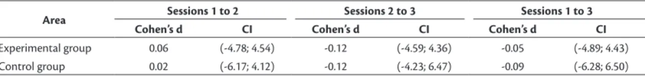
In our study, no signiicant change in the cross-sectional area of the popliteal vein between 25, 30, and 35 weeks of gestation was found in either the control or the experimental group. This finding is consistent with a previous study by Ropacka-Lesiak et al.,16
who also found that the transverse diameter of the popliteal vein remained unchanged between different phases of pregnancy. A different study, focusing on pregnant patients with preexisting unilateral deep venous obstruction, did observe changes in popliteal vein diameter, but those changes were found to be inconsistent.18
A clinically signiicant reduction in the peak systolic velocity of the popliteal vein was observed from week 25 to week 35 of pregnancy in the control group. This inding is consistent with a previous study by Ropacka-Lesiak et al.,1 who also observed
a gradual reduction of blood low in the popliteal vein with advancing phases of pregnancy. In contrast to the decreased PSV found in the control group,
signiicant increases in peak systolic velocity were observed in women in the experimental group who were provided with the special footwear, especially between the irst and second measurements. A similar effect on the venous blood low was observed in pregnant women wearing balanced inclined shoes in a previous study by Jang et al.10 In a study by
Sousa et al.,19 who was testing unstable shoes on
non-pregnant subjects, an increase in popliteal vein blood low velocity was found in participants wearing the footwear tested. The increase in venous return in participants wearing the unstable shoes was associated with the increased gastrocnemius muscle activity in their study. The venous hemodynamics of the lower limb may be improved by muscle pumps, comprising the foot, calf and thigh muscle pump. The calf muscle pump has been found to be the most eficient. Moreover, calf muscle pump strength and ankle mobility training have been reported to improve venous hemodynamics in patients with chronic venous insuficiency.19,20 A complementary mechanism to the
calf muscle pump is a plantar venous muscle pump that plays an important role in venous return by moving the blood in the foot venous reservoir upwards. The plantar muscle pump is activated with each step during walking, especially during the heel contact and stance phases of the gait cycle, when venous return is stimulated by a weight bearing compression of the plantar veins and muscular contraction around the veins.21,22 The footwear tested in our study is assumed
to improve the blood circulation by strengthening the lower limb foot and/or calf muscle pumps, since the design of the insole tested stimulates activation of the foot muscles. Nevertheless, there is a need for further investigation of the beneicial effect on venous
Table 4. Diferences in peak systolic velocity (cm/s) between particular data collection sessions with Cohen’s d values and their conidence intervals.
PSV Sessions 1 to 2 Sessions 2 to 3 Sessions 1 to 3
Cohen’s d CI Cohen’s d CI Cohen’s d CI
Experimental group -0.38 (-0.54; -0.19) 0.17 (-0.01; 0.34) -0.21 (-0.38; -0.05)
Control group 0.21 (-0.02; 0.42) 0.32 (0.11; 0.46) 0.55 (0.32; 0.69)
PSV: peak systolic velocity; CI: conidence interval.
Table 5. Diferences in cross-sectional area (cm2) between particular data collection sessions with Cohen’s d values and their conidence intervals.
Area Sessions 1 to 2 Sessions 2 to 3 Sessions 1 to 3
Cohen’s d CI Cohen’s d CI Cohen’s d CI
Experimental group 0.06 (-4.78; 4.54) -0.12 (-4.59; 4.36) -0.05 (-4.89; 4.43)
Control group 0.02 (-6.17; 4.12) -0.12 (-4.23; 6.47) -0.09 (-6.28; 6.50)
low of the footwear tested and its applications, since there are many factors affecting the pregnant body, demonstrated by a signiicant difference between PSV in the experimental and control groups at the irst data collection session, before the test footwear had been worn.
CONCLUSION
The indings of this study have conirmed the
occurrence of changes in venous flow during uncomplicated pregnancies. With advancing phases of pregnancy, the peak systolic velocity in the popliteal vein decreased signiicantly in the control group, potentially increasing the risk of venous disease development. However, it was demonstrated in the experimental group that wearing the J Hanák R, Ltd. footwear that was tested may prevent venous blood velocity reduction during advanced phases of pregnancy. Notwithstanding, there is a need for further investigation of the possible application of the footwear tested and its beneicial effect on venous low in both pregnant and non-pregnant patients with venous disease
REFERENCES
1. Ropacka-Lesiak M, Kasperczak J, Bręborowicz G. Pregnancy-dependent blood flow velocity changes in lower extremities veins in venous insufficiency. Ginekol Pol. 2015;86(9):659-65. PMid:26665566. http://dx.doi.org/10.17772/gp/59224. 2. Calderwood CL, Jamieson R, Greer IA. Gestational related changes
in the deep venous system of the lower limb on light reflection rheography in pregnancy and the puerperium. Clin Radiol. 2007;62(12):1174-9. PMid:17981165. http://dx.doi.org/10.1016/j. crad.2007.06.003.
3. Rabhi Y, Charras-Arthapignet C, Gris J-C, et al. Lower limb vein enlargement and spontaneous blood flow echogenicity are normal sonographic findings during pregnancy. J Clin Ultrasound. 2000;28(8):407-413. PMid:10993968. http://dx.doi. org/10.1002/1097-0096(200010)28:8<407::AID-JCU5>3.0.CO;2-S. 4. Barros N Jr, Janeiro Perez MC, de Amorim JE, Miranda F Jr. Pregnancy and lower limb varicose veins: prevalence and risk factors. J Vasc Bras. 2010;9(2):29-35.
5. Volejníková H. Cvičení v práci porodní asistentky Exercise in the midwife practice. Brno: Národní Centrum Ošetřovatelství a Nelékařských Zdravotnických Oborů; 2005.
6. Pinto K, Kramer R. Těhotná a fit fit and healthy pregnancy. Praha: Mladá Fronta; 2015.
7. Maršál K. Vznik a vývoj těhotenství Formation and development of pregnancy. In Hájek Z, Čech E, Maršál K. Porodnictví. 3rd ed. Praha: Grada Publishing; 2014.
8. Simočková V. Gynekologicko-pôrodnícke ošetrovateľstvo Gynecology and Obstetrics nursing. Martin: Vydaveteľstvo Osveta; 2011.
9. Chiou W, Chiu H, Chao A, Wang M, Chen Y. The influence of body mass on the foot dimensions during pregnancy. Appl Ergon. 2015;46(Pt A):212-7. PMid:25168196. http://dx.doi.org/10.1016/j. apergo.2014.08.004.
10. Jang SI, Lee YR, Kwak HS, Moon KS, Shin J, Kim J. The effect of balanced incline shoes on walking and feet for the pregnant women. Korean J Obstet Gynecol. 2010;53(11):988-97. http:// dx.doi.org/10.5468/kjog.2010.53.11.988.
11. Botyhanak [cited 2017 mar 23]. http://www.botyhanak.cz/ 12. Gyselaers W, Mesens T, Tomsin K, Peeters L. Doppler assessment
of maternal central venous hemodynamics in uncomplicated pregnancy: a comprehensive review. Facts Views Vis ObGyn. 2009;1(3):171-81. PMid:25489462.
13. Zvonar M, Kolarova K. Case study: verifying the effect of specific orthopaedic insoles and biomechanical shoes on plantar pressure. In Milanović D, Sporiš G. Proceedings of the 7th International Scientific Conference on Kinesiology; 2014; Opatija, Croatia. Opatija: University of Zagreb, Faculty of Kinesiology; 2014. p. 221-226.
14. Cohen J. Statistical power analysis for behavioral sciences. New York: Academic Press; 1977.
15. Wolf FM. Meta-analysis: quantitative methods for research synthesis. Beverly Hills: Sage; 1986.
16. Ropacka-Lesiak M, Kasperczak J, Breborowicz GH. Pregnancy-dependent changes in the vein width of the lower extremities in venous insufficiency. Ginekol Pol. 2012;83(12):922-8. PMid:23488295. 17. Krajcar J, Radakovic B, Stefanic L. Pathophysiology of venous
insufficiency during pregnancy. Acta Med Croatica. 1998;52(1):65-9. PMid:9599818.
18. Cordts PR, Gawley TS. Anatomic and physiologic changes in lower extremity venous hemodynamics associated with pregnancy. J Vasc Surg. 1996;24(5):763-7. PMid:8918321. http://dx.doi. org/10.1016/S0741-5214(96)70010-1.
19. Sousa A, Tavares JM, Macedo R, Rodrigues AM, Santos R. Influence of wearing an unstable shoe on thigh and leg muscle activity and venous response in upright standing. Appl Ergon. 2012;43(5):933-9. PMid:22277098. http://dx.doi.org/10.1016/j. apergo.2012.01.001.
20. Padberg FT Jr, Johnston MV, Sisto SA. Structured exercise improves calf muscle pump function in chronic venous insufficiency: a randomized trial. J Vasc Surg. 2004;39(1):79-87. PMid:14718821. http://dx.doi.org/10.1016/j.jvs.2003.09.036.
21. Corley GJ, Broderick BJ, Nestor SM, et al. The anatomy and physiology of the venous foot pump. Anat Rec. 2010;293(3):370-8. PMid:19957343. http://dx.doi.org/10.1002/ar.21085. 22. Uhl JF, Gillot C. The plantar venous pump: anatomy and
*
Correspondence
Marta Gimunová Masaryk University – FSpS MU, Faculty of Sports Studies, Department of Kinesiology 625 00 - Kamenice 5 Brno, Czech Republic Tel.: +420 549 49 6211 E-mail: [email protected]
Author information
MG - MSc degree in Physical Anthropology from Faculty of Science, Masaryk University. MZ - Professor at the Department of Kinesiology, Faculty of Sports Studies, Masaryk University. KK - PhD degree from Kinanthropology, Faculty of Sports Studies, Masaryk University. ZJ - Professor at the Department of Kinesiology, Faculty of Sports Studies, Masaryk University. OM and RM - MSc degree in sports Science, Faculty of Sports Studies, Masaryk University. PV - Professor at the Department of Gynecology and Obstetrics, Faculty of Medicine, Masaryk University and Faculty Hospital Brno (LF MU and FN Brno). PŠ - Professor at the Department of Physical Education, Health and Recreation, Prince Sultan University (PSU).
Author contributions
Conception and design: MZ Analysis and interpretation: MG, RM Data collection: KK Writing the article: MG, KK Critical revision of the article: ZJ, PV, PŠ Final approval of the article*: MG, MZ, KK, ZJ, OM, RM, PV, PŠ Statistical analysis: MG, OM Overall responsibility: MZ