Pulmonary embolism and stroke associated with
mechanical thrombectomy
Embolia pulmonar e AVC isquêmico associado à trombectomia mecânica
Paulo Bastianetto1, Daniel Mendes Pinto1
Abstract
Mechanical thrombectomy ofers the advantage of rapid removal of venous thrombi. It allows venous obstructions to be removed and requires shorter duration of infusion of thrombolytic agents. However, aspiration of thrombi can lead to complications, particularly pulmonary embolism and hemolysis. he validity of using vena cava ilters during thrombectomy in order to avoid embolism has not yet been established. he authors report a case of massive pulmonary embolism associated with ischemic stroke in a patient with a hitherto undiagnosed patent foramen ovale. he patient developed respiratory failure and neurological deicit after thrombectomy. his case raise questions about the value of the thrombectomy for the treatment of proximal vein thrombosis due to the risks of this procedure. he authors also discuss the need for vena cava ilters and ruling out a patent foramen ovale in patients undergoing thrombectomy. Keywords: thrombectomy; mechanical thrombolysis; pulmonary embolism; stroke.
Resumo
A trombectomia mecânica tem a vantagem de levar à rápida remoção dos trombos. Permite a desobstrução venosa e a redução do tempo de infusão de trombolíticos. A aspiração dos trombos pode levar a complicações, principalmente embolia pulmonar e hemólise. Algo que não está deinido é a validade do uso de iltros de veia cava durante a trombectomia, com o objetivo de evitar a TEP maciça. Os autores relatam um caso de embolia pulmonar maciça associada a AVC isquêmico, em uma paciente que apresentava forame oval patente. Não havia o diagnóstico prévio da PFO. A paciente evoluiu com insuiciência respiratória e déicit neurológico logo após a trombectomia. Neste caso, questiona-se o valor da trombectomia para o tratamento da trombose venosa proximal, devido aos riscos deste procedimento. Os autores discutem sobre a necessidade de iltro de veia cava e sobre a pesquisa de forame oval nos pacientes que serão submetidos à trombectomia.
Palavras-chave: trombectomia; trombólise mecânica; embolia pulmonar; acidente vascular cerebral.
¹ Hospital Mater Dei, Belo Horizonte, MG, Brazil. Financial support: None.
Conlicts of interest: No conlicts of interest declared concerning the publication of this article. Submitted: 03.29.13. Accepted: 07.29.13.
• the thrombosed segment was recanalized with a hydrophilic guide wire;
• an initial bolus of 20 mg alteplase (r-tPA) was ad-ministered via the thrombectomy catheter, using the pulse-spray technique;
• after waiting 15 min, the process of thrombi aspira-tion was started, using an Angiojet device and an
Xpeedior 6F catheter. A volume of 460 mL was
aspirated during a total time of use of 420 seconds, in the cranial-distal direction.
In the middle of the procedure, the patient exhibited intense dyspnea, hypoxemia, bradycardia and central and peripheral cyanosis. She was placed in decubitus dorsal and intubated. After transportation to the intensive care unit, a transthoracic echocardiogram showed considerable enlargement combined with hypocontractility of the right ventricle, and an estimated pulmonary artery pressure of 46 mmHg (normal range: up to 25 mmHg). On the basis of suspected massive pulmonary embolism, treatment was started with systemic r-tPA at a total dose of 100 mg infused over 2 hours.
Chest angiotomography conirmed pulmonary
embolism with involvement of pulmonary arteries and of the inferior lobar branches bilaterally (Figure 1). The patient’s brain natriuretic peptide (BNP, reached 5,296 pg/mL (normal range up to 154 pg/mL), which is associated with heart failure, poor prognosis and increased mortality in pulmonary embolism cases.
After treatment with systemic thrombolytics, on the following day the patient was extubated, respiratory parameters had improved and pulmonary artery pressure had fallen. The patient was kept anticoagulated with venous UFH .
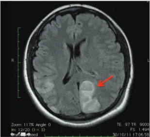
On the third day after thrombectomy, the patient exhibited right-side hemiparesis and loss of vision, somnolence and aphasia. Magnetic resonance
identiied multiple areas of ischemia bilaterally,
following the territory of the middle cerebral artery
INTRODUCTION
The objective of early removal of thrombi after an acute deep venous thrombosis (DVT) event is to reduce the incidence of postthrombotic syndrome. The syndrome occurs in 20 to 59% of cases and is characterized by disorders caused by chronic venous hypertension, such as stasis dermatitis, dermatosclerosis and venous ulcers.1,2 Removal of the
acute thrombi, thrombectomy, can be accomplished using conventional surgery or with percutaneous methods using catheters with a range of different mechanisms of action.
Several cases series and retrospective analyses
have shown the beneits of percutaneous mechanical
thrombectomy for treatment of proximal DVT.1-3
However, certain risks are associated with use of this technology.
The objective of this report is to describe serious complications that occurred after a case of percutaneous thrombectomy: ischemic cerebral vascular accident (stroke) and pulmonary embolism. In addition to the description, we discuss the technique, the risks involved and results reported in the literature.
CASE DESCRIPTION
A female, 29-year-old patient was admitted with considerable pain and voluminous swelling of the left lower limb (LLL). The patient was taking oral contraceptives, was a non-smoker and had a family positive family history of DVT.
Doppler ultrasonography showed acute venous thrombosis in the superficial femoral, common femoral and external iliac veins of the LLL., without signs of vena cava involvement or of pulmonary embolism.
Treatment was initiated with continuous unfractionated heparin (UFH). After 2 days of treatment the symptoms remained and Doppler ultrasonography showed that the thrombotic process had progressed to the popliteal and great saphenous veins. These signs, together with the worsening edema, continual pain, increased muscle tension and cyanosis of the LLL, indicated a case of Phlegmasia Cerulea Dolens. Treatment with percutaneous thrombectomy and infusion of a thrombolytic agent was proposed, and accepted by the patient.
Percutaneous thrombectomy was performed 5 days after onset of symptoms, using the following technique:
• with the patient in decubitus ventral, an ultrasound-guided puncture was made in the left popliteal vein
and left posterior cerebral artery, and bilateral cerebellar infarcts (Figure 2). Treatment with UFH
was withdrawn and a ilter itted in the inferior vena
cava.
Transesophageal echocardiogram showed a patent
foramen ovale with low diverted from the right to the left atrium, conirming the hypothesis of ischemic
stroke due to paradoxical embolism. Five days after the stroke diagnosis, the patient was discharged from intensive care, showing considerable improvement in her neurological symptoms.
After the venous thrombectomy, despite the serious complications, the swelling, pain and cyanosis of the LLL receded. Twenty-eight days after admission, the patient was discharged with a prescription for warfarin and elastic compression stockings and instructed not to take estrogens, with follow-up by the Neurology, Vascular Surgery and
Physiotherapy services. A genetic study identiied
thrombophilia, with a heterozygous mutation of the genes for prothrombin and methylenetetrahydrofolate reductase A1298c.
Six months later the patient had returned to her usual employment and the only remaining neurological deficit was bilateral, regressive, hemianopsia.
Control venous Doppler ultrasonography showed
absence of deep or supericial venous relux, with the
femoropopliteal DVT in recanalization. Clinically, there was no sign of edema of the affected limb.
DISCUSSION
The proportion of iliofemoral DVT that progress to postthrombotic syndrome varies from 20 to 59%, when treated with anticoagulants alone, and from 5 to 31%, when treated using intrathrombal thrombolytics.2,3 When thrombolytics are used,
complete thrombi removal occurs in up to 45% of cases, compared to just 4% of cases when treatment is with anticoagulation alone.3,4
When compared with thrombolysis alone, mechanical thrombectomy devices offer more rapid and complete thrombus removal. Combining thrombectomy with thrombolytics offers certain advantages: shorter stays in intensive care unit and hospital; lower number of control venographies, and lower doses of the thrombolytic agent. Thrombectomy combined with thrombolytics can therefore lead to fewer hemorrhagic complications and reduced morbidity (Table 1).5,6
Several different thrombectomy devices are available on the market, with a range of different mechanisms of action: simple aspiration of thrombi, hydrodynamic (aspiration by negative pressure of saline jets), fragmentation and combination systems.7
The device used in this case, the Angiojet (Possis Medical Inc., Mineapolis, MN), has an action based on the Venturi effect. The catheter employed, an Expeedior, has two lumens: one for infusion of
saline and the other for aspiration of the luid and the
clot. A high-pressure jet of saline solution is directed from the tip of the catheter backwards, thereby creating a vacuum effect, which forces the clot to enter the catheter after maceration. The catheter does not touch the vessel wall and the high-pressure jet does not denude the endothelium, thereby preserving its antithrombotic effect.
The main complications related to mechanical thrombectomy are hemolysis and pulmonary embolization. Hemolysis is the result of fragmentation of red blood cells caused by contact with saline jets.
Table 1. Indications for percutaneous thrombectomy.6
Indication Grade of recommendation and
level of evidence First episode of acute iliofemoral DVT, up to 14 days since onset of symptoms, low risk of bleeding,
good functional capacity and an acceptable life expectancy. 2C Patients with iliofemoral DVT, associated or not with femoropopliteal thrombosis, threatening limb
viability (phlegmasia cerulea dolens) 1A
in addition to deining indications for use of a vena cava ilter for protection .
REFERENCES
1. Nazir SA, Ganeshan A, Nazir S, Uberoi R. Endovascular treatment options in the management of lower limb deep venous thrombosis. Cardiovasc Intervent Radiol. 2009;32(5):861-76. PMid:19641957. http://dx.doi.org/10.1007/s00270-009-9662-z 2. Gloviczki P, Comerota AJ, Dalsing MC, et al. he care of patiens
with varicoses veins and associated chronic venous diseases: clinical practice guidelines of the Society for Vascular Surgery and the American Venous Forum. J Vasc Surg. 2011;53(5):2S-48S. PMid:21536172. http://dx.doi.org/10.1016/j.jvs.2011.01.079 3. Comerota AJ, Gravett MH. Iliofemoral venous thrombosis. J
Vasc Surg. 2007;46(5):1065-76. PMid:17980295. http://dx.doi. org/10.1016/j.jvs.2007.06.021
4. Malgor RD, Gasparis AP. Pharmaco-mechanical thrombectomy for early thrombus removal. Phlebology. 2012;27(Suppl 1):155-62. PMid:22312084. http://dx.doi.org/10.1258/phleb.2012.012S14 5. Bush RL, Lin PH, Bates JT, Mureebe L, Zhou W, Lumsden
AB. Pharmacomechanical thrombectomy for treatment of symptomatic lower extremity deep venous thrombosis: safety and feasibility study. J Vasc Surg. 2004;40(5):965-70. PMid:15557912. http://dx.doi.org/10.1016/j.jvs.2004.08.025
6. Meissner MH, Gloviczki P, Comerota AJ, et al. Early thrombus removal strategies for acute deep venous thrombosis: Clinical Practice Guidelines of the Society for Vascular Surgery and the American Venous Forum. J Vasc Surg. 2012;55(5):1449-62. PMid:22469503. http://dx.doi.org/10.1016/j.jvs.2011.12.081
7. Kim HS, Patra A, Paxton BE, Khan J, Streiff MB. Adjunctive percutaneous mechanical thrombectomy for lower-extremity deep vein thrombosis: clinical and economic outcomes. J Vasc Interv Radiol. 2006;17(7):1099-104. PMid:16868161. http://dx.doi. org/10.1097/01.RVI.0000228334.47073.C4
8. Biuckians A, Meier GH. Treatment of symptomatic lower extremity acute deep venous thrombosis: role of mechanical thrombectomy. Vascular. 2007;15(5):297-303. http://dx.doi. org/10.2310/6670.2007.00070
9. Cynamon J, Stein EG, Dym RJ, Jagust MB, Binkert CA, Baum RA. A new method for aggressive management of deep vein thrombosis: retrospective study of the power pulse technique. J Vasc Interv Radiol 2006;17(6):1043-9. PMid:16778240. http:// dx.doi.org/10.1097/01.RVI.0000221085.25333.40
10. Kasirajan K, Gray B, Ouriel K. Percutaneous AngioJet thrombectomy in the management of extensive deep venous thrombosis. J Vasc Interv Radiol. 2001;12(2):179-85. http://dx.doi. org/10.1016/S1051-0443(07)61823-5
11. Lin PH, Zhou W, Dardik A, et al. Catheter-direct thrombolysis versus pharmacomechanical thrombectomy for treatment of symptomatic lower extremity deep venous thrombosis. Am J Surg. 2006;192(6):782-8. PMid:17161094. http://dx.doi. org/10.1016/j.amjsurg.2006.08.045
12. Vedantham S, horpe PE, Cardella JF, et al. Quality improvement guidelines for the treatment of lower ext.remity deep vein thrombosis with use of endovascular thrombus removal. J Vasc Interv Radiol. 2006;17(3):435-47. PMid:16567668. http://dx.doi. org/10.1097/01.RVI.0000197348.57762.15
13. Augustinos P, Ouriel K. Invasive approaches to treatment of venous thromboembolism. Circulation. 2004;110(Suppl I):I27-34. PMid:15339878. http://dx.doi.org/10.1161/01. CIR.0000140900.64198.f4
Almost all patients exhibit hemoglobinuria.8,9 Venous
hydration is essential, with the objective of avoiding kidney damage. Our patient did not suffer from
signiicant hemolysis.
Several different studies have found evidence of asymptomatic pulmonary embolism at rates of 5.3% to 17%, in addition to a few symptomatic cases.10-13 The vena cava ilter prevents embolization
by larger thrombi, but does not restrict the passage of microthrombi into pulmonary circulation. As a
result, the beneit of using a cava ilter in patients
undergoing aspiration thrombectomy is not well-established in the literature. Certain groups may
beneit from protection with a ilter, such as patients
with little cardiopulmonary reserves or where the thrombus extends to the vena cava.14
Patients with a persistent foramen ovale are at greater than normal risk from venous thrombosis because of the possibility of paradoxical embolism. The foramen ovale can remain patent in 25% of the population. Diagnosis is made by transesophageal echocardiogram. In many cases the foramen initially closes, only to open in situations of right ventricle overload.15 In the case described here, the extensive
cerebral vascular accident was associated with the patent foramen ovale. The increased pressure in the pulmonary artery could have diverted the clots to the left atrium with consequent cerebral embolization. Such a mechanism could explain the fact that cerebral ischemia only had onset on the third day after the pulmonary embolism.
After our experience with this case, and in view of the high prevalence of persistent foramen oval in the population, we now use transesophageal echocardiograms to examine all patients who are to undergo thrombectomy.
14. Kölbel T, Alhadad A, Acosta S, Lindh M, Ivancev K, Gottsäter A. Thrombus embolization into IVC filters during catheter-directed thrombolysis for proximal deep venous thrombosis. J Endovasc her. 2008;15(5):605-13. PMid:18840047. http://dx.doi. org/10.1583/08-2462.1
15. Chamié F, Chamié D, Ramos S, et al. Fechamento Percutâneo do Forame Oval Patente. Rev Bras Cardiol Invas. 2005;13(3):185-19.
Correspondence
Paulo Bastianetto Rua Uberaba, 436, sala 502 - Barro Preto CEP 30180-080 - Belo Horizonte (MG), Brazil Tel.: +55 (31) 32952030 E-mail: [email protected]
Author information
PB é Angiologista e Cirurgião Vascular / Endovascular, Membro Efetivo da SBACV, Hospital Mater Dei e Angiocare. DMP é Angiologista e Cirurgião Vascular, Membro Titular da SBACV, Hospital Mater Dei e Instituto Vascular BH.
Author contributions
Conception and design: PB, DMP Analysis and interpretation: PB, DMP Data collection: PB Writing the article: PB, DMP Critical revision of the article: PB, DMP Final approval of the article*: PB, DMP Statistical analysis: N/A