Two-stage hybrid open-endovascular repair of a Crawford
type IV aortic aneurysm: therapeutic challenge
Tratamento híbrido (debranching) de aneurisma tóraco-abdominal
tipo IV de Crawford: desafio terapêutico
Abdo Farret Neto1,2, Liana Berucia Freire de Oliveira2, Guilherme Tarso de Andrade Alves2,
George Anderson da Penha Andrade2, Eduardo Dantas Baptista de Faria1,2
Abstract
We present a case of a patient with Crawford type IV aortic thoracoabdominal aneurysm. he patient underwent hybrid repair in two stages. Initially a Dacron graft was implanted surgically with revascularization of all visceral branches from the left external iliac artery (debranching). On a later date, the second stage of treatment was with an endovascular technique with bi aorto-iliac endoprosthesis. After 2 years the patient remains asymptomatic and in full working activity.
Keywords: aortic aneurysm; surgery; aorta; aneurysm.
Resumo
Apresentamos o caso de um paciente com aneurisma aórtico tóraco-abdominal tipo IV de Crawford submetido à correção híbrida em dois estágios. Inicialmente submetido a implante cirúrgico de prótese de Dacron com revascularização de todos os ramos viscerais a partir da ilíaca externa esquerda (debranching) e, posteriormente, tratado pela técnica endovascular com endoprótese aorto bi-ilíaca. Após dois anos, o paciente permanece assintomático e em plena atividade laborativa.
Palavras-chave: aneurisma aórtico; cirurgia; aorta; aneurisma.
1 Hospital Universitário Onofre Lopes – HUOL, Natal, RN, Brazil.
2 Faculdade de Medicina, Universidade Federal do Rio Grande do Norte – UFRN, Natal, RN, Brazil.
Financial support: None.
Conlicts of interest: No conlicts of interest declared concerning the publication of this article. Submitted: 01.10.14. Accepted: 05.13.14
he study was carried out at Hospital Universitário Onofre Lopes, Natal-RN, Brazil.
INTRODUCTION
There is still no consensus on the best form of surgery for Crawford type IV aortic aneurysms (AA), whether open surgery, endovascular treatment or a hybrid technique.1-5 In 1999,
Quiñones-Baldrich et al.6 described the irst case of treatment
of type IV AA in a combination approach using both conventional surgery and endovascular surgery, during the same operation. A few years later, Chiesa et al.7 described a two-stage hybrid approach
to treating the same type of AA.
We describe the case of a patient with Crawford type IV AA treated using a hybrid technique in two separate operations. In the irst operation, a Dacron graft was placed in the left external iliac and revascularization of the visceral branches accomplished. In the second operation, endovascular repair of the aneurysm was performed, followed by repair of stenosis at the anastomosis of the right renal artery.
PART I – CLINICAL PRESENTATION
The patient was a 49-year-old male ex-smoker with diabetes and high blood pressure. He reported a history of severe abdominal pains 3 years previously. On that occasion he had had an abdominal tomography without contrast, which had shown an abdominal aorta aneurysm (AAA) of unknown size. An angiotomography (ACT) conducted in October of 2011 showed that the patient had a thoracoabdominal aneurysm (TAA) measuring 8.47 cm at its largest diameter, involving the celiac trunk and abdominal visceral branches and extending to the aortic bifurcation (Crawford IV) (Figures 1 and 2).
PART II – WHAT WAS DONE
The patient underwent open surgery for visceral revascularization on 12 December, 2011, and a 14 × 7 mm bifurcated Dacron Hemashield graft was implanted (InterVascular, La Ciotat Cedex, France). Access was via a median xiphopubic incision, with medial retraction of the retroperitoneal space from the left paracolic gutter (Mattox maneuver).
The main body of the graft was anastomosed end-to-side (ETS) to the left external iliac. Its left branch was anastomosed ETS to the left renal and end-to-end (ETE) to the celiac trunk. The right branch of the bifurcated graft was anastomosed ETE to the right renal artery. A straight segment of 7 mm Dacron graft was anastomosed ETS to the right branch of the prosthesis and ETE to the superior mesenteric (Figure 3). The inferior mesenteric had a small
Figure 1. Angio CT in 3D reconstruction, showing the TAA.
caliber and was not revascularized, but was ligated at its origin.
The patient remained stable during surgery, with adequate diuresis and with no need for transoperative blood transfusion. However, during the irst 24 hours after surgery, the patient developed signs of hypovolemic shock, with reduced diuresis, hypotension and abdominal distension. An ACT showed a large quantity of liquid free in the cavity and good low through the grafts (Figure 4). The patient was transfused and underwent abdominal laparotomy, which found around 2,000 mL of blood in the cavity. Visceral perfusion was adequate and all branches implanted were patent. No sites of active bleeding were identiied during reoperation.
The patient continued to exhibit hemodynamic stability, plus acute renal failure due to hypovolemic shock. Renal failure was treated with hemodialysis sessions for 7 days. On the ninth day, after return of adequate diuresis and improvement in renal function with normalization of creatinine levels, the patient underwent surgery to place an endoprosthesis for aneurysm repair.
Transoperative angiography showed stenosis at the anastomosis of the right renal artery, which was treated by placement of a Viabahn 9 × 50 mm endoprosthesis (W.L. Gore & Associates, Arizona, USA) followed by angioplasty with a Passeo-35 6 × 40 mm balloon (Biotronik Ag, Bulach, Switzerland) (Figures 5 and 6).
After repair of the renal stenosis, a 31 mm × 26 mm × 10 cm conical TAG thoracic endoprosthesis was implanted starting at the diaphragmatic segment of the abdominal aorta. Fitted to this, an Excluder C3 endoprosthesis with a main body measuring
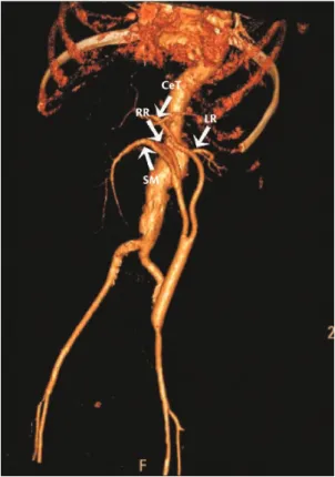
Figure 4. Angio CT in immediate postoperative period showing visceral branches celiac trunk (CeT), right renal (RR), left renal (LR) and superior mesenteric (SM).
28.5 mm × 14.5 mm × 16 cm and an extension measuring 14.5 mm × 10 cm (W.L. Gore & Associates, Arizona, USA) was implanted. Segments
were itted to each other using a Tri-Lobe balloon
(W.L. Gore & Associates, Arizona, USA). The procedure was conducted with no intercurrent
Figure 3. Transoperative image showing revascularization of visceral branches, such as celiac trunk (CeT), right renal (RR), left renal (LR) and superior mesenteric (SM).
grafts remained patent, with no evidence of stenosis or kinking in the branches (Figure 8).
At a recent outpatients follow-up consultation, in December 2013, the patient had returned to full employment, was asymptomatic, except for a complaint of retrograde ejaculation. Renal function was normal and he was still on antihypertensive, antidiabetic and antiplatelet drugs (losartan, amlodipine, metformin and aspirin).
DISCUSSION
In AA cases with involvement of visceral branches, the possibility of employing endovascular techniques such as multilayer stents,8 chimney
stents,9 fenestrated stents10 and, particularly, branched
endoprostheses, should be considered, although they present therapeutic challenges, with a chance of visceral ischemic complications, endotension and type I endoleaks,11 and their long-term durability is
unproven.12
In this case we chose a combination of conventional surgery with debranching followed by placement of the endoprosthesis, necessary because of complete compromise of the visceral branches. The relatively young age of the patient and his good physical condition were also decisive factors in choosing this technique. These factors suggest good prospects for recovery after conventional by-pass surgery, in addition to a long life expectancy after repair of an AA. We therefore chose the approaches described, because we believed that the visceral bypasses conditions and the inal angiography did not detect
any endoleaks (Figure 7).
The patient recovered well, with progressive improvement in renal function and was discharged, asymptomatic, on 30 December, 2011, on his 18th day in hospital.
A control ACT performed on 6 September, 2013, showed that the aneurysm sac was excluded and the
Figure 6. Final appearance after implantation of Viabahn followed by angioplasty.
Figure 7. Control angiography after implantation of endoprostheses, showing good visceral low and absence of endoleaks.
6. Quiñones-Baldrich WJ, Panetta TF, Vescera CL, Kashyap VS. Repair of type IV thoracoabdominal aneurysm with a combined endovascular and surgical approach. J Vasc Surg. 1999;30(3):555-60. http://dx.doi.org/10.1016/S0741-5214(99)70084-4
7. Chiesa R, Melissano G, Civilini E, Setacci F, Tshomba Y, Anzuini A. Two-stage combined endovascular and surgical approach for recurrent thoracoabdominal aortic aneurysm. J Endovasc her. 2004;11(3):330-3. PMid:15174918. http://dx.doi. org/10.1583/03-1145.1
8. Pieper CC, Meyer C, Verrel F, Schild HH, Wilhelm KE. Using the multilayer stent as a supplement to EVAR in combined abdominal aortic aneurysm and iliac artery aneurysm with inadequate distal landing zone: a case report. Vasc and Endovasc Surg. 2012;46(7):565-9. http://dx.doi. org/10.1177/1538574412456306
9. Ohrlander T, Sonesson B, Ivancev K, Resch T, Dias N, Malina M. he chimney graft: A technique for preserving or rescuing aortic branch vessels in stent-graft sealing zones. J Endovasc Ther. 2008;15(4):427-32. PMid:18729550. http://dx.doi. org/10.1583/07-2315.1
10. Verhoeven EL, Vourliotakis G, Tielliu IF, et al. Fenestrated Stent grafting for short-necked and justarenal abdominal aortic aneurysm: an 8-year single-centre experience. Eur J Vasc Endovasc Surg. 2010;39(5):529-36. PMid:20202868. http://dx.doi. org/10.1016/j.ejvs.2010.01.004.
11. Amato ACM, Abraham FA, Kraide HD, Rocha LT, Santos RV. Endotensão: ruptura de aneurisma de aorta abdominal. J Vasc Bras. 2012;11(2):162-65. http://dx.doi.org/10.1590/ S1677-54492012000200016
12. Criado FJ, Duson S. he history of snorkel, chimney, periscope and sandwich grafting compared with fenestrated and branched endografting. In: Ferris M, editor. Vascular and endovascular challlenges update. 1th ed. London: BIBA; 2013. p. 449. 13. Huynh TT, Miller CC 3rd, Estrera AL, Sheinbaum R, Allen SJ, Sai HJ.
Determinants of hospital length of stay after thoracoabdominal aortic aneurysm repair. J Vasc Surg. 2002;35(4):648-53. PMid:11932657. http://dx.doi.org/10.1067/mva.2002.121566 14. Q u i n o n e s - B a l d r i c h W J . D e s c e n d i n g t h o r a c i c a n d
thoracoabdominal aortic aneurysm repair: 15 year results using a uniform approach. Ann Vasc Surg. 2004;18(3):335-42. PMid:15354636. http://dx.doi.org/10.1007/s10016-004-0033-6 15. Roselli EE, Greenberg RK, Pfaf K, Francis C, Svensson LG, Lytle BW.
Endovascular treatment of thoracoabdominal aortic aneurysms. J horac Cardiovasc Surg. 2007;133(6):1474-82. PMid:17532942. http://dx.doi.org/10.1016/j.jtcvs.2006.09.118
16. Greenberg RK, Lu Q, Roselli EE, et al. Contemporary analysis of descending thoracic and thoracoabdominal aneurysm repair: a comparison of endovascular and open techniques. Circulation. 2008;118:808-17. PMid:18678769. http://dx.doi. org/10.1161/CIRCULATIONAHA.108.769695
17. Verhoeven E, Tielliu IF, Zeebregts CJ, et al. Bericht über die ersten 50 endovaskulären thorakoabdominellen Behandlungen mit Seitenastprothesen. Zentrabl Chir. 2011;136(5):451-57. PMid:21766273. http://dx.doi.org/10.1055/s-0031-1271550 18. Guillou M, Bianchini A, Sobocinski J, et al. Endovascular
treatment of thoracoabdominal aortic aneurysms. J Vasc Surg . 2012;56(1):65-73. PMid:22560310. http://dx.doi. org/10.1016/j.jvs.2012.01.008
would offer greater durability if constructed using conventional surgery. We also chose to avoid conventional endoaneurysmorrhaphy surgery because of the longer duration of visceral ischemia, and greater blood loss and mortality.
Mortality for conventional surgery was around 60% before 1990, and current estimates are 4-21%.13,14 When hybrid repair techniques, branched
and fenestrated prostheses are used, mortality rates of 5.5 to 8.9% are reported,15-18 although the fact
that long-term follow-up is lacking should be borne in mind.
The choice of retroperitoneal access via the left paracolic gutter allowed us to place the main body of the Dacron graft in the left external iliac artery. The choice of the external iliac also facilitated implantation of the endoprosthesis, which was anchored distally at the common iliac artery.
The stenosis at the anastomosis of the right renal artery was treated using a Viabahn graft to avoid the bleeding that would occur if the recent anastomosis were to be dilated. The angioplasty balloon employed was smaller and shorter than the Viabahn because we did not wish to injure the unprotected renal artery.
Concluding, hybrid repair of a Crawford type IV aneurysm is an option that should be considered for patients with good surgical risk proiles and reasonable life expectancy.
REFERENCES
1. Patel R, Conrad MF, Paruchuri V, Kwolek CJ, Chung TK, Cambria RP. horacoabdominal aneurysm repair: Hybrid versus open repair. J Vasc Surg. 2009;50(1):15-22. PMid:19563950. http:// dx.doi.org/10.1016/j.jvs.2008.12.051
2. Wong DR , Parenti JL, Green SY, et al. Open repair of thoracoabdominal aortic aneurysm in the modern surgical era: contemporary outcomes in 509 patients. J Am Coll Surg. 2011;212(4):569-81. PMid:21463791. http://dx.doi. org/10.1016/j.jamcollsurg.2010.12.041
3. Hughes GC, Barield ME, Shah AA, et al. Staged total abdominal debranching and thoracic endovascular aortic repair for thoracoabdominal aneurysm. J Vasc Surg. 2012;56(3):621-9. PMid:22575483. http://dx.doi.org/10.1016/j.jvs.2011.11.149
4. Patel HJ, Upchurch GR, Eliason JL, et al. Hybrid debranching w i t h e n d o v a s c u l a r r e p a i r f o r t h o r a c o a b d o m i n a l aneurysms: a comparison with open repair. Ann Thorac Surg. 2010;89(5):1475-81. PMid:20417763. http://dx.doi. org/10.1016/j.athoracsur.2010.01.062
Correspondence
Abdo Farret Neto Clínica Endovasc, Hospital do Coração Rua Auris Coelho, 235, 2º andar – Lagoa Nova CEP 59075-050 – Natal (RN), Brazil Tel.: +55 (84) 41410941 Fax: +55 (84) 3234-0125 E-mail: [email protected]
Author information
AFN é Angiologista, Cirurgião Vascular e Angiorradiologista. Titular do Colégio Brasileiro de Cirurgiões (TCBC) e Titular da Sociedade Brasileira de Angiologia e Cirurgia Vascular (TSBACV). Angiorradiologista e Cirurgião Vascular do Hospital Onofre
Lopes. Professor de Doenças do Sistema Cardiovascular na Universidade Federal do Rio Grande do Norte (UFRN). Doutor em Desenvolvimento e Inovação Tecnológica em Medicamentos pela UFRN. LBFO é Angiologista, Cirurgiã Vascular e Angiorradiologista. Cirurgiã Vascular do Hospital Onofre Lopes. GTAA é Residente em Cirurgia Geral do Hospital Onofre Lopes. GAPA é Acadêmico de Medicina da Universidade Federal do Rio Grande do Norte (UFRN). EDBF é Angiologista e Cirurgião Vascular do Hospital Onofre Lopes. Professor de Doenças do Sistema Cardiovascular na Universidade Federal do Rio Grande do Norte (UFRN).
Author contributions
Conception and design: AFN Analysis and interpretation: AFN Data collection: GTAA, GAPA Writing the article: AFN Critical revision of the article: EDBF, LBFO Final approval of the article*: AFN, GTAA, GAP, EDBF, LBFO Statistical analysis: N/A Overall responsibility: AFN Obtained funding: None.