Brachial index does not relect upper extremity functionality
following surgery for vascular trauma
O índice tornozelo-braço não reflete a funcionalidade da extremidade
superior após cirurgia para o trauma vascular
Erdal Simsek1, Kasim Karapinar2, Ilker Ince3, Ahmet Tulga Ulus1
Abstract
Objectives: Vascular injuries to the upper extremities requiring surgical repair are common after accidents. However, neither postoperative functionality nor hemodynamic status of the extremity are routinely described. We evaluated the postoperative functional and hemodynamic status of patients with vascular traumas in the upper extremities.
Methods: 26 patients who sufered penetrating vascular traumas in the upper extremities from November 2008 to December 2011 were retrospectively evaluated. Data on irst approach, surgical technique employed and early postoperative outcomes were recorded. Further data on the post-discharge period, including clinical functional status of the arm, Doppler ultrasonography and brachial-brachial index were also evaluated. Results: Average follow up was 33.5±10.8 months. Right (1.05±0.09) and left (1.04±0.08) brachial indexes were measured during follow up,. Doppler ultrasonography showed arterial occlusion in 4 patients (15%). Near-normal brachial-brachial indexes was observed in all four of these patients with occlusion of one of the upper extremity arteries, even though they exhibited limited arm function for daily work. Conclusions: Evaluation of the postoperative outcomes of this small series of patients with penetrating vascular traumas in the upper extremity revealed that 15% of them sufered occlusion of one artery of the upper extremity. Artery occlusion did not correlate with brachial-brachial Doppler index, probably due to rich collateral circulation, but occlusion was associated with an extremity that was dysfunctional for the purposes of daily work. he result of the brachial-brachial index does not therefore correlate with functionality.
Keywords: arterial injury; trauma; upper extremity; brachial-brachial index; multidisciplinary approach.
Resumo
Objetivos: Os vasos das extremidades superiores são frequentemente lesados em razão de acidentes. Nós pesquisamos a funcionalidade dos membros superiores com base nos resultados cirúrgicos, além do acompanhamento de lesões de outros órgãos e tecidos. Métodos: Entre novembro de 2008 e dezembro de 2011, 26 pacientes foram encaminhados ao nosso serviço de emergência em razão de lesões vasculares traumáticas da extremidade superior, sendo avaliados retrospectivamente. O atendimento inicial aos pacientes, assim como os diagnósticos e as variedades de tratamento, foram avaliados. Após a alta hospitalar, os pacientes foram acompanhados pelo eco-Doppler e pelo índice tornozelo-braço. Resultados: O tempo médio de seguimento foi de 33,5±10,8 meses. Durante o seguimento, os índices braquiais direito (1,05±0,09) e esquerdo (1,04±0,08) foram medidos. Na ultrassonograia Doppler, foi observada obstrução arterial em quatro pacientes. Devido à circulação colateral, o índice tornozelo-braço foi próximo do normal nesses pacientes; entretanto, eles apresentavam limitações funcionais dos membros superiores acometidos, para o trabalho diário. Conclusões: Traumas penetrantes são frequentemente observados em adultos jovens. Esses pacientes devem ser rapidamente tratados para que retornem ao seu trabalho. O planejamento do tratamento deve ser multidisciplinar, para abranger também as lesões de outros órgãos e tecidos. Nosso estudo mostrou que não há correlação entre o índice tornozelo-braço e a funcionalidade da extremidade superior, após cirurgia para o trauma vascular.
Palavras-chave: lesão arterial; trauma; membro superior; índice tornozelo-braço; abordagem multidisciplinar.
1 Turkiye Yuksek Ihtisas Training and Research Hospital, Department of Cardiovascular Surgery, Ankara 2 Diskapi Yildirim Beyazit Training and Research Hospital, Department of Cardiovascular Surgery, Ankara 3 Tokat Gaziosmanpaşa University, Department of Cardiovascular Surgery, Tokat
Financial support: None.
Conlicts of interest: No conlicts of interest declared concerning the publication of this article. Submitted: 10.04.13. Accepted: 03.18.14.
Physical examination, Doppler ultrasonography (dUSG) and digital subtraction angiography (DSA) were available for all vascular trauma patients. However, patients with radial and ulnar arterial injuries were treated immediately after physical examination.
Patients with brachial artery, axillary artery and vein, jugular vein, basilar vein and subclavian vein injuries were diagnosed using dUSG.
Patients who had injured tissues other than vascular structures were operated by a multidisciplinary team. Local or general anesthesia were chosen depending on patients’ status and the areas damaged. All vascular injuries were managed by vascular surgeons. During surgical repair, end to end anastomoses were conducted using one of two different sewing techniques depending on vascular diameter (single sewing technique for thin vessels and continuous sewing technique for larger vessels). Saphenous vein,
polytetraluoroethylene (PTFE) or Dacron grafts
were used where end to end anastomosis was not possible. Simple vessel ligation was used in cases in which there was no chance of repairing radial and
ulnar arterial injuries. For venous injuries, saphenous vein or PTFE grafts were used if primary repair could
not be accomplished.
Upper extremity vascular trauma patients were evaluated in terms of injury features, surgical procedures performed and concomitant extravascular tissue injuries. Postoperatively, they were evaluated in terms of loss of capacity to work and functional status of the arm for daily work.
Length of hospital stay was recorded.
Follow-ups were conducted at intervals of 3 to 6 months
with physical examination, dUSG and the
brachial-brachial index (upper extremity systolic pressure ratio to the lower extremities). Depending on
follow-up indings, patients with disabilities affecting
physical activities were prescribed physical therapy and rehabilitation.
RESULTS
We evaluated 26 patients with upper limb vascular traumas, 80.8% of whom were male and 19.2% of whom were female.
Complementary diagnostic methods could not be used for patients presenting with hemodynamic instability. In one patient with an axillary arterial injury and a huge hematoma, physical examination was not sufficient to establish diagnosis and so DSA was used despite the hemodynamic instability. Another patient with injuries to the left subclavian artery and vein and the internal mammary artery
INTRODUCTION
Traumas often lead to upper extremity injuries because of people’s relex to protect themselves. The
brachial artery is one of the arteries most prone to injury from bone fragments, sharp and penetrating
objects and blunt traumas. The frequency of vascular
injury is higher in penetrating traumas compared to blunt injuries.1 Clavicle fractures are responsible for the 50% of subclavian arterial injuries.2 The incidence of vascular system damage is 48% in blunt thorax traumas.3 Vascular injuries can result from upper extremity traumas and in 28% of these the brachial artery is affected.4
Upper extremity vascular injuries are common
in emergency rooms. The majority of them are immediately treated by vascular surgeons. The
frequency of venous injuries is lower than arterial injuries. Nerve tissues runs parallel to the vascular system and as a result nerves are also at a high risk of injury.
Early transport of patients, early diagnosis and
early surgical treatment have together contributed to increased postoperative success rates in recent years. However, it is important to determine, whether these injuries impact the capacity to work and cause motor
deicit of the arm, in order to prescribe specialized
treatment to avert major dysfunctionality. It is
therefore important to follow-up these patients after surgery. Trauma patients are usually young active
individuals and should be able to go back to work as soon as possible with effective treatment.
METHODS
Twenty-six patients who suffered upper extremity
vascular traumas from November 2008 to December
2011 were evaluated retrospectively. This study was
approved by internal review board at the hospital (No: 297; 07.01.2013).
Patients presenting at our center were operated according to certain principles. In accordance with
the Advanced Trauma Life Support Guidelines,5 all patients with emergency upper extremity vascular traumas were evaluated with reference to presence of at least two of the following symptoms, accepted as
a strong signs of arterial damage: inability to ind a
pulse or weak distal pulse, arterial bleeding, pulsatile hematoma, growing hematoma and/or presence of spreading thrill.
Tetanus vaccine and antibiotic prophylaxis were administered after stabilization of vital signs (blood transfusion, volume expanders). Temporary
and presenting with hemorrhagic shock underwent immediate surgery without any examination or complementary methods.
Overall, 3.8% of vascular traumas were due to
irearm injuries, 3.8% due to trafic accidents and 92.3% due to penetrating injuries (Figure 1). The
distribution of types of injuries, such as isolated arterial injuries (76.9%), isolated vein injuries
(3.8%), artery-vein injuries (7.7%), and artery-vein-nerve injuries (11.6%), is illustrated in Figure 2. Bone
(4%), muscle (69.2%) and tendon injuries (7.7%)
were also evaluated (Table 1).Vascular repair (end
to end anastomosis [73.1%], ligation [7.7%], use of saphenous vein graft [15.4%], or use of synthetic graft [3.8%]) was performed under general anesthesia
(46.2 %) or local anesthesia (53.8%) (Figure 3).
During the postoperative follow up period, one patient (3.8%) had infection due to chylothorax, and
was hospitalized for 30 days. He was discharged from
the hospital with complete recovery. One patient with subclavian arterial injuries developed upper
extremity disability due to atrophy. The remainder
of this sample of patients did not have any serious
problems related to nerve damage. Thereafter,
no further complications related to surgery were
observed in any of the patients. The average length
of hospital stay was 4.5±5.6 days. No amputations were performed on these patients.
Brachial-brachial index (BBI) measurements
were 1.05±0.09 on the right side and 1.04±0.08
on the left side. The average follow-up time was
Figure 1. Variety of upper extremity trauma.
Figure 2. Distribution of damaged tissues.
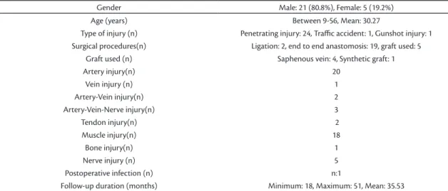
Table 1. Demographic and clinical features of patients.
Gender Male: 21 (80.8%), Female: 5 (19.2%)
Age (years) Between 9-56, Mean: 30.27
Type of injury (n) Penetrating injury: 24, Traic accident: 1, Gunshot injury: 1
Surgical procedures(n) Ligation: 2, end to end anastomosis: 19, graft used: 5
Graft used (n) Saphenous vein: 4, Synthetic graft: 1
Artery injury(n) 20
Vein injury (n) 1
Artery-Vein injury(n) 2
Artery-Vein-Nerve injury(n) 3
Tendon injury(n) 2
Muscle injury(n) 18
Bone injury(n) 1
Nerve injury (n) 5
Postoperative infection (n) n:1
Follow-up duration (months) Minimum: 18, Maximum: 51, Mean: 35.53
n: number of patients.
The surgeon’s decision on managing artery
injuries is very important during the operation. If there is a healthy intima which will not cause
stretching to the anastomosis line, then end-to-end
anastomosis should be preferred at all times.12 If
end-to-end anastomosis is not possible, vein grafts should be preferred irst, due to better patency rate and
resistance to infection.13 End-to-end anastomosis is
the irst method of choice for small diameter arteries. For anastomosis of these arteries, single stitches
should be used instead of a continuous sewing technique, in order to avoid spasms and tightening that could occur along the anastomosis line.
In our follow-up we found occlusion in one patient
who had gunshot injuries and whose radial artery had
been anastomosed in an end-to-end fashion. Although
an anastomosis line seems suitable in such injuries, vascular damage can occur due to the thermal effect and this reduces the success rate. In such patients, anastomosis has to be performed as distant from the injured area as possible, and, if possible, autogenic grafts should be used. Amputation rarely occurs in upper extremity injuries because of the rich collateral circulation.14
Doppler ultrasonography and BBI were used for patient follow-up. With regard to evaluation of lower extremity low indings, those with an BBI <1 should be accepted as having low limitation.15
In our series we didn’t find any difference
between the BBI of normal upper extremities and BBI for upper extremities with traumatic vascular occlusion after surgical intervention. The BBI was
even found to be 0.97 in a patient with left subclavian
occlusion. This is proof that the collateral circulation
is of much importance in the upper extremity and the reason why amputation is rarely needed. In our series, no patients with arterial occlusion required
amputation. On the other hand, functional deicits
related to nerve and vessel damage may develop.
This is why long-term follow-up is important in
these patients.
In the literature the rate of functional deicits
following nerve injuries in upper extremities is
27-44%.3 Traumatic nerve damages mostly cause deformations in extremities.7 It is still open to debate whether the nerves should be treated primarily or secondarily.11 During follow-up, complaints of numbness of an extremity were observed in 71.4% of patients although artery and vein damage was only
seen in 15.4% of patients. Throughout the follow-up period we observed that although enough blood low
is provided, problems related to loss of the capacity
33.5±10.8 months. Functional status was evaluated
according to the following: neurological assessment,
arm movements, muscle atrophy, indings due to arterial occlusion. Evidence of sensory and motor
injury was found in patients as follows: numbness (71.4%), muscle weakness (26.9%), sensations of coldness (15.4%), paleness (23%) after hard work and swelling in the arms (3.9%). While 71.4% of the patients had no functional complaints about upper extremity, 7.7% of the patients were functionally impaired for work.
According to the dUSG utilized during the
follow-up period, vascular occlusion was observed in 15.4% of the patients. Arterial occlusions were observed in 2 patients with subclavian arterial injury and radial
arterial injury (radial artery was treated with end-to-end anastomosis). BBI were detected as normal
or close to normal in four patients diagnosed with artery occlusion on dUSG. In two patients with
subclavian artery occlusion BBI was 0.97 and 0.98,
while two patients with radial artery occlusion had
BBI of 1 and 1.1.
DISCUSSION
Artery injuries are always the easiest upper extremity vascular injuries to diagnose quickly and
accurately. Physical examination is the irst and the
most important tool for diagnosis of upper extremity vascular injuries. Active bleeding, paleness,
coldness, inability to ind a pulse in an extremity
and growing pulsatile masses are major criteria for diagnosis. Diagnosis can be made by dUSG with 99% sensitivity, and 98% accuracy.6 Routine use of angiography should be discussed.7 For subclavian arterial injuries, computed tomography angiography stands out.4 Many of the patients in our series were diagnosed with dUSG and DSA was necessary in only one patient, due to axillary arterial injury.
Further discussion is still needed of whether the irst approach should be to treat vascular systems or
the bone system if there is bone damage in addition to arterial injuries. One of our patients had bone injuries and, as supported by the literature, the orthopedic
intervention was performed irst.8 Shunts can be used in order to avoid increasing tissue ischemia time while orthopedic interventions are being performed.9
There are also some authors who support the idea that vascular systems should be treated irst, to shorten
the duration of ischemia.10,11 Our opinion is that if orthopedic intervention is conducted after vascular systems have been treated, the reconstructed vascular
8. McHenry TP, Holcomb JB, Aoki N, Lindsey RW. Fractures with major vascular injuries form gunshot wounds: implications of surgical sequence. J Trauma. 2002;53:717-21. PMid:12394872. http://dx.doi.org/10.1097/00005373-200210000-00016
9. Franz RW, Goodwin RB, Hartman JF, Wright ML. Management of upper extremity arterial injuries at an urban level 1 trauma center. Ann Vasc Surg. 2009;23:8-16 PMid:18640813. http://dx.doi. org/10.1016/j.avsg.2008.04.012
10. Hunt CA , Kingsley JR . Vascular injuries of the upper extremity. South Med J. 2000;93:466-68. PMid:10832942. http:// dx.doi.org/10.1097/00007611-200005000-00004
11. Hardin WD Jr, O’Connell RC, Adinolfi MF, Kerstein MD. Traumatic arterial injuries of the upper extremity: determinants of disability. Am J Surg. 1985;150:266-70. http://dx.doi. org/10.1016/0002-9610(85)90134-5
12. Ekim H, Tuncer M. Management of traumatic brachial artery injuries: a report on 49 patients. Ann Saudi Med. 2009;29:105-9. PMid:19318753 PMCid:PMC2813640. http://dx .doi. org/10.4103/0256-4947.51797
13. Reynolds RR, McDowell HA, Diethelm AG. The surgical treatment of arterial injuries in the civilian population. Ann Surg. 1979;189:700-8. PMid:453941 PMCid:PMC1397245. http://dx.doi. org/10.1097/00000658-197906000-00005
14. McCroskey BL, Moore EE, Pearce WH, Moore FA, Cota R, Sawyer JD. Traumatic injuries of the brachial artery. Am J Surg.1988;156:553-5. http://dx.doi.org/10.1016/S0002-9610(88)80551-8
15. Hafez HM, Woolgar J, Robbs JV. Lower extremity arterial injury: results of 550 cases and review of risk factors associated with limb loss. J Vasc Surg. 2001;33:1212-9. PMid:11389420. http://dx.doi. org/10.1067/mva.2001.113982
16. Timberlake GA, O’Connell RC, Kerstein MD. Venous injury: to repair or ligate, the dilemma. J Vasc Surg. 1986;4:553-8. PMid:3783830.
Correspondence
Erdal Simsek Ceyhun Atıf Kansu street 166/4 Cevizlidere, Balgat/Ankara, Turkey Tel.: +905323280217 E-mail: [email protected]
Author information
ES is a cardiovascular surgeon at Turkiye Yuksek Ihtisas Training and Research Hospital. KK is a cardiovascular surgeon at Diskapi Yildirim Beyazit Training and Research hospital. II is a cardiovascular surgeon at Tokat Gaziosmanpaşa University. ATU is a cardiovascular surgeon at Turkiye Yuksek Ihtisas Training and Research Hospital.
Author contributions
Conception and design: ES, KK, II Analysis and interpretation: ES, ATU Data collection: KK, II Writing the article: ES Critical revision of the article: ATU Final approval of the article*: ES, KK, II, ATU Statistical analysis: ATU Overall responsibility: ES
* All authors have read and approved of the inal version of the article submitted to J Vasc Bras.
to work and inability to use the extremity persist as long as nerve injury remains.
Related venous injuries are often seen intraoperatively. Small venous vessels can be ligated, but larger venous injuries should be reconstructed.
Thrombosis occurs in 39-59% after venous repairs.16 We did not observe venous thrombosis in our patients.
In the course of repairing vascular injuries surgeons generally concentrate on arterial damage.
It is obvious that attention must irst be directed
to the arterial injury. However, other concomitant injuries to veins, bones, tendons and nerves should not be ignored, so a multidisciplinary approach is
important. This group of young patients should be
sent back to their work lives and social lives right away and should be kept under strict surveillance and given help in the form of physical therapy and rehabilitation.
In summary, BBI did not correlate with functional
disability in patients who had undergone upper extremity vascular surgery for trauma.
Evaluation of the postoperative outcomes of this
small series of patients with penetrating vascular traumas in the upper extremity revealed that 15% of them suffered occlusion of one artery of the upper extremity. Artery occlusion did not correlate with
brachial-brachial Doppler index, probably due to rich
collateral circulation, but occlusion was associated with an extremity that was dysfunctional for the purposes of daily work.
REFERENCES
1. Milas ZL, Dodson TF, Ricketts RR. Pediatric blunt trauma resulting in major arterial injuries. Am Surg. 2004;70:443- 7. PMid:15156954.
2. Kendall KM, Burton JH, Cushing B. Fatal sub clavian a r ter y tr a ns e c ti o n f ro m is o l ate d cl av i cl e f r a c tu re . J Trauma. 2000;48:316-8. PMid:10697095. http://dx.doi. org/10.1097/00005373-200002000-00022
3. Shalhub S, Starnes BW, Hatsukami TS, Karmy-Jones R, Tran NT. Repair of blunt thoracic outlet arterial injuries: an evolution from open to endovascular approach. J Trauma. 2011,71:114-21. PMid:22071946. http://dx.doi.org/10.1097/TA.0b013e31822cf359 4. Zellweger R, Hess F, Nicol A, Omoshoro-Jones J, Kahn D, Navsaria P. An analysis of 124 surgically managed brachial artery injuries. Am J Surg. 2004;188:240-5. PMid:15450827. http://dx.doi.org/10.1016/j. amjsurg.2004.02.005
5. Bennett JR, Bodenham AR, Berrıdge JR. Advanced trauma life support: A time for reappraisal. Anaesthesia. 1992;47:798-800.
6. Bynoe RP, Miles WS, Bell RM, Greenwold DR, Sessions G, Haynes JL. Non-invasive diagnosis of vascular trauma by duplex ultrasonography. J Vasc Surg. 1991;14:346-52. http://dx.doi. org/10.1016/0741-5214(91)90087-B