IntroductIon
Obesity is an isolated risk factor for cardiovascular disease; it can lead to cardiac hypertrophy followed by dilated cardiomyopathy, predisposing to fatal arrhyth-mias. (1) Obesity also causes organ-speciic changes, which are due to a direct mechanical effect of the adipose tissue or occur systemically through humoral mediators and metabolic adjustments that change heart hemodynamics and geometry, as well as possibly lung function.(1-3)
A recent epidemiological study(4) found a relationship between lung function and left ventricular mass (LVM), although the relationship differed between genders. Functional effects of obesity that are associated with being male or female are common in pulmonary function testing, since fat concentrated in the chest (android obesity) could lead to deeper changes in lung function than could fat concentrated in the hips (gynecoid obesity). In contrast, another study, which had a case-control design and involved children with and without metabolic syndrome, found no relationship between lung function and LVM.(5)
Recent evidence suggests that inlammatory mediators act independently of confounding variables on cardiac remodeling in obese individuals,(2,6) as well as on lung
function,(7,8) primarily at the level of the small airways. In contrast, there is evidence of purely mechanical cardiopulmonary effects, with no effect of inlammatory mediators,(9,10) or an interaction of mechanical and inlammatory factors in cases of lung function and asthma associated with obesity.(11)
Therefore, the primary objective of this exploratory study was to look for correlations between lung function and cardiac dimension variables, in order to test the hypothesis that regardless of being a purely mechanical or gender-related factor, the relative size of the small airways (dysanapsis), as measured indirectly by the ratio of FEF25-75% to FVC (FEF25-75/FVC, %), is correlated with left ventricular hypertrophy (remodeling), since the small airways are especially susceptible to mechanical-inlammatory interactions and bronchial hyperreactivity. Similarly, we aimed to determine the degree of association between lung function and ventricular mass in the study population, with the goal of guiding future studies related to common mechanisms of cardiac and pulmonary impairment in morbid obesity. To the best of our knowledge, based on our review of currently available databases (Bireme, SciELO, PubMed, Cochrane Library, and Google Academic), this is the irst study to investigate this relationship in morbidly obese individuals. AbstrAct
Objective: To look for correlations between lung function and cardiac dimension variables in morbidly obese patients, in order to test the hypothesis that the relative size of the small airways is independently correlated with left ventricular hypertrophy. Methods: This was a retrospective study involving 192 medical records containing a clinical protocol employed in candidates for bariatric surgery between January of 2006 and December of 2010. Results: Of the 192 patients evaluated, 39 (10 males and 29 females) met the inclusion criteria. The mean BMI of the patients was 49.2 ± 7.6 kg/ m2, and the mean age was 35.5 ± 7.7 years. The FEF
25-75/FVC, % correlated signiicantly
with left ventricular posterior wall thickness and relative left ventricular posterior wall
thickness, those correlations remaining statistically signiicant (r = −0.355 and r = −0.349, respectively) after adjustment for weight, gender, and history of systemic arterial
hypertension. Stepwise multivariate linear regression analysis showed that FVC and FEV1 were the major determinants of left ventricular mass (in grams or indexed to body
surface area). Conclusions: A reduction in the relative size of the small airways appears to be independently correlated with obesity-related cardiac hypertrophy, regardless of factors affecting respiratory mechanics (BMI and weight), gender, or history of systemic arterial hypertension. However, FEV1 and FVC might be important predictors of left ventricular mass in morbidly obese individuals.
Keywords: Obesity; Spirometry; Echocardiography; Body mass index.
Lung function and left ventricular
hypertrophy in morbidly obese candidates
for bariatric surgery
Paulo de Tarso Müller1,2, Hamilton Domingos3, Luiz Armando Pereira Patusco1,2, Gabriel Victor Guimarães Rapello1
Correspondence to:
Paulo de Tarso Müller. Avenida Senador Filinto Müller, S/N, Vila Ipiranga, Campus da Universidade Federal de Mato Grosso do Sul, Faculdade de Medicina, CEP 79070-900, Campo Grande, MS, Brasil.
Tel.: 55 67 3345-3149. E-mail: [email protected] Financial support: None.
1. Laboratório de Fisiopatologia Respiratória – LAFIR – Universidade Federal de Mato Grosso do Sul, Campo Grande, Brasil.
2. Disciplina de Pneumologia, Faculdade de Medicina, Universidade Federal de Mato Grosso do Sul, Campo Grande, Brasil.
3. Disciplina de Cardiologia, Faculdade de Medicina, Universidade Federal de Mato Grosso do Sul, Campo Grande, Brasil.
Submitted: 23 February 2015.
Accepted: 5 July 2015.
Methods
We planned this study based on secondary data obtained from a bariatric surgery outpatient clinic, which is a state referral center for this type of surgery. In this retrospective study, we reviewed the medical records of all obese individuals who were candidates for bariatric surgery between January of 2006 and December of 2010, with the total number of individuals being 192. Individuals underwent a standardized clinical assessment, which was based on an instrument designated “clinical assessment form for obese patients” (a protocol at the department of bariatric surgery), in which detailed information was obtained on anthropometric parameters, degree of obesity, comorbidities, and pressure levels, among other clinical data and relevant tests, assessed by physicians, nutritionists, and nurses working in that department.
The aim of the medical record review was to system-atically collect the following clinical data on and test results for morbidly obese individuals (BMI ≥ 40 kg/ m2)(12): (i) anthropometry; (ii) simple spirometry; (iii) M-mode echocardiography; (iv) reporting of bronchial asthma (diagnosis and/or treatment); (v) reporting of current or former smoking; and (vi) reporting (diagnosis and/or treatment) of systemic arterial hypertension (SAH). Asthma was deined as reporting of a current or prior medical diagnosis. Current or former smoking refers to a longer than 1-year history of smoking, regardless of the number of pack-years. SAH was deined as a current drug treatment for SAH or at least two arterial blood pressure measurements ≥ 140/90 mmHg. Data on the forms, as well as test results, were only accepted if they had been properly recorded within 1 year before surgery. Only 45 patients met all of the above inclusion criteria, of whom 3 were excluded because of an echocardiographic report of acoustic window impairment caused by obesity and 3 were excluded because they did not meet the spirometry quality criteria. The main reasons for exclusion of the remaining cases were not being diagnosed with morbid obesity (136 individuals) and being a morbidly obese patient with no spirometric data (4 individuals), no echocardiographic data (5 individuals), or neither (2 individuals).
Anthropometric data were obtained by using a stadiometer and a scale for obese individuals, and BMI was calculated by the formula weight/height2 (in kg/ m2). Waist circumference values were not collected, since such values were lacking in many cases. The study was approved by the Human Research Ethics Committee of the Federal University of Mato Grosso do Sul and was in compliance with the Declaration of Helsinki.
Spirometry with forced expiratory maneuver was performed in the pulmonary function section of the department of pulmonology of the university. All tests met the acceptability criteria established in the Brazilian guidelines for pulmonary function testing,(13) and
values were corrected to body temperature, pressure saturated. All tests were performed with a Vitatrace VT 130 spirometer (Pró Médico Ltda., Rio de Janeiro, Brazil), with individuals in a sitting position and wearing a nose clip. The equipment was always calibrated in the morning, in accordance with the manufacturer instructions, and the tests were always conducted by one of two trained spirometry technicians belonging to the department of pulmonology. At least three acceptable maneuvers were always performed, and instantaneous low was expressed as that obtained from the one maneuver with the highest sum of FVC (in L) and FEV1 (in L/min). The reference values used were those of Pereira et al.,(14) and the mean ± SD of the study population was recorded.
Echocardiography was performed by two cardiologists, who used a Nemio 17-2005 echocardiograph (Toshiba, Tokyo, Japan). Those two cardiologists, one of whom is one of the present authors, work speciically in the echocardiography section of the hospital. For the purposes of this study, only data acquired in M-mode were collected; left ventricle end-diastolic volume (LVDV) and LV end-systolic volume (LVSV) were indirectly measured using the Teicholz formula, and LVM was calculated using the formula by Devereux et al.(15) The measurements were obtained from parasternal LV cross-sections at the level of the papillary muscles. Left atrial (LA) dimensions, LV end-diastolic diameter (LVDD), and LV end-systolic diameter (LVSD) were also measured. We used as criteria for LVM indexation both height to the power of 1.7 (LVM/m1.7), which is a criterion recommended for obese individuals, and height squared (LVM/m2). Relative LV posterior wall thickness (RLVPWT) was obtained by dividing LV posterior wall thickness (LVPWT) by LVDD.
are traditionally the most important determinants of LV hypertrophy in obesity. In order to determine which variable would be the best predictor of LVM (dependent variable), we tested a stepwise multivariate linear regression model, in which the independent variables were only the variables showing a signiicant correlation (p< 0.05) with LVM, whether indexed or not. For all calculations and graphs, we used the IBM SPSS Statistics software package, version 20.0 (IBM Corp., Armonk, NY, USA). Results were considered signiicant at the level of p ≤ 0.05.
results
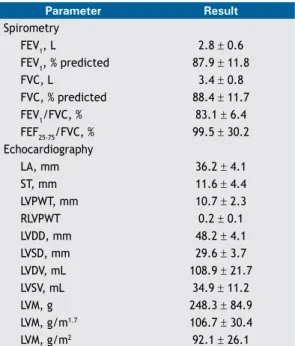
Of the 45 patients for whom all data were originally available for inclusion, 6 were excluded because of reported failures in M-mode echocardiography or in pulmonary function testing, which did not meet quality criteria. The anthropometric and demographic data of the 39 patients included in the study are shown in Table 1. Females predominated in the study (74.3%), the mean age of the participants was 35.5 ± 7.7 years, and 8 participants were considered super-obese (BMI > 55 kg/m2). Asthma and ever smoking were reported by 7 and 6 individuals, respectively. SAH (diagnosis and/or treatment) was reported to be present in 21 individuals (54%). Only 1 individual reported both asthma and smoking. The main spirometric and echocardiographic parameters are shown in Table 2. The mean spirometric values were above 80% of predicted. The mean values of LA diameter, LVM, and LVM/m2 were increased relative to normal mean values for the Brazilian population.(16)
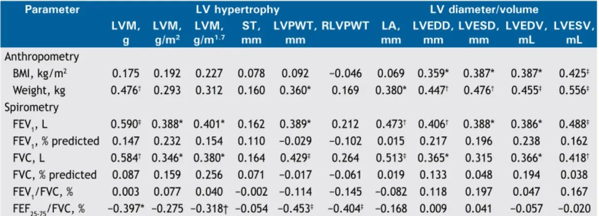
The various correlations between the echocardio-graphic and spirometric variables are shown in Table 3. No correlation was found between BMI and any of the spirometric variables studied. Chief among the signiicant correlations is the weak direct relationship between BMI and the variables LVDD(r = 0.359; p < 0.05), LVSD (r = 0.387; p < 0.05), LVDV (r = 0.387; p < 0.05), and LVSV (r = 0.425; p< 0.01). A moderate inverse correlation was found between FEF25-75/FVC, % and the variables of LV remodeling (Table 3) in the univariate analysis, and this correlation remained statistically signiicant for LVPWT (r = −0.355; p < 0.05) and RLVPWT (r = −0.349; p < 0.05) even after adjustment for weight, gender, and SAH (Figures 1A and 1B, respectively). The correlation between indexed LVM (in g/m1.7) and FEF
25-75/FVC, % was borderline for statistical signiicance (p = 0.05; Figure 1C). Interestingly, FEF25-75/FVC, % did not correlate with the variables of LV internal diameter or LV volume. No correlation was found between FEF25-75% and any of the echocardiographic variables. Fischer’s exact test showed no statistically signiicant association between FEF25-75/FVC, %, below or above the lower limit of the normal range, and reporting of asthma or smoking (p > 0.05 for both).
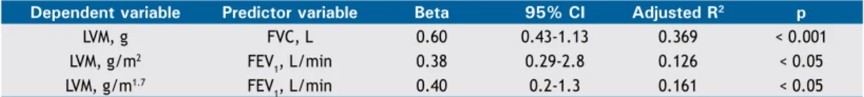
Stepwise multiple linear regression analysis (Table 4) showed that the variation in LVM among morbidly
obese individuals is better predicted by FVC (in L), which explained 36.9% (p < 0.0001) of the variation in LVM in the study population. The best predictor of LVM indexed to height squared and LVM indexed to height to the power of 1.7 was FEV1 (in L/min; p < 0.05 for both; Table 4).
dIscussIon
This retrospective study shows there is an independent association between the relative size of the small airways (FEF25-75/FVC,%) and echocardiographic parameters of ventricular hypertrophy in morbidly obese individuals.
table 1. Characteristics of the individuals included in the
study (N = 39).a
Characteristic Result
Age, years 35.5 ± 7.7 Height, cm 163.1 ± 9.1 Weight, kg 131.4 ± 25.9 BMI, kg/m2 49.2 ± 7.6 BS, m2 2.3 ± 0.3 Gender (M/F), n/n 10/29 Asthma (Y/N), n/n 7/32 Smoking (Y/N), n/n 6/33 SAH (Y/N), n/n 21/18
BS: body surface; M: male; F: female; Y: yes; N: no; and SAH: systemic arterial hypertension. aValues expressed as mean ± SD, except where otherwise indicated.
table 2. Spirometric and echocardiographic parameters
(N = 39).
Parameter Result
Spirometry
FEV1, L 2.8 ± 0.6 FEV1, % predicted 87.9 ± 11.8 FVC, L 3.4 ± 0.8 FVC, % predicted 88.4 ± 11.7 FEV1/FVC, % 83.1 ± 6.4 FEF25-75/FVC, % 99.5 ± 30.2 Echocardiography
In addition, FVC (in L) and FEV1 (in L/min) were found to be important predictors of LVM in grams or indexed to body surface area.
Both LVM and LV internal diameters are increased in obesity, regardless of SAH.(17) In our study, we found
table 3. Univariate correlations of anthropometric and spirometric variables with transthoracic echocardiography
variables (M-mode) in the sample as a whole (N = 39).
Parameter lV hypertrophy lV diameter/volume
lVM, g
lVM, g/m2
lVM, g/m1.7
St, mm
lVPWt, mm
RlVPWt lA,
mm
lVeDD, mm
lVeSD, mm
lVeDV, ml
lVeSV, ml Anthropometry
BMI, kg/m2 0.175 0.192 0.227 0.078 0.092 −0.046 0.069 0.359* 0.387* 0.387* 0.425‡ Weight, kg 0.476† 0.293 0.312 0.160 0.360* 0.169 0.380* 0.447† 0.476† 0.455‡ 0.556‡ Spirometry
FEV1, L 0.590‡ 0.388* 0.401* 0.162 0.389* 0.212 0.473† 0.406† 0.388* 0.386* 0.488‡ FEV1, % predicted 0.147 0.232 0.154 0.110 −0.029 −0.102 0.015 0.217 0.196 0.238 0.162 FVC, L 0.584† 0.346* 0.380* 0.164 0.429‡ 0.264 0.513‡ 0.365* 0.315 0.366* 0.418† FVC, % predicted 0.087 0.159 0.256 0.071 −0.017 −0.061 0.019 0.133 0.048 0.194 0.038 FEV1/FVC, % 0.003 0.077 0.040 −0.002 −0.114 −0.145 −0.082 0.118 0.197 0.047 0.167 FEF25-75/FVC, % −0.397* −0.275 −0.318† −0.054 −0.453‡ −0.404‡ −0.168 0.009 0.041 −0.057 −0.020 LV: left ventricular; LVM: LV mass; ST: septal thickness; LVPWT: LV posterior wall thickness; RLVPWT: relative LV posterior wall thickness; LA: left atrium; LVDD: LV end-diastolic diameter; LVSD: LV end-systolic diameter; LVDV: LV end-diastolic volume; and LVSV: LV end-systolic volume. *p < 0.05; †p = 0.05; ‡p < 0.01.
Figure 1. Correlation of FEF25-75/FVC, %, adjusted for the
variables weight, gender, and systemic arterial hypertension, with left ventricular posterior wall thickness, in A; with relative left ventricular posterior wall thickness, in B; and with left ventricular mass adjusted for body size in obese individuals, in C.
Left ventricular posterior
wall thickness. mm
R
elative left ventricular posterior
wall thickness
Left ventricular mass,
g/m
1.7
r =−0.355 / p < 0.05
r =−0.349 / p < 0.05
r =−0.318 / p = 0.05 18.0
16.5 15.0 13.5 12.0 10.5 9.0 7.5 6.0
.350 .325 .300 .275 .250 .225 .200 .175 .150
A
B
C
40 60 80 100 120 140 160 180 250
200
150
100
50
FEF25-75/FVC, %
no relationship of BMI with LVM in grams or indexed to body surface area or with LVPWT, but we found a relationship of BMI with LV internal diameters and LV volumes, which is consistent with the literature. (2,17) This positive association is likely to be due to increased preload and increased cardiac output, causing ventricular dilatation, which could later progress to LV remodeling.(18)
The literature shows inconsistent associations between BMI and spirometric results, with some studies showing an association(19) and others not showing any.(20) This is probably due to differences in gender ratio among studies, since the android type of obesity favors the correlation between BMI and lung function, unlike the gynecoid phenotype, which does not produce excessive accumulation of fat mass in the chest. In our study, females predominated (74%), which partly explains the lack of correlation between any spirometric variable and BMI.
The relationships of FVC, FEV1, and FEF25-75% with LVM and LVPWT have been studied in (non-obese) elderly individuals with cardiovascular disease, and the results differ among studies in regard to the direction of the correlation, with correlations being positive(21) or negative,(22) which more strongly relects loss of lung function associated with advanced age and with the effects of SAH and pulmonary hypertension, as well as with the pulmonary restrictive effects of cardiomegaly. One study(4) showed an inverse relationship between LVM and FVC in nonsmoking females and a direct relationship between LVM and FVC in nonsmoking males under 60 years of age. The fact that most of our sample consists of females (74%) suggests that, in morbidly obese individuals, the direction of the correlation may be different for some lung function variables.
to body surface area (in g/m1.7) and with LVPWT/ RLVPWT, correlations that remained after adjustment for weight, gender, and SAH, possibly relect the direct mechanical effects of obesity, but may also suggest that there are other independent (inlammatory or lipotoxic) mechanisms.
Since research on the subject is scarce, there is limited evidence that the small airways are independently affected by obesity, as has been reported in nonsmoking males.(23) The hypotheses raised in that study(23) include an increase in blood volume in obese individuals, causing bronchial vessel congestion; the presence of increased levels of very-low-molecular-weight lipoproteins, which could trigger the release of histamine; and altered lipoprotein metabolism in obesity, which could elicit and amplify these effects.
Recent data in the literature also indicate that obesity is characterized by hyperresponsiveness to methacho-line, predominantly in the small airways,(24) and that this hyperresponsiveness correlates better with FEF25-75/ FVC, %.(25) On this point, a recent study suggests that groups of obese individuals with hyperresponsiveness are associated with greater LVM.(26) Small airway hyperresponsiveness in obese individuals also could be partially explained by dysanapsis (which is assessed indirectly by FEF25-75/FVC, %), a term coined by Green et al.(27) to explain the large interindividual variability in airway size, regardless of lung parenchyma size. An important predictor of LVM, BMI correlated directly with cardiac size and mass in several studies, although lean body mass remains a better predictor of LVM. (2,28,29) This retrospective study found no correlations between BMI and LVM (in grams or indexed to body surface area), which is in agreement with another study,(30) and this is possibly due to differences in obesity phenotypes, prevalence of SAH, and number of individuals studied.
Of note in our study is the fact that FVC (in L) was the best predictor of variation in LVM (in g), explaining 37% of this variation in the study population, which suggests that reduced lung volume may be an important variable in establishing a predictive model for LVM in obese individuals in future studies. In turn, LVM correlates with cardiovascular morbidity and mortality.
Among the limitations of our study is the fact that our sample was small, consisting of candidates for bariatric surgery, was limited by the criterion of including only morbidly obese and super-obese individuals, and was based on criteria that were unclear in the medical charts, such as the diagnosis of asthma or SAH. In addition, data on diabetes were not collected, although the relationship between diabetes and LVM is inconsistent in the literature. Other important limitations were the limited acoustic window in the analysis of M-mode echocardiographic variables in obese individuals and the lack of a speciic, standardized protocol for M-mode echocardiography, since we did not obtain data on inter-rater agreement for the two echocardiographers. In this regard, because this was a retrospective study, we sought not to use echocardiographic data for which accuracy is signiicantly decreased by the effects of obesity on the acoustic window, such as ejection fraction and Doppler echocardiography data.
We therefore conclude that the small airways in morbidly obese individuals have a correlation with cardiac hypertrophy, regardless of usual anthropometric variables, gender, or SAH. This study reveals that factors other than mechanical and/or hemodynamic limitations imposed by increased body mass may be important in the joint changes seen in the small airways and in cardiac hypertrophy. In addition, further studies are needed to examine the impact that lung function parameters have on predictive equations for LVM in obese individuals.
table 4. Stepwise multiple linear regression for the dependent variable left ventricular mass (in grams or indexed to
body surface area; N = 39).
Dependent variable Predictor variable Beta 95% Ci Adjusted R2 p
LVM, g FVC, L 0.60 0.43-1.13 0.369 < 0.001 LVM, g/m2 FEV
1, L/min 0.38 0.29-2.8 0.126 < 0.05 LVM, g/m1.7 FEV
1, L/min 0.40 0.2-1.3 0.161 < 0.05 LVM: left ventricular mass.
reFerences
1. Poirier P, Giles TD, Bray GA, Hong Y, Stern JS, Pi-Sunyer FX, et al.
Obesity and cardiovascular disease: pathophysiology, evaluation, and effect of weight loss. Arterioscler Thromb Vasc Biol.
2006;26(5):968-76. http://dx.doi.org/10.1161/01.ATV.0000216787.85457.f3 2. Ashraian H, Athanasiou T, le Roux CW. Heart remodelling and
obesity: the complexities and variation of cardiac geometry. Heart .2011;97(3):171-2. http://dx.doi.org/10.1136/hrt.2010.207092
3. Salome CM, King GG, Berend N. Physiology of obesity and effects
on lung function. J Appl Physiol (1985). 2010;108(1): 206-11. http://
dx.doi.org/10.1152/japplphysiol.00694.2009
4. Charles LE, Burchiel CM, Andrew ME, Gu JK, Petrini MF, Butler
KR Jr. Pulmonary function and left ventricular mass in African Americans: the Atherosclerosis Risk in Communities (ARIC) study.
Echocardiography. 2012;29(2):131-9. http://dx.doi.org/10.1111/ j.1540-8175.2011.01550.x
5. Del Río-Camacho G, Domínguez-Garrido MN, Pita J, Aragón I,
Collado R, Soriano-Guillén L. Left ventricular mass, forced baseline
spirometry and adipocytokine proiles in obese children with and
without metabolic syndrome [Article in Spanish]. An Pediatr (Barc).
2013;78(1):27-34. http://dx.doi.org/10.1016/j.anpedi.2012.05.010 6. Lai YH, Liu CC, Kuo JY, Hung TC, Wu YJ, Yeh HI, et al. Independent
effects of body fat and inlammatory markers on ventricular
geometry, midwall function, and atrial remodeling. Clin Cardiol.
2014;37(3):172-7. http://dx.doi.org/10.1002/clc.22242
7. Hickson DA, Burchiel CM, Petrini MF, Liu J, Campbell-Jenkins BW,
in African Americans, independent of adiposity: the Jackson Heart
Study. Obesity (Silver Spring). 2011;19(5):1054-61. http://dx.doi.
org/10.1038/oby.2010.240
8. Lecube A, Sampol G, Mu-oz X, Ferrer R, Hernández C, Simó R.
TNF-α system and lung function impairment in obesity. Cytokine.
2011;54(2):121-4. http://dx.doi.org/10.1016/j.cyto.2011.01.010
9. Held M, Mittnacht M, Kolb M, Karl S, Jany B. Pulmonary and cardiac
function in asymptomatic obese subjects and changes following a
structured weight reduction program: a prospective observational
study. PLoS One. 2014;9(9):e107480. http://dx.doi.org/10.1371/ journal.pone.0107480
10. Hickson DA, Liu J, Bidulescu A, Burchiel CM, Taylor HA, Petrini
MF. Pericardial fat is associated with impaired lung function and a restrictive lung pattern in adults: the Jackson Heart Study. Chest.
2011;140(6):1567-73. http://dx.doi.org/10.1378/chest.11-0258
11. Santamaria F, Montella S, Pietrobelli A. Obesity and pulmonary
disease: unanswered questions. Obes Rev. 2012;13(9):822-33.
http://dx.doi.org/10.1111/j.1467-789X.2012.01008.x
12. World Health Organization. Obesity: preventing and managing the global epidemic. Report of a WHO consultation. Geneva: WHO;
1998.
13. Sociedade Brasileira de Pneumologia e Tisiologia. Diretrizes para
testes de função pulmonar. J Pneumol. 2002; 28(Suppl 3):S1-S238.
14. Pereira CA, Sato T, Rodrigues SC. New reference values for forced
spirometry in white adults in Brazil. J Bras Pneumol.
2007;33(4):397-406. http://dx.doi.org/10.1590/S1806-37132007000400008 15. Devereux RB, Alonso DR, Lutas EM, Gottlieb GJ, Campo E, Sachs I,
et al. Echocardiographic assessment of left ventricular hypertrophy:
comparison to necropsy indings. Am J Cardiol. 1986;57(6):450-8. http://dx.doi.org/10.1016/0002-9149(86)90771-X
16. Angelo LC, Vieira ML, Rodrigues SL, Morelato RL, Pereira AC,
Mill JG, et al. Echocardiographic reference values in a sample of asymptomatic adult Brazilian population. Arq Bras Cardiol. 2007;89(3):168-73, 184-90.
17. Lauer MS, Anderson KM, Levy D. Separate and joint inluences of
obesity and mild hypertension on left ventricular mass and geometry: the Framingham Heart Study. J Am Coll Cardiol. 1992;19(1):130-4.
http://dx.doi.org/10.1016/0735-1097(92)90063-S
18. Rider OJ, Petersen SE, Francis JM, Ali MK, Hudsmith LE, Robinson
MR, et al. Ventricular hypertrophy and cavity dilatation in relation
to body mass index in women with uncomplicated obesity. Heart. 2011;97(3):203-8. http://dx.doi.org/10.1136/hrt.2009.185009 19. Wei YF, Wu HD, Chang CY, Huang CK, Tai CM, Hung CM, et al.
The impact of various anthropometric measurements of obesity on pulmonary function in candidates for surgery. Obes Surg.
2010;20(5):589-94. http://dx.doi.org/10.1007/s11695-009-9961-0
20. Gabrielsen AM, Lund MB, Kongerud J, Viken KE, Røislien J,
Hjelmesæth J. The relationship between anthropometric measures, blood gases, and lung function in morbidly obese white subjects. Obes Surg. 2011;21(4):485-91.
http://dx.doi.org/10.1007/s11695-010-0306-9
21. Ricart S, Casan P, Bellido-Casado J, González M, Cotes C, López L,
et al. Lung function in cardiac dysfunction [Article in Spanish]. Arch
Bronconeumol. 2004;40(2):62-6.
http://dx.doi.org/10.1016/S0300-2896(04)75474-5
22. Enright PL, Kronmal RA, Smith VE, Gardin JM, Schenker MB,
Manolio TA. Reduced vital capacity in elderly persons with hypertension, coronary heart disease, or left ventricular hypertrophy. The Cardiovascular Health Study. Chest. 1995;107(1):28-35. http://
dx.doi.org/10.1378/chest.107.1.28
23. Rubinstein I, Zamel N, DuBarry L, Hoffstein V. Airlow limitation
in morbidly obese, nonsmoking men. Ann Intern Med.
1990;112(11):828-32.
http://dx.doi.org/10.7326/0003-4819-112-11-828
24. Skloot G, Schechter C, Desai A, Togias A. Impaired response to deep
inspiration in obesity. J Appl Physiol (1985). 2011;111(3):726-34.
http://dx.doi.org/10.1152/japplphysiol.01155.2010
25. Zerah-Lancner F, Boyer L, Rezaiguia-Delclaux S, D’Ortho MP, Drouot
X, Guilloteau-Schoennagel I, et al. Airway responsiveness measured by forced oscillation technique in severely obese patients, before and
after bariatric surgery. J Asthma. 2011;48(8):818-23. http://dx.doi.org/
10.3109/02770903.2011.613508
26. Gagnon-Audet AA, Poirier P, Turcotte H, Martin J, Bastien M, Simard
S, et al. Inluence of cardiac dysfunction and systemic inlammation on pulmonary function and airway responsiveness in obese subjects.
Clin Invest Med. 2013;36(5):E255-63.
27. Green M, Mead J, Turner JM. Variability of maximum expiratory
low-volume curves. J Appl Physiol. 1974;37(1):67-74.
28. Rocha IE, Victor EG, Braga MC, Barbosa e Silva O, Becker Mde M.
Echocardiography evaluation for asymptomatic patients with severe
obesity. Arq Bras Cardiol. 2007;88(1):52-8. http://dx.doi.org/10.1590/
S0066-782X2007000100009
29. Ballo P, Motto A, Mondillo S, Faraguti SA. Impact of obesity on
left ventricular mass and function in subjects with chronic volume overload. Obesity (Silver Spring). 2007;15(8):2019-26. http://dx.doi.
org/10.1038/oby.2007.241
30. Ribeiro Filho FS, Rosa EC, Faria AN, Lerário DD, Ferreira SR, Kohlmann O,
et al. Obesidade, hipertensão arterial e suas inluências sobre a massa e
função do ventrículo esquerdo. Arq Bras Endocrinol Metab.