CASE REPORT
606 Bras J Rheumatol 2010;50(5):603-08
Received on 02/18/2010. Approved on 08/26/2010. We declare no conflict of interest. Centro de Pesquisa Clínica Hospital Abreu Sodré- AACD.
1. Rheumatologist - Hospital Abreu Sodré-AACD and Hospital Israelita Albert Einstein. 2. Nurse – Clinical Research Specialist
3. Clinical Pathologist - Laboratory of Clinical Chemistry of Hospital Israelita Albert Einstein 4. Hematologist at the Clinical Chemistry Laboratory - Hospital Israelita Albert Einstein
5. Director Laboratory of Clinical Chemistry - Doctor in Pathology - Universidade de São Paulo (USP); Physician Hospital Albert Einstein
6. Rheumatologist and Immunologist – PhD in Immunology - Boston University; Free Professor of Immunology - USP; Clinician and Rheumatologist - Hospital Israelita Albert Einstein; Scientific Director of Hospital Abreu Sodré - AACD (Hospital Unit Specialized in Musculoskeletal Diseases)
Correspondence to: Ricardo Prado Golmia. Av. Prof. Ascendino Reis, 724, Bloco C- Pesquisa Clínica - 1º Subsolo, São Paulo, SP, Brazil. CEP: 04027-000. E-mail: [email protected].
AL amyloidosis in a young adult: remission
with autologous stem cell transplantation
Ricardo Prado Golmia1, Isabel Clapis Bello2, Eliane Rosseto3,
João Carlos Campos Guerra4, Cristóvão Mangueira5, Morton Aaron Scheinberg6
ABSTRACT
Autologous stem cell transplant is one of the therapies employed in the treatment of primary amyloidosis or AL. The authors report on a 46-year-old patient with bilateral periorbital hematomas, macroglossia who presented, during the
investigation, IgG-Kappa paraprotein in serum. The diagnosis of primary amyloidosis or AL was conirmed and the
treatment proposed consisted of high-dose melphalan as conditioning regimen before autologous stem cell transplant, which determined complete remission of the disease, along with the disappearance of clinical signs and absence of the monoclonal component.
Keywords: amyloidosis, autologous transplantation, melphalan.
INTRODUCTION
AL Amyloidosis is a deposition disease in tissues of fragments of light chain, with spatial and tinctorial disposition of amyloid
ibrillar material.1 The amyloid deposition affects several
or-gans and both the deposition extension and the affected organ are related with worse prognosis, such as cardiovascular
in-volvement. The therapeutic dificulty in these cases motivated
the authors to report the results obtained with autologous stem cell transplant.
We recently assessed a young patient. For 2 years, the cli-nical picture consisted of periorbital hematomas, carpal tunnel syndrome and presence of serum monoclonal protein at the protein electrophoresis. After the assessment, AL amyloidosis
was identiied. The proposed treatment consisted of chemothe -rapy associated with autologous stem cell transplant.
CASE REPORT
M.M.V.F., a 46-year-old male patient, sought medical attention complaining of hand paresthesia, myalgia, periorbital hemato-mas and macroglossia. The symptoms had been ongoing for 2
years, without a deinitive diagnosis. At physical examination,
he presented periorbital hematomas, macroglossia, signs
sug-gestive of carpal tunnel syndrome in both hands and unspeciic
muscular pains.
The protein electrophoresis identiied a monoclonal peak
at the gamma-globulin region, which corresponded to 31.3% of the total proteins. The erythrocyte sedimentation rate (ESR) after the 1st hour was altered, at 39 mm (normal value ≤ 15 mm).
The whole blood count did not present any alterations at the following parameters: HB = 13.9 g/dL; HT =41.9%, red blood cells = 4.91 106 µ/L, leukocytes = 9,100 µ/L and platelet count
AL amyloidosis in a young adult: remission with autologous stem cell transplantation
607 Bras J Rheumatol 2010;50(5):603-08
and creatinina = 1.53 mg/dL. No alterations were identiied at
the partial urinalysis (urine I). The patient presented IgM level of 30 mg/dL (within normal range) and monoclonal IgG-Kappa light chain measurement of 3.6 g/100 mL at the serum protein
immunoixation test. The echocardiogram with Doppler was
considered normal.
The aspiration myelogram showed normal cellularity in the erythrocytic, monocytic and granulocytic series, with preserved cell maturation progress. The lymphoplasmacytic series presented 14% of mature plasmacytes. The patient was submitted to a rectal biopsy, of which result showed hyaline amorphous material positive for amyloid material with
speci-ic staining methods. Eighteen months later, the patient is in
complete remission, with no clinical signs and the monoclonal component has disappeared.
The patient received, for a consecutive week, 0.4 mg of granulokine via subcutaneous route, followed by high-dose Melphalan via intravenous route, 200 mg/m2. After the third
day, he was submitted to apheresis, to obtain CD34-positive cells (stem cells) (Figures 1 and 2).
DISCUSSION
Amyloidosis is not a single disease. The term is currently used generically to refer to any pathological condition where there
is extracellular deposition of ibrillar proteins, with special
birefringence characteristics. The binding to certain stains such
as Congo red or thiolavin is a common denominator in cases
of amyloidosis. There are currently more than twenty proteins with these characteristics and each one of them must be seen as a distinct clinical entity.
The three most common forms, with a multiorgan system presentation, are the light-chain amyloidosis (AL), formed
by kappa and lambda chain ibrils, reactive-form amyloido -sis (AA), derived from the serum protein precursors and the hereditary form, caused by mutations in the plasma proteins.
Although the amyloidosis form linked to Alzheimer’s should
be the most prevalent form of amyloidosis, it is not mentioned in the context of systemic disease, as it is a disease located in a single organ.2
In the AA and hereditary forms of amyloidosis, the course of the disease is slow and periods of survival present conside-rable variation.3 In the AL form, a combination of symptoms
that affect kidneys, heart, gastrointestinal, neurological and hematological systems, or liver alterations are part of the clinical picture.
Before the 1970s, in a series of 42 patients from Boston
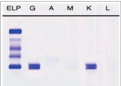
University, nineteen patients who presented AL amyloidosis Figure 2 (B) – Disappearance of Kappa monoclonal protein.
Figure 1 – Presence of periorbital hematoma at diagnosis.
Golmia et al.
608 Bras J Rheumatol 2010;50(5):603-08
were compared with 23 that presented AA amyloidosis. Approximately two-thirds of the patients presented Nephrotic Syndrome (NS) and cardiac involvement, the latter more fre-quently observed in AL amyloidosis4.
In another study, a review of 229 cases of AL amyloidosis from the Mayo Clinic assessed from 1970 to 1980 showed that one third of the patients presented NS, a little less presented heart failure and 10% presented orthostatic hypotension. Approximately half of the patients presented neuropathy and carpal tunnel syndrome.5
The present case went undiagnosed, in spite of the presence of the carpal tunnel syndrome, bilateral periorbital hematomas and presence of high levels of monoclonal protein in the peri-pheral blood. Periorbital hematomas, as seen in our case, can be the single isolated manifestation for several years, according to previous reports.6 The absence of anemia, bone lesions and
presence of moderate plasmacytosis with mature forms, led to the hypothesis is amyloidosis, which was established by rectal biopsy. As the patient was young (in the fourth decade of life), it became necessary to demonstrate that it was not a case involving some type of hereditary component, which was per-formed by attaining the diagnosis of AL amyloidosis through immunohistochemical techniques.
The treatment of AL amyloidosis depends in part of the clinical presentation and the involvement of visceral organs that are essential for survival.4 Although the conventional therapy
with melphalan and prednisone leads to partial remissions, the duration of these depend on the intensity of the cardiac, hepatic and renal involvement.
The use of chemotherapy with high doses of melphalan and autologous stem cell transplant in the absence of cardiac involvement can result in prolonged remissions, which some-times last longer than 2 years.
Survival in the AL form, in the absence of cardiac involve-ment, usually varies from around one year with conventional chemotherapy and 6 months with the opposite situation; that
seems to be the situation of the present case, as our patient has 24 months post-stem-cell transplant.7,8,9
In brief, we reported on a case of a young male adult with
AL amyloidosis, where the irst-choice conventional therapy
(melphalan and prednisone) was replaced by high doses of melphalan, followed by autologous stem cell transplant, with very favorable results, currently at 24 months post-transplant. No studies on the regression of the amyloid material were performed, as scintigraphic techniques for this purpose are not available in our country.
REFERÊNCIAS
REFERENCES
1. Scheinberg MA. Current aspects of the etiopathogenesis and therapy of amyloid disease: future trends. Rev Paul Med 1980; 96:108-11. 2. Sanchorawala V. Light-chain (AL) amyloidosis: diagnosis and
treatment. Clin J Am Soc Nephrol 2006; 1:1331-41.
3. Gertz MA, Lacy MQ, Dispenzieri A, Hayman SR. Amyloidosis. Best Pract Res Clin Haematol 2005; 18:709-27.
4. Sanchorawala V, Skinner M, Quillen K, Finn KT, Doros G, Seldin DC. Long-term outcome of patients with AL amyloidosis treated with high-dose melphalan and stem-cell transplantation. Blood 2007; 110:3561-3.
5. Scheinberg MA. Another look at serum amyloid protein SAA. Clin Rheumatol 1983; 2:431-3.
6. Passos RH, Pereira A, Neto AC, Ribeiro AF, Kutner J, Hamerschlak N et al. Clinical imagine: bilateral black eyes (raccons eyes) in AL amyloidosis. Arthritis Rheum 2006; 54:3724.
7. Perz JB, Rahemtulla A, Giles C, Szydlo RM, Davis J, Gopaul D et al. Long-term outcome of high-dose melphalanand autologous stem cell transplantation for AL amyloidosis. Bone Marrow Transplant 2006; 37:937-43.
8. Hawkins PN, Richardson S, MacSweeney JE, King AD, Vigushin DM, Lavender JP et al. Scintigraphic quantiication and serial monitoring of human visceral amyloid deposits provide evidence for turnover and regression. Q J Med 1993; 86:365-374.