Received on 26/07/2010. Accepted on 09/30/2010. We declare no conflicts of interest. This work was supported by a Specialized Center of Research (SCOR) in SLE grant awarded by the National Institutes of Health (Grant No. 5 P50 AR048940).
1. Arthritis & Immunology Program, Oklahoma Medical Research Foundation, Oklahoma City, Oklahoma, USA 2. Department of Medicine, University of Oklahoma Health Sciences Center, Oklahoma City, Oklahoma, USA
Correspondence to: James Fesmire. 825 N.E. 13th Street, MS #10, Oklahoma City, Oklahoma 73104, Phone: (405) 271-7107, Fax: (405) 271-7391. Email: [email protected].
Effects of autoimmune antibodies anti-lipoprotein
lipase, anti-low density lipoprotein, and anti-oxidized
low density lipoprotein on lipid metabolism and
atherosclerosis in systemic lupus erythematosus
James Fesmire, BS1, Marianne Wolfson-Reichlin, MS1, Morris Reichlin, MD1,2
ABSTRACT
Introduction: Premature development of atherosclerosis in systemic lupus erythematosus has been widely reported. Anti-lipoprotein lipase antibody may be one cause contributing to this disorder. Objective: To assess the extent of coronary risk due to autoimmune antibodies in terms of carotid plaque in lupus patients. Patients and Methods: We compared 114 documented lupus patients with 111 normal controls matched for sex and age. Anti-lipoprotein lipase (A-LPL), anti-oxidized low density lipoprotein (A-OXLDL), and anti-low density lipoprotein (A-LDL) were measured by enzme-linked immunoabsorbent assay. Low density triglyceride (LDL-Trig) and high density lipoprotein-triglyceride (HDL-Trig) were also measured. Plaque was measured by bilateral carotid ultrasound. Results: 45.6% of patients tested positive for A-LPL, and 34.4% for A-OXLDL. 44% of normal controls tested positive for A-LPL, and 20% for A-OXLDL. Risk increased sharply in subgroups with increased antibody levels. Patients with A-LPL and A-OXLDL > 0.40 (n = 12) showed coronary risk correlations of: A-LPL x LDL-Trig = 0.7008, P = 0.0111; bilateral ultrasound vs total cholesterol = 0.62205, P = 0.0308; LDL-Trig vs myocardial infarction (MI) = 0.76562, P = 0.0037; total triglycerides vs MI = 0.78191, P = 0.0027); LDL-Trig/LDL-cholesterol vs MI = 0.80493, P = 0.0016; A-OXLDL
vs USBL = 0.71930, P = 0.0084. Correlations of SLEDAI with risk variables were highly signiicant only in subgroups
of elevated antibody levels (SLEDAI x A-OXLDL = 0.70366, P = 0.0107). Conclusion: A-LPL initiates the develop-ment of LDL mutations, followed by antibody production, plaque formation and coronary risk in some SLE patients.
Keywords: systemic lupus erythematosus, dyslipidemia, triglycerides, lipoprotein lipase, atherosclerosis.
INTRODUCTION
Previous studies have shown a relationship between antibodies to lipoprotein lipase and elevated triglycerides in patients with systemic lupus erythematosus (SLE).1-4 The Pearson
correla-tion 0.84 (P = 0.0001) of anti-lipoprotein lipase (A-LPL) was reported by this laboratory in 2002.1 A-LPL also correlated at
0.85 (P = 0.0001) with low density lipoprotein-triglyceride
(LDL-Trig), and 0.85 (P = 0.001) with Apo B respectively. Apo E correlated with a-LPL at 0.87 (P = 0.0002). These lipid-lipoprotein particles are representative of the very low density lipoprotein (VLDL) and LDL density classes which play central roles in lipid metabolism.
these antibodies may be part of the mechanism underlying the premature atherosclerosis characteristic of SLE.
It has been reported that circulating triglycerides alone present a direct risk for the development of atherosclerosis.5,6
LDL-Trig is also reported to be an independent risk factor of coronary artery disease and inlammatory agent.7 This
accen-tuates the overall risk of excess triglyceride in circulation, particularly in combination with other developing mutant lipoprotein forms that inhibit lipid metabolism. Our focus was to relate those effects on the established lipid transport mechanism in terms of plaque formation and coronary events.
The presence of plaque measured by ultrasound of the carotid arteries has been shown to be a useful predictor of coronary artery disease and is associated with clinical risk of coronary artery disease events such as angina and myocardial infarction.8,9 In our present study, we used carotid plaque scores
as a measure of atherosclerosis in 114 SLE patients and 111 normal subjects. Additionally, complete lipid proiles on serum, LDL and HDL were performed, as well as antibody levels for A-LPL, anti-oxidized low density lipoprotein (A-OXLDL), and anti-low density lipoprotein (A-LDL) on all subjects. The collected data was used to evaluate the role of these autoimmu-ne antibodies in the development of premature atherosclerosis characteristic of SLE patients.
PATIENTS AND METHODS
This study was approved by the Institutional Review Board of the Oklahoma Medical Research Foundation and all sub-jects signed informed consent forms. The patient population was predominantly female totaling 114 subjects including 10 males matched for sex and age ranging from age 16 to 87 with an average age of 43. The majority of patients had a long term duration of disease, and consequently were on the usual variety of autoimmune medications. However, none of the study subjects were taking lipid lowering medications. No lipid exclusion limits were imposed on patients or controls. All patients were tested and met the diagnostic criteria for SLE of the American College of Rheumatology.10 They also tested positive for “The Reichlin
Proile”, anti-nuclear antibody (ANA), anti-double strand DNA (dsDNA) and extractable nuclear antibodies (ENA). Controls were recruited from within our local health science center, selected to match the patient group for sex and age, and otherwise were in a healthy state of well being, taking no lipid lowering medications.
Carotid ultrasound
Plaque scores (measured on a scale from 0 to 10) were per-formed and provided by the Cardiovascular section of the Department of Medicine, University of Oklahoma Health Sciences Center. Study subjects were given a duplex carotid screen (both arteries) by Doppler sonography. The atheros-clerotic burden of plaque was expressed as the sum of values measured in both arteries. Only study subjects having a value entry of the ultrasound test were included for statistical corre-lations of study groups.
Assays of autoimmune antibodies
The same standard enzyme-linked immunoabsorbent assay (ELISA) methodology for measurements of the anti-LPL, anti-OXLDL, and anti-LDL was used in this study. Only the reactant antigens and antibodies differ. This consisted of coa-ting the plate with the antigen then adding the subject serum at a 100 fold dilution with incubation overnight followed by two washes, then adding the anti-human IgG with the O.D. read at 280 nm. This laboratory reference method was applied for measurement of all patients and controls.1
Antibody/Lipid determination
The measurements of cholesterol and triglycerides were perfor-med in accordance with the manual of Laboratory Operations, Lipid and Lipoprotein Analysis, Revised 1982 Methodology, Lipid Research Clinics Program.11 Enzymatic lipid reagents
used in the study were purchased from Roche Laboratories and used on a Cobas Mira Plus autoanalyzer manufactured by Roche Laboratories as described in Reichlin, et al.1
The accuracy and precision of the assays were maintained by an ongoing successful performance in a national analytical surveillance program.
Analytical HDL/LDL isolation
loat the LDL supernate. The LDL supernate was removed and stored with the HDL to be analyzed by routine cholesterol and triglyceride serum assays.Measured and derived methods using the 1:5 VLDL-cholesterol:triglyceride ratio of the Friedwald estimation for LDL-cholesterol (LDL-Chol) determination were applied for all samples in the study. A comparison of 1,100 samples analyzed for veriication of LDL-Chol values by the measured method with the derived method yielded a correla-tion coeficient of 0.82 with a signiicant P value of < 0.0001.
Preparative LDL isolation
Isolation of the low density lipoprotein was performed ac-cording to the method of Lee et al. 12 with two additional
centrifugation washes at density 1.070 Na Cl to exclude albu-min. Ultracentrifugation runs were performed for 26 hours at 45,000 rpm in a Beckman J-25 ultracentrifuge. The isolated LDL fractions were dialyzed for two days in .05% EDTA, PBS prior to use. LDL Oxidation
For the oxidation of LDL, a slightly modiied version of the copper oxidation method by Palinski et al, 1990, was used.13
200 µL of the intact LDL at 3.80 mg/mL was added with 5 µL of 1 M copper sulfate in 1 mL of PBS for each preparation of the study. The mixture was incubated overnight in a water bath at 37°C, then centrifuged at 10,000 rpm in a Sorvall RC5C centrifuge. The supernate was removed and dialyzed in 0.10% EDTA PBS for two days.
The oxidized sample was tested by immunodiffusion against anti-Apo B as compared with the intact LDL and sho-wn to present a comparatively faint disrupted precipitin line.
Data management
Data ile storage, statistical analysis and regression plots were performed using SAS software, Version 9.1, purchased from SAS Institute Inc. of Cary, North Carolina.
RESULTS
Table 1 shows the comparison of A-LPL (+) and (-) patients with controls and the stages of associated risk variables. The majority of signiicant variables were comprised of triglyce -ride containing particles. Total cholesterol, LDL-Chol and HDL-Chol were not dramatically different among groups. HDL-Chol was mildly so.
Statistical review of a subgroup of A-OXLDL (+) patients (n = 41) also showed a very similar mean A-LPL value for the A-LPL antibody (average A-LPL = 0.41; A-OXLDL = 0.41). However, a subgroup of patients selected for an A-LPL
Table 1. A-LPL positive/negative levels in patient and control groups
Variable Mean SD Mean SD t(P value)-test
Normal Control
A-LPL (-) (n = 62) Normal Control A-LPL (+) (n = 9)
Plaque 0.37 0.87 0.40 0.90 0.43 Total Chol 187.7 38.8 195.8 33.3 0.0499 Total Trigl 120.4 53.5 129.9 27.5 0.4669 LDL-Chol 105.9 31.2 112.6 23.5 0.0718 LDL-Trig 73.4 73.4 84.4 59.8 0.3881 Trig/Chol 0.65 0.31 0.65 0.34 0.4075 LDL-Trig/LC 0.70 0.34 0.72 0.43 0.4144 HDL-Trig/HC 0.79 0.74 0.76 0.32 0.3932 A-OXLDL 0.14 0.12 0.17 0.11 0.0145 A-LPL 0.28 0.17 0.54 0.19 <0.0001 HDL-Chol 53.8 53.8 56.4 15.4 0.2244 HDL-Trig 39.5 39.5 40.5 14.3 0.2058
Lupus A-LPL (-) (n = 62)
Lupus A-LPL (+) (n = 52)
Plaque 0.74 1.18 1.19 1.95 0.2223 Total Chol 196.8 59.0 202.1 50.6 0.2446 Total Trig 149.9 79.3 191.2 118.6 0.0099 LDL-Chol 111.4 42.3 121.3 35.8 0.0668 LDL-Trig 98.8 69.3 126.3 70.6 0.0083 Trig/Col 0.78 0.42 0.94 0.44 0.0141 LDL-Trig/LC 0.88 0.55 1.04 0.50 0.0333 HDL-Trig/HC 0.85 0.38 0.93 0.39 0.0949 A-OXLDL 0.19 0.15 0.28 0.13 0.0108 A-LPL 0.25 0.13 0.51 0.16 <0.0001 HDL-Chol 54.1 20.5 50.8 16.6 0.1943 HDL-Trig 42.0 12.4 44.2 16.4 0.3338
Normal Control A-LPL (-) (n = 62)
Lupus A-LPL (+) (n = 52)
Plaque 0.37 0.87 1.19 1.95 0.0032 Total Chol 187.7 38.8 202.1 50.6 0.0521 Total Trig 120.4 53.5 191.2 118.6 <0.0001 LDL Chol 105.9 31.2 121.3 35.8 0.0209 LDL Trig 73.4 73.4 126.3 70.6 <0.0001 Trig/Chol 0.65 0.31 0.94 0.44 0.0001 LDL-Trig/LC 0.70 0.34 1.04 0.50 0.0002 HDL-Trig/HC 0.79 0.74 0.93 0.39 0.0025 A-OXLDL 0.14 0.12 0.28 0.20 <0.0001 A-LPL 0.28 0.17 0.51 0.16 <0.0001 HDL-Chol 53.8 53.8 50.8 16.6 0.0728 HDL-Trig 39.5 39.5 44.2 16.4 0.0237 Total Chol, total cholesterol; Total Trig, total triglyceride; LDL-Chol, low density lipoprotein-cholesterol; LDL-Trig, low density lipoprotein-triglyceride; Trig/Chol, triglyceride/lipoprotein-cholesterol; LDL-Trig/LC, low density lipoprotein-triglyceride/low density lipoprotein cholesterol; HDL-Trig/HC, high density lipoprotein-triglyceride/high density lipoprotein-cholesterol; A-OXLDL, anti-oxidized lipoprotein lipase; A-LPL, anti-lipoprotein lipase; HDL-Chol, high density lipoprotein-cholesterol; HDL-Trig, high density lipoprotein-triglyceride.
The highest O.D. levels of autoimmune antibodies measu-red were from anti-LPL. The most signiicant impact on lipid proiles was shown by elevated triglycerides in whole serum and among the measured density classes LDL and HDL.
Bilateral measurements of carotid plaque done by ul-trasound (USBL) were highest in patient groups measuring positive for A-LPL.
In Table 2 the highest correlation with plaque is sho-wn with triglyceride measured in the HDL fraction of patients with A-LPL (+), in contrast to subjects of both SLE and control groups having low levels of A-LPL for which a correlation with plaque is not shown. Patients which were negative for the A-LPL antibody showed a significant negative correlation for the USBL x HDL-TG variable by comparison. Patients measuring > 0.45 for the A-LPL antibody (n = 37) showed significant correlations of incidence of MI with lipid distribution ratios of serum density classes.
We found the greatest percent distribution in favor of triglyceride in the LDL fraction of patients. Comparison by
t test of the LDL-Trig and the LDL distribution ratio were highly signiicant compared to the normal group (P = 0.0001), although LDL-Chol was not signiicant.
Table 2. Correlation of ultrasound carotid plaque score and incidence of myocardial infarction in lupus subgroups of anti-LPL levels with lipid variables
Correlation variables Lupus A-LPL (-) (n = 67) P value
USBL x Total Chol 0.0749 0.5467
USBL x Total Trig 0.0044 0.9716
USBL x HDL-Trig/HDL-Col -0.1560 0.2148
USBL x HDL-Trig -0.2866 0.0206
Lupus A-LPL (+) (n = 52)
USBL x Total Chol 0.2659 0.0567
USBL x Total Trig 0.2927 0.0352
USBL x HDL-Trig/HDL-Chol 0.3130 0.0239
USBL x HDL-Trig 0.4372 0.0012
Lupus A-LPL > 0.45 O.D. (n = 37)
MI x Trig/Chol 0.4231 0.0091
MI x LDL-Trig 0.3822 0.0196
IM x LDL-Trig/LDL-Chol 0.5052 0.0014
MI x HDL-Trig/HDL-Chol 0.4358 0.0076
USBL, bilateral ultrasound; Total Chol, total cholesterol; Total Trig; total triglyceride; HDL-Trig/HDL-Chol, high density triglyceride/high density lipoprotein-cholesterol; MI, myocardial infarction; A-LPL, anti-lipoprotein lipase.
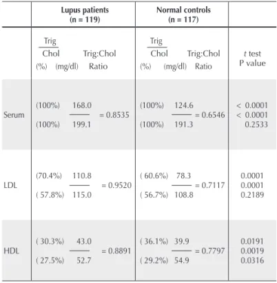
Table 3. Average lipid distribution ratios of lupus patients and normal controls
Lupus patients
(n = 119) Normal controls(n = 117)
Trig
Chol Trig:Chol (%) (mg/dl) Ratio
Trig
Chol Trig:Chol (%) (mg/dl) Ratio
t test P value
Serum
(100%) 168.0 = 0.8535 (100%) 199.1
(100%) 124.6 = 0.6546 (100%) 191.3
< 0.0001 < 0.0001 0.2533
LDL
(70.4%) 110.8
= 0.9520 ( 57.8%) 115.0
( 60.6%) 78.3 = 0.7117 ( 56.7%) 108.8
0.0001 0.0001 0.2189
HDL
( 30.3%) 43.0
= 0.8891 ( 27.5%) 52.7
( 36.1%) 39.9 = 0.7797 ( 29.2%) 54.9
0.0191 0.0019 0.0316 Beyond the increasing risk for coronary events with increasing A-LPL shown in Table 2, a subset of 21 patients containing antibodies to both A-LPL and A-OXLDL higher than baseline at > 0.3500 O.D. units showed a correlation coeficient of MI x LDL-Trig/LDL-Chol = 0.75897, P ≤ 0.0001 (data not shown).
Given that LPL activity is a major point at issue, the density classes LDL and HDL were isolated to permit cho-lesterol and triglyceride, including the distribution within and among patients and normals. Table 3 shows the t test
comparative results of the Trig/Chol ratios of SLE patients with normal controls at the right margin of the table. These data relect the percent distribution of Trig:Chol within serum, LDL and HDL and the t test comparison of each
Table 4. Correlations of autoimmune antibodies with coronary risk variables
Correlation variables (n = 67) (range 0.16 – 0.83)Lupus A-OXLDL (+) P value
LDL-Chol x A-LPL 0.4190 0.0064
LDL-Chol x A-LDL 0.4774 0.0018
LDL-Chol x A-OXLDL 0.4267 0.0068
%LDL-Trig x A-LPL 0.4014 0.4126
HDL-Trig x A-LDL 0.4126 0.0091
%LDL-Trig x MI 0.3158 0.0430
Normal control A-OXLDL (-) (n = 88)
LDL-Chol x A-LPL 0.2159 0.0433
LDL-Chol x A-LDL -0.0244 0.8212
LDL-Chol x A-OXLDL 0.1245 0.2480
%LDL-Trig x A-LPL 0.0727 0.5008
HDL-Trig x A-LDL 0.0354 0.7464
%LDL-Trig x MI No MI No MI
Autoimmune antibodies normal control A-OXLDL (+) (n = 23)
(range 0.16 – 0.61)
LDL-Chol x A-LPL 0.1974 0.3665
LDL-Chol x A-LPL 0.2672 0.2178
LDL-Chol x A-OXLDL 0.5258 0.0100
%LDL-Trig x A-LPL -0.4080 0.0533
HDL-Trig x A-LDL 0.4740 0.0223
%LDL-Trig x IM No MI No MI
Lupus patients with A-LPL and A-OXLDL > 0.400 O.D. (n = 12) Total Chol x Carotid
Plaque (USBL)
0.6225 0.0380
Total Serum Trig x MI 0.7819 0.0027
LDL-Trig/LDL-Chol x MI 0.8049 0.0016
LDL-Trig x MI 0.7656 0.0037
HDL-Trig/HDL-Chol x MI 0.7031 0.0108
LDL-Chol x CVA 0.6825 0.0145
Anti-LPL x LDL-Trig 0.7480 0.0081
Anti-OXLDL x Carotid
Plaque (USBL) 0.7193 0.0084
LDL-Chol, low density lipoprotein-cholesterol; A-LPL, lipoprotein lipase; A-LDL, anti-low density lipoprotein; A-OXLDL, anti-oxidized anti-low density lipoprotein; %LDL-Trig, percent of low density lipoprotein of total triglyceride; USBL, bilateral ultrasound; MI, myocardial infarction; HDL-Trig/HDL-Chol, high density lipoprotein-triglyceride/high density lipoprotein cholesterol; CVA, cerebrovascular accident; LDL-Trig, low density lipoprotein-triglyceride.
Table 5. Correlations of risk variables in SLEDAI subgroups
Correlation variables SLEDAI ≥ 25 (n = 30) P value
SLEDAI x Total Trig 0.4440 0.0140
Plaque x Total Chol 0.5091 0.0041
SLEDAI x HDL-Trig 0.5948 0.0005
Plaque x LDL-Chol 0.5452 0.0018
Plaque x LDL-Trig 0.3899 0.0331
SLEDAI x MI 0.3693 0.0446
CVA x MI 0.5649 0.0011
SLEDAI < 25 (n = 81)
SLEDAI x Total Trig -0.0094 0.9350
Plaque x Total Chol 0.0807 0.4739
SLEDAI x HDL-Trig 0.0874 0.4527
Plaque x LDL-Chol 0.0721 0.5224
Plaque x LDL-Trig 0.1086 0.3345
SLEDAI x MI 0.2997 0.0077
CVA x MI -0.0362 0.7479
SLEDAI ≥ 25 + A-LPL > .35 O.D. (n = 10)
SLEDAI x Plaque 0.8586 0.0015
SLEDAI x LDL-Chol 0.7387 0.0147
Plaque x LDL-Chol 0.6837 0.0293
Plaque x Trig/A-LPL 0.7962 0.0054
Plaque x Total Trig 0.6395 0.0465
SLEDAI, systemic erythematosus disease activity index; Total Trig, total triglyceride; Total Chol, total cholesterol; HDL-Trig, high density lipoprotein-triglyceride; LDL-Chol, low density lipoprotein-cholesterol; LDL-Trig, low density lipoprotein-triglyceride; MI, myocardial infarction; CVA, cerebrovascular accident; A-LPL, anti-lipoprotein lipase. Correlations of LDL lipids (Table 4) with all three
autoimmune antibodies measured in subject samples abo-ve baseline show an increased risk in both patients and controls where a combination of antibodies are positive. The subset of patients in Table 4 having both A-LPL and A-OXLDL > 0.40 (n = 12) shows the highest correlations among LDL associated antibodies, LDL risk variables and coronary risk.
with variable activity management eficiencies. The overall ave -rage SLEDAI is elevated at 19.0, although does not signiicantly correlate with risk variables and events as a group. However, as shown in Table 5, subgroups of the population of SLEDAI > 25 x < 25 begin to show consistent patterns of risk according to these groups. Further, in a subgroup of more severe conditions having SLEDAI > 25 combined with A-LPL above the background baseline of 0.35 OD, the correlations of SLEDAI with plaque and other known coronary risk variables become more dramatic. We conclude these data are supportive of previous reports3,18 of
increased lipid dysfunction during high disease activity. Results of this study support closer attention being focu-sed on autoimmune antibodies as a potentially increafocu-sed risk for premature development of atherosclerosis in some SLE patients. Elevated levels and distribution of triglyceride in serum and density classes may be an indication, of autoimmune activity on lipid transport. However, equally important, the results also emphasize the need for further study to explore the presence and roles of other autoimmune antibodies con-tributing to this risk.
While only the higher concentrations of antibodies showed strong correlations with coronary risk variables, we suggest that constant exposure to low levels of speciic antibodies also present a risk for development of vascular disease in SLE patients and may also develop as a source of coronary risk in normal subjects over time.
REFERÊNCIAS REFERENCES
1. Reichlin M, Fesmire J, Quintero-Del-Rio AI, Wolfson-Reichlin M. Autoantibodies to lipoprotein lipase and dyslipidemia in systemic lupus erythematosus. Arthritis Rheum 2002; 46:2957-63.
2. de Carvalho JF, Borba EF, Viana VST, Bueno C, Leon EP, Bonfá E. Anti-Lipoprotein Lipase Antibodies. Arthritis Rheum 2004; 50(11):3610-5.
3. de Carvalho JF, Viana VST, Neto EFB, Leon EP, Bueno C, Bonfá E. Longitudinal luctuation of anti-lipoprotein lipase antibody is related with disease activity in systemic lupus erythematosus patients without anti-dsDNA antibodies. Rev. Bras. Reumatol 2009; 49(1):39-47.
4. Kodera M, Hayakawa I, Komura K, Yanaba K, Hasegawa M, Takehara
K et al. Anti-lipoprotein lipase antibody in systemic sclerosis:
Association with elevated serum triglyceride concentrations. J Rheumatol 2005; 32(4):629-36.
5. Nordestgaard BG, Benn M, Schnohr P, Tybjaerg-Hansen A. Nonfasting triglycerides and risk of myocardial infarction, ischemic heart disease, and death in men and women. JAMA 2007; 298:299-308.
6. Patsch JR, Miesenbock G, Hopferwieser T, Muhlberger V, Knapp E, Dunn JK et al. Relation of triglyceride metabolism and coronary
artery disease, studies in the postprandial state. Arterioscler Thromb 1992; 12(11):1336-45.
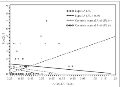
Figure 1. Relationship of anti-oxidized low density lipoprotein (A-OXLDL) with carotid artery plaque among subgroups of patients and controls at different levels of anti-lipoprotein lipase (A-LPL).
PLA
QUE
9
8
7
6
5
4
3
2
1
0
A-OXLDL (O.D.)
0.25 0.35 0.45 0.55 0.65 0.75 0.85 0.95 1.05 1.15 1.25 Lupus A-LPL (-)
Lupus A-LPL > 0,40 Controle normal Anti-LPL (+) Controle normal Anti-LPL (-)
Figure 1 shows the highest relationship with measured plaque formations to be in lupus patients with high levels of both A-LPL and A-OXLDL, compared with all other groups.
DISCUSSION
A general review of the data in this study shows approximately 47% of patients to be A-LPL positive, with approximately 36% positive for the A-OXLDL antibody. The levels and distribution of antibodies observed are consistently higher for A-LPL than A-OXLDL in the antibody positive group.
It is well established that lipid-lipoprotein particles among density classes are metabolically processed forming a sequen-ce of diminishing size and lipid distribution, beginning with LPL activity on triglyceride in chylomicron and continuing through HDL, resulting in increasing density and loss of lipid due to enzymic activities during the course of normal lipid transport.14-17 LPL being irst in the sequence of transport,
combined with the observations of antibody levels and dis-tribution as described, we interpret that the A-LPL antibody develops irst, with A-OXLDL developing later as a result of A-LPL impeding transport and promoting mutation, which subsequently produces mutant autoimmune antibodies which collectively contribute greatly to the lipid dysfunction and lipid excess in circulation in some SLE patients.
13. Palinski W, Ylä-Herttuala S, Rosenfeld ME, Butler SW, Socher SA, Parthasarathy S et al. Antisera and monoclonal antibodies speciic
for epitopes generated during oxidative modiication of low density lipoprotein. Arteriosclerosis 1990; 10(3):325-35.
14. Havel RJ, Eder HA, Bradgon JH. The distribution and chemical composition of ultracentrifugally separated lipoproteins in human serum. J Clin Invest 1955; 34(9):1345-53.
15. Blackett PR, Kittredge D. Hyperlipidemia in children. South Med J 1993; 86:1083-92.
16. Friedwald WT, Levy RI, Fredrickson DS. Estimation of the concentration of low-density lipoprotein cholesterol in plasma, without use of the preparative ultracentrifuge. Clin Chem 1972; 18(6):499-502.
17. Warnick GR, Knopp RH, Fitzpatrick V, Branson L. Estimating low-density lipoprotein cholesterol by the Friedewald equation is adequate for classifying patients on the basis of nationally recommended cutpoints. Clin Chem 1990; 36(1):15-9.
18. Borbá EF, Bonfá E. Dyslipoproteinemias in systemic lupus erythematosus: Inluence of disease activity and anticardiolipin antibodies. Lupus 1997; 6:533-9.
7. März W, Scharnagl H, Winkler K, Tiran A, Nauck M. Low-density lipoprotein triglycerides associated with low-grade systemic inlammation, adhesion molecules and angiographic coronary artery disease: The Ludwig Shafen Risk and Cardiovascular Health Study. Circulation 2004; 110: 3068-3074.
8. Zanolla CN, Franceschini L, Cacici G, De Cristan B, Arieti M, Vassanelli C. Usefulness of ultrasonographic markers of carotid atherosclerosis (intima-media thickness, unstable carotid plaques and severe carotid stenosis) for predicting presence and extent of coronary artery disease. J Cardiovasc Med 2009; 10(12):906-12. 9. Johnsen SH, Mathiesen EB. Carotid plaque compared with intima-medial thickness as a predictor of coronary and cerebrovascular disease. Curr Cardiol Rep 2009; 11(1):21-7.
10. Tan EM, Cohen AS, Fries JF, Masi AT, McShane DJ, Rothield NF
et al. The revised criteria for the classiication of systemic lupus
erytematosus. Arth Rheum 1982; 25:1271-7.
11. Lipid Research Clinics Program. Manual of Laboratory Operations: Lipid and Lipoprotein Analysis. Second Edition, Bethesda, MD: National Heart, Lung and Blood Institute, NIH, U.S. Dept. of Health and Human Services, 1982.