ISSN/$–see front matter © 2013 Sociedade Brasileira de Ortopedia e Traumatologia. Published by Elsevier Editora Ltda. All rights reserved. Rev Bras Ortop. 2013;48(1):114-117
www.rbo.org.br/
*Corresponding author at: Rua Dr. Cesário Motta Júnior, 112, Prédio Ortopedia, 2° andar, Sala Quadril, CEP: 01221-020, São Paulo, SP, Brazil.
E-mail: [email protected] article info
Article history:
Received November 28 2011 Approved December 4 2012
Keywords:
Piriformis Muscle Syndrome Diagnosis
Magnetic Resonance Imaging Endoscopy
Case Report
Anatomical variation of piriformis muscle as a cause of deep
gluteal pain: diagnosis using MR neurography and treatment
Giancarlo Cavalli Polesello,
1*Marcelo Cavalheiro Queiroz,
2João Paulo Tavares Linhares,
3Denise Tokechi Amaral,
4Nelson Keiske Ono
51PhD. Assistant Professor and Head of the Hip Group, School of Medical Sciences, Santa Casa de São Paulo, São Paulo, SP, Brazil. 2Orthopedist and Attending Physician in the Hip Group, School of Medical Sciences, Santa Casa de São Paulo, São Paulo, SP, Brazil.
3Orthopedist and Trainee in the Hip Group, School of Medical Sciences, Santa Casa de São Paulo, São Paulo, SP, Brazil. 4Radiologist at the Syrian-Lebanese Hospital, São Paulo, SP, Brazil.
5PhD. Adjunct P and Attending Physician in the Hip Group, School of Medical Sciences, Santa Casa de São Paulo, São Paulo, SP, Brazil.
Work performed in the Department of Orthopedics and Traumatology, School of Medical Sciences, Santa Casa de São Paulo, São Paulo, SP, Brazil.
a b s t r a c t
Female patient, 42 years old with a history of low back pain on the left for seventeen years in which the definitive diagnosis of the etiology of pain was evident after the completion of neurography magnetic resonance imaging of the sciatic nerve. In this test it was identified the presence of an anatomical variation in the relationship between the piriformis muscle and sciatic nerve. We discuss details of this imaging technique and its importance in the frames of refractory low back pain. We also describe the treatment given to the case.
© 2013 Sociedade Brasileira de Ortopedia e Traumatologia. Published by Elsevier Editora Ltda. All rights reserved.
Rev Bras Ortop. 2013;48(1):114-117
115
Introduction
In 1928, Yeoman was the first to describe the piriformis muscle as an etiological factor in sciatic pain and low back pain.1 Between the sciatic nerve and the piriformis muscle,
there are many anatomical variations and some authors have correlated this condition with piriformis syndrome2 and deep
gluteal pain syndrome.3
Because of the similarity between this condition and disorders of the lumbar region, there is no consensus regarding its diagnosis and no specific criteria for this.4 In
this context, in addition to clinical criteria and a directed physical examination, magnetic resonance can be used for most patients, especially for evaluating the lumbosacral spine. However, when performed using traditional techniques, this diagnostic method is not always capable of identifying the origin of the problem, particularly when there is involvement of the lumbosacral plexus or the sciatic nerve. In these cases, magnetic resonance neurography is the preferred examination for defining the intrinsic abnormalities of the sciatic nerve and the possible anatomical variations, and for characterizing the extrinsic compression of the nerve bundle, thereby enabling better surgical planning.5
In this patient, with a history of left-side low back pain for 17 years, the definitive diagnosis of the etiology of the pain was only achieved through magnetic resonance neurography. Subsequently, endoscopic treatment of the abnormalities found was performed.
Case report
The patient was a 42-year-old woman who was a physiotherapist. At the age of 25 years, she started to present a condition of left-side low back pain that, at that time, was diagnosed as L5-S1 spondylolisthesis, with an associated disc hernia. After six months of treatment with analgesics and physiotherapy, she evolved with a deficit of hallux extension strength and then underwent L5-S1 arthrodesis without instrumentation. Even with this treatment, she often felt recurrent pain in the left gluteal region. Two years before she came to our clinic (end of 2009), her condition evolved with worsening of the pain, without any factor that would improve it, which prevented her from continuing to work. The pain worsened when she was seated or was standing upright for a long time. Over the latter two years, she underwent treatment with cortisone infiltration into the gluteal region, with fleeting improvements. Also over that period, she underwent five magnetic resonance examinations on the lumbosacral spine and left hip, without any diagnostic elucidation (Fig. 1). In February 2010, she underwent botulinum toxin application to the piriformis muscle and thereafter remained asymptomatic for four months. In May 2011, with recurrence of the symptoms, she sought our clinic for diagnostic investigation and treatment.
Diagnosis
The Friberg test4 (adduction and forced passive internal
rotation of the lower limb affected) and Pace test4 (abduction
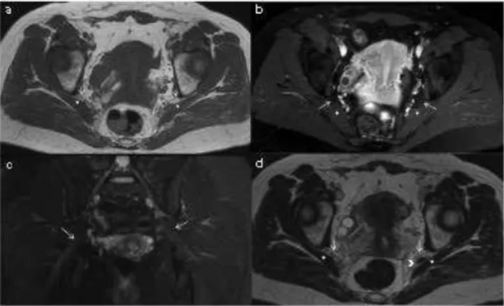
and external rotation against resistance) were positive. The patient presented pain in the gluteal region, which worsened with palpation in the region of the external hip rotators, at approximately 1 cm laterally to the ischium. Taking together the physical examination with the report of temporary improvement of the symptoms through application of botulinum toxin to the piriformis muscle, a diagnosis of deep gluteal pain syndrome was suspected. After analysis on magnetic resonance imaging of the hips and lumbosacral spine, which showed normal results (Fig. 1), magnetic resonance neurography was performed over the left hip region. The equipment used was the Philips Achieva, with the following specifications: 1.5 Tesla, phased array coil, an isotropic volumetric sequence named Vista, T2 with TR/TE of 4500/90 ms, thickness of 1 mm and FOV of 28 cm. Following this, multiplanar reformatting was done along the major axis of the sciatic nerve (Fig. 2).
An accessory muscle belly of the left piriformis was identified (anatomical variation), and the fibular branch of the sciatic nerve passed between the fibers of this accessory belly and the standard piriformis muscle (Fig. 2). This corresponded to a type B variation (Fig. 3), according to the classification of Beaton and Anson.5 Because of this, compression and
tensioning of the sciatic nerve was occurring, with consequent pain that was refractory to the treatments previously instituted. The tendon of the piriform muscle was in reality acting as a dart, transfixing the nerve.
116
Rev Bras Ortop. 2013;48(1):114-117Treatment
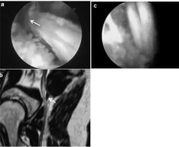
In July 2011, using anterior and posterior peritrochanteric portals and an auxiliary portal (Fig. 5), endoscopic release of the piriformis was performed by means of its muscle mass, in association with tenotomy (Fig. 4).
Fig. 2 - Magnetic resonance neurography: reformatting to the major axis of the sciatic nerve (arrow) on the right side (a) which passes anteriorly to the piriformis muscle. On the left side (b), the accessory muscle belly of the piriformis (*) passes between the common fibular and tibial bands of the sciatic nerve.
Fig. 3 - Beaton and Anson classification for the
anatomical variations between the piriformis muscle and the sciatic nerve.
Fig. 4 - (a) Transoperative image of the anatomical variation of the piriformis muscle and (b) correlation with the image from magnetic resonance neurography (arrow). Postoperative image (c) after surgical release of the piriformis muscle.
Fig. 5 - Anterior paratrochanteric arthroscopic portal (PTA), posterior paratrochanteric arthroscopic portal (PTP) and auxiliary portal (PAX).
After the operation, a specific rehabilitation protocol for such cases was used, consisting of stretching exercises, cryotherapy and muscle reinforcement. Nine months after the surgery, the patient was asymptomatic.
Discussion
The first descriptions of the piriformis muscle acting as an etiological factor on sciatic pain and low back pain date from 1928, published by Yeoman.1 The presence of anatomical
variations between the sciatic nerve and the piriformis muscle have been reported with the appearance of the piriformis syndrome,2 characterized by sensory, motor and trophic
disorders in the area of innervation of the sciatic nerve. The incidence of this syndrome in the population is only 6%, and it is more common among females than among males.6
In around 83% of the population, the sciatic nerve leaves the pelvis in a single stem and passes below the piriformis muscle, maintaining a descending path towards the popliteal fossa, where it divides into two terminal branches: the common fibular nerve and the tibial nerve.5 Among patients who present
anatomical variations, 81% have an appearance similar to what was presented by our patient, in whom the sciatic nerve emerged already divided, with the common fibular part crossing the middle of the belly of the piriformis muscle and the tibial part passing below the lower margin of this muscle.5 Pecina8
emphasized that anatomical variations of this type could lead to development of the piriformis syndrome, in which stretching of the piriformis muscle might compress the common fibular branch between the tendinous parts of this muscle.
Rev Bras Ortop. 2013;48(1):114-117
117
patients with complaints of refractory sciatic pain and low back that remain unexplained through subsidiary examinations such as magnetic resonance, given that the latter examination does not have the capacity to evaluate pain originating in the lumbosacral plexus or sciatic nerve.5
Clinical tests such as the Friberg, Pace and FADIR tests4
(flexion, adduction and internal rotation of the affected hip) that show deep gluteal pain irradiating through the region of the sciatic nerve reinforce the hypothesis of compression of this nerve in the deep gluteal region. However, this diagnosis is controversial and it has been difficult to obtain objective evidence for the existence of this entity.9 In this context,
magnetic resonance neurography is an important diagnostic option. In this technique, high-resolution 1 mm slices are used, with T1 and T2-weighted sequences and fat suppression.5 The
T1 sequence enables anatomical evaluation of the muscle layers and better definition of the peripheral nerves, which present a fascicular pattern with an intermediate signal, surrounded by perineural fat with hypersignal. With the T2 sequence with fat saturation, the objective is to define whether there is any thickening and hypersignal of the sciatic nerve, along with signs of denervation of the piriformis muscle characterized by increased signal.9
Isotropic volumetric data acquisition enables post-processing of the images on a workstation with distortion of the images, thus making it possible to reformat the data along the major axis of the sciatic nerve in the sagittal and oblique coronal planes.10 Lewis et al.11 evaluated 14 patients
with low back pain and normal results from magnetic resonance who underwent magnetic resonance neurography of the lumbosacral plexus and sciatic nerve. Of these, 12 presented signal abnormalities relating to the sciatic nerve, and in eight cases these alterations were located at the ischial incisure or in the piriformis muscle. The patient of the present case underwent five magnetic resonance examinations on the left hip and lumbosacral spine, all with normal results. The definitive diagnosis, made following clinical suspicion, was only achieved through magnetic resonance neurography, from which a Beaton and Anson type B variation was identified.
The initial treatment for these cases is eminently clinical, with use of nonsteroidal anti-inflammatory drugs, physiotherapy, stretching and strengthening exercises, massage, local application of heat, cryotherapy, muscle relaxants, perisciatic injection of corticoids and injection of botulinum toxin guided by computed tomography.12 Fanucci
et al.13 evaluated 30 patients with 12 months of follow-up
after injection of intramuscular botulinum toxin into the piriformis guided by means of computed tomography, which was found to provide symptom relief in all the patients. Despite the good results presented in the literature, the patient of the present case only achieved an improvement for a period of approximately four months, with subsequent recurrence of the symptoms.
After failure of conservative treatment, surgical treatment is indicated. Reports on open exploration have shown good results, although the endoscopic method has shown lower morbidity and achieved extensive exploration of the path of
the nerve, along with identifying anatomical abnormalities.13
Martin et al.3 reported good results from endoscopic treatment
on a series of 35 patients with deep gluteal pain, mean follow-up of 23 months and use of a technique similar to that of the present case.
Therefore, it seems to us that it is important to perform magnetic resonance neurography on the sciatic nerve, in cases in which the etiology of the pain has not been elucidated through conventional magnetic resonance, in order to obtain diagnostic clarification of deep gluteal pain and identify anatomical variations that give rise to compressive effects on the sciatic nerve.
Conflicts of interest
The authors declare that there was no conflict of interests in conducting this study.
R E F E R E N C E S
1. Yeoman W. The relation of arthritis of the sacro-iliac joint to sciatica: with one analysis of 100 Cases. Lancet. 1928;2:1119-23. 2. Fishman LM, Dombi GW, Michaelsen C, Ringel S, Rozbruch
J, Rosner B, et al. Piriformis syndrome diagnosis, treatment and outcome a 10 years study. Arch Phys Med Rehabil. 2002;83(3):295-301.
3. Martin HD, Shears SA, Johnson JC, Smathers AM, Palmer IJ. The endoscopic treatment of sciatic nerve entrapment deep gluteal syndrome. Arthroscopy. 2011;27(2):172-81.
4. Parziale JR, Hudgins TH, Fishman LM. The piriformis syndrome. Am J Orthop. 1996;25(12):819-23.
5. Beaton LE, Anson BJ. The relation of the sciatic nerve and of its subdivisions to the piriformis muscle. Anat Rec. 1937;70(1):1-5.
6. Smoll NR. Variations of the piriformis and sciatic nerve with clinical consequence: a review. Clin Anat. 2010;23(1):8-17. 7. Pace JB, Nagle D. Piriformis syndrome. West J Med.
1976;124(6):435-9.
8. Pecina M. Contribution to the ethiological explanation of the piriformis syndrome. Acta Anat. 1979;105(2):181-7.
9. Petchprapa CN, Rosenberg ZS, Sconfienza LM, Cavalcanti CF, Vieira RL, Zember JS. MR imaging of entrapment neuropathies of the lower extremity. Part I. The pelvis and hip. Radiographics. 2010;30(4):983-1000.
10. Chhabra A, Williams EH, Wang KC, Dellon AL, Carrino JA. MR neurography of neuromas related to nerve injury and entrapment with surgical correlation. AJNR 2010;38(1):1363-8. 11. Lewis AM, Layzer R, Engstrom JW, Barbaro NM, Chin CT.
Magnetic resonance neurography in extraspinal ciatica. Arch Neurol. 2006;63(10):1469-72.
12. Polesello GC, Rosa JM, Queiroz MC, Honda EK, Guimarães RP, Junior WR, et al. Dor glútea profunda: problema comum no consultório – revisão da literatura e relato do tratamento endoscópico de 3 casos. Rev Bras Ortop. 2011;46(Suppl 2):56-63. 13. Fanucci E, Masala S, Sodani G, Varrucciu V, Romagnoli A,