SUMMARY
BACKGROUND AND OBJECTIVES: The sciatic
nerve is often involved in conditions of pain. It is a long nerve, prone to injuries that are the consequence of trauma, inlammation and entrapment. One possible cause of sciatic pain derives from the piriformis muscle, which maintains a very close anatomical relationship with the sciatic nerve. The objective of the present study was to evaluate the char -acteristics of the sciatic nerve and its relationship to the piri-formis muscle in a group of Brazilian cadavers.
METHOD: Anatomical dissection of 40 human limbs
with detailed studies of the sciatic nerve and the piri-formis muscle.
RESULTS: Anatomical variations of the relationship
between the sciatic nerve and the piriformis muscle were rare. Data on the sciatic nerve length and width showed similar results to those from the literature.
CONCLUSION: The piriformis syndrome is a painful
condition considered by many to be associated to ana -tomical variations of the relationship between the sciatic nerve and the piriformis muscle. The rarity of such vari-ations, in the present series and in the studies published by other groups, suggests that the painful syndrome may often occur without the anatomical variations.
Keywords: Lower limbs, Pain, Piriformis muscle, Sciatic
nerve.
Anatomical variations of the sciatic nerve in a group of Brazilian
cadavers*
Variações anatômicas do nervo ciático em um grupo de cadáveres brasileiros
Joseph Bruno Bidin Brooks
1, Cristiano Augusto Cruz Silva
2, Sônia Aparecida Soares
1, Margareth
Reiko Kai
2, Richard Halti Cabral
1, Yara Dadalti Fragoso
1* Received from Universidade Metropolitana de Santos. Santos, SP.
1. Medical Lectures, Departments of Physiology, Anatomy and Neurology, Universidade Metropolitana de Santos. San
-tos, SP, Brazil.
2. Departments of Neurology and Neurophysiology, Hospital do
Servidor Publico Estadual de São Paulo. São Paulo, SP, Brazil.
Correspondence to:
Joseph Bruno Bidin Brooks, MD.
Rua da Constituição 374 11015-470 Santos, SP.
Phone-Phax: +55 13 3226-3400 E-mail: [email protected]
RESUMO
JUSTIFICATIVA E OBJETIVOS: O nervo ciático fre
-quentemente se encontra envolvido em situações de dor. É um nervo longo, propenso a lesões que podem ser con-sequência de trauma, inlamação e aprisionamento. Um causa possível de dor ciática deriva do músculo piriforme, que mantém estreita relação anatômica com o nervo ci -ático. O objetivo do presente estudo foi avaliar as carac -terísticas do nervo ciático e de sua relação com o músculo piriforme em um grupo de cadáveres brasileiros.
MÉTODO: Dissecção anatômica de 40 membros infer
-iores com estudo detalhado do nervo ciático e do músc -ulo piriforme.
RESULTADOS: As variações de relação anatômica en
-tre o nervo ciático e do músculo piriforme foram raras. Dados de comprimento e largura do nervo ciático foram semelhantes àqueles descritos na literatura.
CONCLUSÃO: A síndrome do piriforme é uma
con-dição dolorosa, considerada por muitos como sendo relacionada às variações anatômicas entre o nervo ciático e o músculo piriforme. A raridade de tais variações, tanto no presente estudo como em publicações por outros gru -pos, sugere que a síndrome dolorosa possa ocorrer fre-quentemente sem que existam variações anatômicas.
Descritores: Dor, Membros inferiores, Músculo piri -forme, Nervo ciático.
INTRODUCTION
The sciatic nerve is frequently involved in daily medical practice of neurology, orthopedics, rehabilitation and anesthesia. The anatomy of the sciatic nerve and its re-lationship with the piriformis muscle are better studied in cadavers1,2. Recording the indings of such anatomical
Piriformis syndrome is an underdiagnosed cause of glu -teus and leg pain, but according to some authors it is vastly overdiagnosed4. The piriformis muscle is closely related to the sciatic nerve, which makes it possible that trauma and inlammation in the piriformis muscle might be clinically represented by sciatic pain5. Identiication of the syndrome and accurate diagnosis are usually dificult, especially if the regional anatomy is not known by the physician. Although originally described in 19476, the existence of the piri -formis syndrome is still contested by some authors4.
How-ever, a very comprehensive recent review of the literature on piriformis syndrome5 has pointed towards conirming the existence of this syndrome as a clinical entity, albeit still somewhat unknown in the medical world.
The piriformis muscle is lat and pear-shaped, originating from the anterior border of the second to fourth sacral seg -ment, from the upper margin of the greater sciatic notch, and from the sacrotuberous ligament7. With the leg ex -tended, the piriformis is mainly an external rotator for the hip, but when the leg is lexed, it is a hip abductor8.
The long and thick sciatic nerve is prone to injuries, and a variety of conditions may originate sciatic pain. One of them seems to be entrapment by the piriformis muscle3. The rela-tionship between the piriformis muscle and the sciatic nerve is variable, since the undivided nerve may emerge above the muscle or through the muscle. The major divisions of the nerve may lie on either side, above or below the muscle9. The anatomical relationship between the sciatic nerve and the piriformis muscle has been classiied using a six-cat -egory classiication system10. The type “A” relationship is considered to the normal one between the sciatic nerve and piriformis muscle, while types ‘‘B’’ to ‘‘F’’ are vari-ants that may lead to piriformis syndrome. A clear schem-atic representation of these types A-F can be found in the recent and detailed work2. This work included a
system-atic review and meta-analysis of the literature, assessing the prevalence of anatomical variations from 18 anatom-ical studies on over 6,000 limbs2.
It is important not to confound the A-F classiication of the relationship between the sciatic nerve and the piriformis muscle with the A-F classiication of sciatic nerve division into tibial and common peroneal nerves1. To make matters more confusing, in approximately 12% of the cases, the common ibular and tibial divisions of the nerve separate proximally to or at the level of the piriformis7. In brief, re -garding sciatic nerve division, Group A consists of sciatic nerve division proximally to its exit in the gluteal region. In Group B, it divides in the gluteal region. In Groups C, D and E, it divides in the upper, middle and lower regions of the back of the thigh, respectively. In Group F, the sci
-atic nerve divides in the popliteal fossa. Once these two classiications are well understood, it is clear that they are totally independent, despite the initial idea that the A-F categories could be the same.
In order to recognize the piriformis syndrome and to be able to address it with proper knowledge, it is important to have good understanding of the anatomy of the region and its varia-tions. Several studies on cadavers have been carried out2 but,
unfortunately, data from Brazilian studies are still scarce11.
The aim of the present study was to report on the anatom-ical indings from Brazilian cadavers and to compare these variations with those reported in the literature.
METHOD
After approved by the Ethics Committee of Universi -dade Metropolitana de Santos, SP, Brazil, on October 13, 2009 (2009-18), forty limbs from 20 adult cadav -ers of mixed ethnic origins were studied (16 males and four females). The cadavers belonged to the Anatomy Department of Universidade Metropolitana de Santos, SP, Brazil. No clinical data on them was available, and therefore, it was not known whether any of them had presented piriformis syndrome when alive.
The cadavers had been kept in 10% formalin, and their glu -teal region was dissected using precise surgical instruments. After dissection, the gluteal regions were photo-docu-mented and the following measurements were made using a 0.05 mm precision pachymeter: 1. Relationship between the sciatic nerve and the piriformis muscle; 2. Width of the sciatic nerve at the lower margin of the piriformis muscle; 3. Extra-pelvic length of the piriformis muscle, taking the reference points of the sacrotuberous ligament and the apex of the major femoral trochanter; 4. Extra-pelvic width of the piriformis muscle at the midpoint of the muscle; 5. Dis-tance between the lateral margin of the sacrotuberous liga -ment and the sciatic nerve margin. For this measure-ment, the reference point was the lateral margin of the ligament, close to the point of ixation into the sciatic tuber and 6. Distance between the apex of the greater femoral trochanter and the lateral margin of the sciatic nerve.
RESULTS
80%) of the type A variation, i.e. the “normal” relation -ship between the sciatic nerve and the piriformis muscle. Table 2 presents the width of the sciatic nerve at the lower margin of the piriformis muscle on the right and left sides, showing a signiicant difference in nerve width between the two sides. Table 3 shows the extra-pelvic length of the piriformis muscle in the right and left limbs, taking the reference points of the sacrotuberous ligament and the apex of the greater femoral trochanter. Table 4 presents
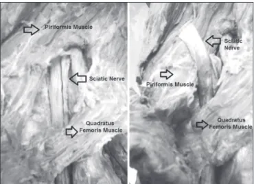
the distance between the lateral margin of the sacrotuber -ous ligament and the sciatic nerve margin, using the lat-eral margin of the ligament, close to the point of ixation into the sciatic tuber, as the reference point. Table 4 also shows the distance between the apex of the greater fem -oral trochanter and the lateral margin of the sciatic nerve. Figure 1 shows dissection images of the normal and variant relationships between the sciatic nerve and the piriformis muscle.
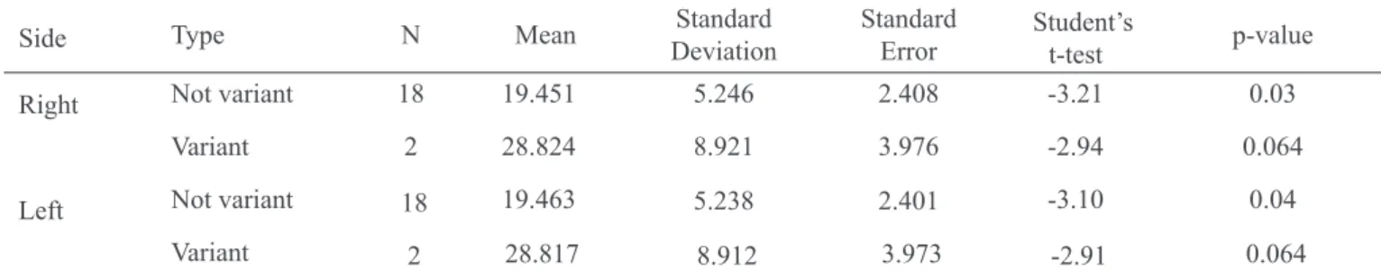
Table 2 – Width of the sciatic nerve (mm) at the lower margin of the piriformis muscle.
Side Type N Mean Standard
Deviation
Standard
Error Student’s t-test p-value
Right Not variant 18 19.451 5.246 2.408 -3.21 0.03
Variant 2 28.824 8.921 3.976 -2.94 0.064
Left Not variant 18 19.463 5.238 2.401 -3.10 0.04
Variant 2 28.817 8.912 3.973 -2.91 0.064
Table 3 – Extra-pelvic length and width of the piriformis muscle in the right and left limbs, taking the reference points of the sacrotuberous ligament and the apex of the greater femoral trochanter.
Type N Mean Standard
Deviation
Standard
Error T test p-value
Length Right 20 74.518 7.251 1.464 1.39 0.129
Left 20 78.454 9.223 1.932
Width Right 20 23.19 6.112 1.228 1.24 0.164
Left 20 22.37 5.24 1.194
Table 4 – Distance between the lateral margin of the sacrotuberous ligament and the sciatic nerve margin, using the lateral margin of the liga
-ment, close to the point of ixation into the sciatic tuber, as the reference point.
Side N Mean Standard
Deviation
Standard
Error Student’s t-test p-value
Medial margin of sciatic nerve D 20 17.974 4.955 1.114 -0.27 0.712
Lateral margin of sacro tuberous ligament E 20 18.42 5.161 1.317
Medial margin of sciatic nerve D 20 18.121 7.942 1.734 0.22 0,794
Apex of greater trochanter E 20 30.264 6.437 1.536
Table 1 – Relationship between the sciatic nerve and the piriformis muscle.
Types Variations Smoll 2010 Present Study
A The sciatic nerve emerges below the piriformis muscle (“normal”) 5,038 (83.1%) 36 (90%)
B The sciatic nerve divisions pass through and below the piriformis muscle 829 (13.8%) 0
C The sciatic nerve divisions pass through and above the piriformis muscle 78 (1.4%) 0
D The sciatic nerve passes above the piriformis muscle 32 (0.7%) 4 (10%)
E The sciatic nerve divisions pass above and below the piriformis muscle 5 (< 0.1%) 0
F The sciatic nerve emerges through the piriformis muscle 5 (< 0.1%) 0
DISCUSSION
It is unclear whether the anatomical variations in the relationship between the sciatic nerve and the piri -formis muscle are responsible for the pain experienced in piriformis syndrome, since asymptomatic patients might have these variations, while symptomatic pa-tients might not have them7. In fact, there is still a tendency to envisage parallel existence of “sciatic-piriformis anomalies” and ““sciatic-piriformis syndrome”. Whether the relationship between nerve and muscle is really cause and consequence of the pain remains to be deined2. However, it is only through detailed study of the regional anatomy that these doubts may one day be clariied.
The present study has conirmed the data in the world -wide literature, with regard to the “normal” type A relationship between the sciatic nerve and the piri -formis muscle. This type of relationship, with the sciatic nerve emerging below the piriformis muscle was prevalent in our population, just as it was in all other similar studies that were systematically 2.
Vari-ants of this “normal” type A relationship are indeed so rare that hundreds of limbs would have to be stud -ied in order to identify variations B-F in a population. Variations in sciatic nerve width and length are not unknown11, and possibly do not represent an anomaly. The relatively small number of limbs studied in the present work does not allow for statistical comparisons and conclusions between normal and variant relation -ships between nerve and muscle. The essence of this study was descriptive, in order to obtain more informa
-Figure 1 - A) The sciatic nerve emerges below the piriformis muscle (“normal”); B) The sciatic nerve passes above the piriformis muscle (“variation”).
tion on variations of the sciatic nerve anatomy. However, taking into consideration data from the present work and from the literature on the subject, it is fair to say that the piriformis syndrome possibly does not depend on abnormal relationships between the sciatic nerve and the piriformis muscle, or it would be an extremely rare pain-generating condition. In fact, even in a situation of normal relationship between the sciatic nerve and the piriformis muscle, any condition affecting the muscle (e.g., inlammation or trauma) could indirectly affect the nerve. This idea seems to be particularly supported by the good pain relief results achieved when low doses of botulinum toxin are injected into the piriformis muscle of patients with typical signs and symptoms of the piri-formis syndrome12.
CONCLUSION
Piriformis syndrome is a rare pain-generating condi -tion, and only detailed study of sciatic nerve anatomy and its anatomical relationship with the piriformis muscle is likely to shed light on the questions re-garding the syndrome. Anatomical variations in the relationship between the sciatic nerve and the piri -formis muscle do not seem to be solely responsible for the piriformis syndrome.
REFERENCES
1. Prakash, Bhardwaj AK, Devi MN, et al. Sciatic nerve division: a cadaver study the Indian population and review of the literature. Singapore Med J 2010;51(9):721-3. 2. Smoll NR. Variations of the piriformis and sciatic nerve with clinical consequence: a review. Clin Anat 2010;23(1):8-17.
3. Halpin RJ, Ganju A. Piriformis syndrome: a real pain in the buttock? Neurosurgery 2009;65(4 Suppl):A197-202. 4. Stewart JD. The piriformis syndrome is overdiag -nosed. Muscle Nerve 2003;28(5):644–6.
5. Hopayian K, Song F, Riera R, et al. The clinical fea -tures of the piriformis syndrome: a systematic review. Eur Spine J 2010;19(12):2095-109.
6. Robinson DR. Pyriformis syndrome in relation to sci -atic pain. Am J Surg 1947;73(3):355-8.
7. Kirschner JS, Foye PM, Cole JL. Piriformis syndrome, diagnosis and treatment. Muscle Nerve 2009;40(1):10-8. 8. Rodrigue T, Hardy RW. Diagnosis and treat-ment of piriformis syndrome. Neurosurg Clin N Am 2001;12(2):311-9.
considerations and the relationship between the piri -formis muscle and the sciatic nerve. Surg Radiol Anat 2008;30(6):467-74.
10. Beaton LE, Anson BJ. The sciatic nerve and the piri -formis muscle: Their interrelation a possible cause of coccygodynia. J Bone Joint Surg 1938;20:686-88. 11. Vicente EJD, Viotto MJS, Barbosa CAA, et al. Study on anatomical relationships and variations be -tween the sciatic nerve and piriform muscle. Rev Bras
Fisioter 2007;11(3):227-32.
12. Yoon SJ, Ho J, Kang HY, et al. Low-dose botulinum toxin type A for the treatment of refractory piriformis syndrome. Pharmacotherapy 2007;27(5):657-65.
Presented in August 15, 2011.
Accepted for publication in November 25, 2011.