Epidemiological profile and prognostic factors in patients with
lung cancer
DAMILA CRISTINA TRUFELLI1*, THAYLES VINICIUS MORAES2, ALINE ANGÉLICA PORTO ROCHA LIMA3, AURO DEL GIGLIO4
1MD, Oncologist/MSc Student – Medical Oncologist. Assistant Physician of the Department of Hematology and Oncology, Faculdade de Medicina do ABC, Santo André, SP, Brazil
2MSc Student – Resident Physician of the Department of Hematology and Oncology, Faculdade de Medicina do ABC, Santo André, SP, Brazil
3MSc in Oncology – Medical Oncologist. Assistant Physician of the Department of Hematology and Oncology, Faculdade de Medicina do ABC, Santo André, SP, Brazil
4Habilitation (BR: Livre-docência) – Full Professor of the Department of Hematology and Oncology, Faculdade de Medicina do ABC, Santo André, SP, Brazil
S
UMMARYStudy conducted at Faculdade de Medicina do ABC, Disciplina de Hematologia e Oncologia, Santo André, SP, Brazil
Article received: 4/21/2015
Accepted for publication: 5/16/2015
*Correspondence:
Address: Av. Príncipe de Gales, 821, Anexo III Santo André, SP – Brazil Postal code: 09060-650 [email protected]
http://dx.doi.org/10.1590/1806-9282.62.05.428
Objective: To describe the epidemiological proile of patients with lung cancer treated at a public tertiary referral hospital specializing in oncology, and to ex-plore variables that may be related to the overall survival (OS) of these patients.
Method: Data from the medical records of all patients with invasive lung can-cer consecutively seen at the Oncology Department of Hospital Estadual Mário Covas between August 2008 and December 2013 were extracted. The informa-tion obtained was submitted to statistical analysis.
Results: Of the total 210 patients, 39 were excluded from analysis due to lack of information in the medical record. The most common histological type was adenocarcinoma, representing 39.41% of the sample, followed by squamous cell carcinoma with 25.29% and small-cell carcinoma with 13.53%. Other histologi-cal types were responsible for the remaining 21.76%. There was a statistihistologi-cally sig-niicant association between Karnofsky performance status (KPS) ≤ 70%, palli-ative chemotherapy lines performed and stage at diagnosis, and OS. Additionally, administration of target therapy to patients with EGFR mutation
was associated with signiicantly better overall survival. However, analysis of lab-oratory variables (hemoglobin, albumin and LDH) as possible prognostic fac-tors for survival showed no statistically signiicant relationship. Among patients with stage III and IV, the median OS was 10.1 months.
Conclusion: The risk factors for shorter OS were KPS score ≤ 70%, less than two lines of palliative chemotherapy, and stage III and IV at diagnosis. The imple-mentation of CT screening for risk patients may allow earlier diagnosis of cas-es and improve thcas-ese rcas-esults.
Keywords: lung neoplasms, epidemiology, survival, prognosis, risk factors.
I
NTRODUCTIONLung cancer was considered a rare disease until the early twentieth century.1 Since then, its occurrence increased
rapidly and is currently one of the most prevalent can-cers in Brazil and in the world, with high mortality rate and increasing incidence especially among women. In Brazil, for 2014, according to the National Cancer Insti-tute (Instituto Nacional do Câncer, INCA), 16,400 new cases of lung cancer are estimated among men and 10,930 among women. These numbers correspond to an estimat-ed risk of 16.79 new cases per 100,000 men and 10.75 per 100,000 women.1 The US estimate far exceeds the
Brazil-ian; for 2014, just over 222,000 new cases of lung cancer are expected.2,3 It is responsible for the highest
percent-age of mortality from malignant neoplasms, approach-ing 30% of total deaths,2 so that only 16.6% of patients
will be alive 5 or more years after diagnosis.4
Although genetic and environmental factors are in-volved in the pathogenesis, smoking persists as the pri-mary trigger for most cases.4,5 Other risk factors include
exposure to asbestos, arsenic, chromium, nickel, cadmi-um and silica.6,7
carci-noma (oat-cell), and large-cell carcicarci-noma. During the last decades there has been a decrease in squamous cell car-cinomas, while adenocarcinomas increased. This is prob-ably due to changes in the composition of tobacco prod-ucts, as well as the change in people’s behavior regarding smoking.1
In the last decade there has been a revolution in the understanding of this cancer’s pathogenesis, molecular biology and treatment. Several gene mutations have been discovered (e.g. EGFR and EML4-ALK), culminating in the
development of the so-called molecularly targeted drugs.8
The treatment, which for many years drew heavily on plat-inum-based chemotherapy has been incremented by add-ing such drugs, which caused an increase in response rate, improved quality of life, a more favorable toxicity proile, and longer progression-free survival.9-12
Despite the current staging system and new tech-nologies developed in recent years, most patients (75 to 80%) are still diagnosed with advanced or metastatic disease, and even those treated with curative intent (stag-es I to III) develop distant metastas(stag-es during disease progression. Some factors such as old age, performance status, and stage at diagnosis are directly related to over-all survival (OS).13
Our study aims to trace the epidemiological proile of patients with lung cancer treated at a public tertiary referral hospital specializing in oncology, as well as ex-plore some prognostic variables that may be involved with OS.
M
ETHODThis is a single-center retrospective epidemiological study that included data from the medical records of all pa-tients with invasive lung cancer consecutively seen at the Oncology Service of Hospital Estadual Mário Covas (HEMC) in August 2008 to December 2013. The HEMC is the largest referral center for oncology in the Greater ABC Area in São Paulo, treating only patients of the pub-lic Uniied Health System.
For the entire sample, clinical, epidemiological and pathological characteristics were analyzed. However, vari-ables considered as possible prognostic factors were an-alyzed only in patients with non-small cell lung cancer, having in mind that this entity has peculiarities if com-pared to small-cell tumors.
The qualitative variables were described as absolute and relative frequency, and the quantitative variables, since they do not have a normal distribution (Shapiro-Wilk, p>0.05), were presented as medians and conidence intervals, at 25 and 75 percentiles.
In order to analyze the association between qualita-tive variables, we used a chi-square test. The analysis of quantitative variables between two categories and between groups was performed using the Mann-Whitney and Krus-kal-Wallis, respectively. As for OS analysis, we used Cox regression with log-rank test and Kaplan-Meyer curves; OS was deined as the time between diagnosis and death. We adopted a conidence level at 95% and data analysis was performed using Stata software, version 11.0. Statis-tical analyzes of prognostic factors were calculated only for non-small-cell tumors.
R
ESULTSFrom August 2008 to December 2013, 210 patients with invasive lung cancer were identiied. Of these, 39 were ex-cluded from the analysis due to lack of necessary infor-mation in the medical records or loss of follow-up. In 171 patients analyzed, the average age was 64 years, ranging from 33 to 90 years. As for gender, 114 (66.67%) were men and 57 (33.33%), women. 119 patients were smokers (69.59%), and the remaining 52 (30.41%), non-smokers.
Regarding histology, 67 (39.41%) had adenocarcino-ma, 43 (25.29%) squamous histology, 37 (21.76%) had other histology types, and 23 (13.53%) small-cell carcino-mas. Among the patients with adenocarcinomas, 15 sam-ples were subjected to mutation analysis of the EGFR gene
(epidermal growth factor receptor), with nine showing mutation and six wild-type EGFR. The other histological
types included: carcinoid tumor, large-cell, unspeciied or undeined non-small-cell, and undifferentiated carci-noma.
Most patients were diagnosed at stage IV (63.74%). The main metastatic site was the lung (35.09%), followed by the bones with 32.16%; noting that patients could have more than one metastatic site.
We observed that chemotherapy was the predomi-nant type of treatment, given to 80.12% of patients. 11 patients (6.43%) were treated with palliative support alone. Targeted drugs were used in 11 patients, nine of them as irst line, and two as second line medication (provided in the clinical research protocol).
The median follow-up was 9.9 months (range 4.2 to 20 months) and, by the end of the study, 80.12% of the patients died due to lung cancer. The median OS was 11.2 months for non-small-cell tumors, and 7.8 months for small-cell (oat-cell) tumors.
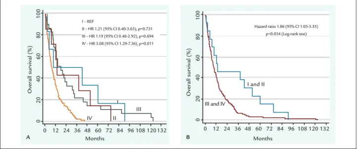
differences in the OS with p=0.034. The corresponding Kaplan-Meier curves are shown in Figure 1.
Regarding Karnofsky performance status (KPS) 77 patients (45.03%) belonged to the ≥ 70% group, favored in the median OS, with hazard ratio of 0.32 (95CI 0.21-0.50) and p<0.001. Another inding was that patients who underwent more than two lines of palliative chemother-apy had longer OS, with p<0.001. The corresponding Ka-plan-Meier curves are shown in Figure 2.
Three laboratory variables were analyzed (hemoglo-bin, albumin and lactate dehydrogenase – LDH). He-moglobin is stratiied into < 11 g/dL and > 11 g/dL,
com-prising 31.2 and 68.6% of patients, respectively. Albumin was stratiied into < 3,0 g/dL and ≥ 3,0 g/dL, and in-cluded 26 and 47 patients, respectively. Last, we strati-ied the LDH variable into < 480 (U/L), which included 48 patients, and LDH ≥ 480 (U/L) found in 60 patients. However, we did not ind any relationship between these variables and OS.
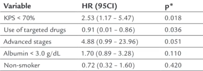
Multivariate analysis by Cox regression showed that KPS score < 70% is a risk factor for shorter OS and the use of targeted drugs is a protective factor for increased OS. Hazard ratio values and their respective conidence intervals are shown in Table 1.
Over
all sur
vival (%)
Months I
III II IV
0
20 40
60
80
100
0 12 24 36 48 60 72 84 96 108 120 132 I – REF
II – HR 1.21 (95% CI 0.40-3.63), p=0.731 III – HR 1.19 (95% CI 0.48-2.92), p=0.694 IV – HR 3.08 (95% CI 1.29-7.36), p=0.011
Over
all sur
vival (%)
Months I and II
III and IV
0
20 40
60
80
100
0 12 24 36 48 60 72 84 96 108 120 132 Hazard ratio 1.86 (95% CI 1.03-3.35)
p=0.034 (Log-rank test)
A B
Over
all sur
vival (%)
Over
all sur
vival (%)
Months Months
KPS>70 KPS<70
> 2 lines 2 lines
0
20 40
60
80
100
0
20 40
60
80
100
0 12 24 36 48 60 72 84 96 108 120 132 0 12 24 36 48 60 72 84 96 108 120 132 Hazard ratio 0.32 (95% CI 0.21-0.50)
p<0.001 (Log-rank test)
Hazard ratio 0.37 (95% CI 0.20-0.66) p<0.001 (Log-rank test)
A B
FIGURE 1 Kaplan-Meier curves. (A) Overall survival for individual staging, and (B) Overall survival grouped by stage I and II versus III and IV. HR: hazard ratio.
TABLE 1 Multivariate analysis of factors associated with overall survival.
Variable HR (95CI) p*
KPS < 70% 2.53 (1.17 – 5.47) 0.018
Use of targeted drugs 0.91 (0.01 – 0.86) 0.036 Advanced stages 4.88 (0.99 – 23.96) 0.051 Albumin < 3.0 g/dL 1.70 (0.89 – 3.28) 0.110 Non-smoker 0.72 (0.32 – 1.60) 0.420
KPS: Karnofsky performance status; HR: hazard ratio; 95CI: 95% confidence interval; *Logistic regression.
D
ISCUSSIONOur study is a retrospective, observational and epidemi-ological analysis of cases of lung cancer in a Brazilian on-cological reference hospital in the public health system, as well as its relationship to the literature. The Brazilian estimate for the number of cases of lung cancer is very low compared to global and US epidemiology. It is worth mentioning that US statistics include more than 224,000 new cases of lung cancer in 2015, while Brazilian statis-tics for 2014 predicted only 27,330 new cases. This reveals strong discrepancy comparing the total population of the two countries.1,2
According to data released by the National Cancer Comprehensive Network (NCCN),4 85% of lung cancers
belong to the class of non-small-cell tumors (NSCLC) and the remaining 15% to the group of tumors known as small-cell or oat-cell lung carcinoma (SCLC). This result was similar to that found in our study, which detected 86.47% of non-small-cell tumors and 13.53% of small-cell carcinomas. As for histological subtype, 39.41% of ade-nocarcinomas, 25.29% of squamous cell carcinomas, 13.53% of small-cell tumors and 21.76% of unspeciied non-small-cell tumors were detected, which is in line with the literature with respectively 38, 20, 13 and 18%.14 In a
study published by Caires-Lima et al.,15 conducted at
In-stituto do Câncer do Estado de São Paulo, also a referral center for oncology, epidemiology of 232 patients point-ed out the adenocarcinoma subtype with 61% of cases, followed by squamous cell carcinoma and large-cell car-cinoma, with 30 and 2%, respectively. In 7% of patients determining the subtype was not possible, and 7.6% rep-resented small-cell tumors.15
As the study was conducted in a service that works with the public Uniied Health System exclusively, only 22% of patients with adenocarcinoma underwent muta-tion analysis of the EGFR gene, which was possible after
inclusion of patients in clinical research protocols. Of the patients tested, 60% had mutations of the EGFR gene, a
number well above that found in the literature, which is
around 10 to 30% for the general population, but reach-ing levels of up to 60% among non-smokreach-ing Asian wom-en.16-18 Therefore, the higher prevalence of EGFR
muta-tion observed in our study might be due to bias in the selected patients, once the majority of them were treated in the context of international clinical trials with rigid inclusion criteria.
NSCLC is usually diagnosed in advanced stages of the disease, rarely in early stages. Even in developed coun-tries like the US, only 15% of patients have cancer stages I and II at diagnosis, according to the SEER (Surveillance, Epidemiology, and End Results).2 Statistics for stage III
and IV are 22 and 57%, respectively; the other 6% are un-known.2 Our data were similar to those observed in the
literature, for example the stage IV, which was present in 63.74% of our sample, similar to the data observed in the study of Caires-Lima et al., in which 71% of patients were diagnosed in stage IV.15
With the exception of rare oligometastatic cases, pa-tients diagnosed with NSCLC stage IV typically die of this disease, and have a median OS of 10 to 12 months,19,20 which
is corroborated in our study. The ive-year survival rate of patients in advanced stages of disease is around 2%.20
Prognostic factors in lung cancer have been studied for a few decades, both in non-small-cell and small-cell tumors.13,21 The SWOG (Southwest Oncology Group)
an-alyzed data from 2,531 patients and found the following variables as predictive of treatment response: good per-formance status, female gender, small tumor volume, nor-mal LDH levels, and hemoglobin higher than 11 g/dL.22
Another study, conducted by a European group, exam-ined 1,052 patients and found that low tumor volumes, a good KPS score (≥ 80), female gender and older age (≥ 70 years) were all associated with a more favorable treat-ment response.23 Even in patients with stage III receiving
deinitive treatment with chemotherapy and radiothera-py, KPS score ≤ 70% has been shown to be an indepen-dent prognostic factor of OS.24 Our study conirms that
adverse prognostic factors for survival include low per-formance status (KPS ≤ 70%) and more advanced stages (III and IV), and these factors are also deined as predic-tive by Caires-Lima et al.15 and Debiasi et al.25
chemotherapy had longer OS. In line with the literature, there are studies that show OS beneit using second and third lines, especially with molecularly targeted drugs.26-29
As the laboratory variables, hemoglobin and LDH were related to survival in previous studies.17,18 Our study
did not establish this association as statistically signii-cant; however, our sample is small compared with the lit-erature and, also, we did not have data on all patients, and these factors may be responsible for any negative data. Al-bumin showed a small statistical trend as a prognostic factor, with a value of p=0.061 and may be related to an-other factor, the weight loss, which in a study by Hoang et al.30 was deined as an adverse prognostic factor. The
latter author also identiied in a multivariate analysis oth-er ive independent factors of worse prognosis: cutane-ous metastases, low performance status (ECOG 1 or 2), liver metastases, ≥ four sites of metastases, and absence of previous surgery.
C
ONCLUSIONRisk factors for shorter OS found in our study were KPS score ≤ 70%, less than two lines of palliative chemother-apy, and stage III and IV at diagnosis. Most of the patients treated in our service had tumors in advanced stages, which probably explains the large number of deaths.
With the advent of low-dose helical CT31 for
screen-ing of smokers and those at high risk for developscreen-ing lung cancer, we may have cases diagnosed earlier and with bet-ter clinical outcomes. Unfortunately, this procedure is not covered by our Public Health System, so that efforts in this direction, parallel to the anti-smoking campaigns should be undertaken if we are to reduce mortality from this devastating disease in our country.
R
ESUMOPeril epidemiológico e fatores prognósticos em pacien-tes com câncer de pulmão
Objetivo: traçar o peril epidemiológico de pacientes com câncer de pulmão atendidos em hospital público terciá-rio de referência em oncologia e explorar variáveis que possam estar relacionadas com a sobrevida global (SG) desses pacientes.
Método: foram extraídos dados dos prontuários de to-dos os pacientes com câncer de pulmão invasivo, entre agosto de 2008 e dezembro de 2013, atendidos consecu-tivamente no Serviço de Oncologia do Hospital Estadual Mário Covas. As informações obtidas foram submetidas à análise estatística.
Resultados: do total de 210 pacientes, 39 foram excluídos da análise pela ausência de informações no prontuário. O tipo histológico mais frequente foi o adenocarcinoma, re-presentando 39,41% da amostra, seguido do carcinoma es-pinocelular com 25,29% e de pequenas células com 13,53%. Outros tipos histológicos foram responsáveis pelos 21,76% restantes. Houve associação com signiicância estatística entre KPS ≤ 70%, linhas de quimioterapia paliativa reali-zadas e estágio ao diagnóstico com SG. A administração de terapia-alvo direcionada para pacientes com mutação do EGFR foi signiicativamente associada à melhor SG. A
análise das variáveis laboratoriais (hemoglobina, albumi-na e desidrogealbumi-nase lática – DHL) como possíveis fatores prognósticos de sobrevida não mostrou relação estatisti-camente signiicativa. Entre os pacientes em estágio III e IV, a SG mediana foi de 10,1 meses.
Conclusão: os fatores de risco para menor SG foram KPS ≤ 70%, menos de duas linhas de quimioterapia paliativa e estágios III e IV ao diagnóstico. A implementação do rastreamento tomográico de pacientes de risco poderá permitir o diagnóstico mais precoce e a melhora desses resultados.
Palavras-chave: neoplasias pulmonares, epidemiologia, sobrevida, prognóstico, fatores de risco.
R
EFERENCES1. Estimativa 2014: incidência de câncer no Brasil / Instituto Nacional de Câncer. Rio de Janeiro: INCA; 2014.
2. Surveillance, Epidemiology, and End Results (SEER). National Cancer Institute [cited 2015 Jan 19]. Available from: http://seer.cancer.gov. 3. Siegel RL, Miller KD, Jemal A. Cancer statistics, 2015. CA Cancer J Clin.
2015; 65(1):5-29.
4. Lung Cancer. Clinical Practice Guidelines in Oncology. National Comprehensive Cancer Network (NCCN) [cited 2015 Jan 19]. Available from: http://www.nccn.org.
5. Alberg AT, Brock MV, Ford JG, Samet JM, Spivack SD. Epidemiology of lung cancer: Diagnosis and management of lung cancer, 3rd ed: American College of Chest Physicians evidence-based clinical practice guidelines. Chest. 2013; 143(5 Suppl):1s-29s.
6. Straif K, Benbrahim-Tallaa L, Baan R, Grosse Y, Secretan B, El Ghissassi F, et al.; WHO International Agency for Research on Cancer Monograph Working Group. A review of human carcinogens--part C: metals, arsenic, dusts, and ibres. Lancet Oncol. 2009; 10(5):453-4.
7. Janerich DT, Thompson WD, Varela LR, Greenwald P, Chorost S, Tucci C, et al. Lung cancer and exposure to tobacco smoke in the household. N Engl J Med. 1990; 323(10):632-6.
8. Herbst RS, Heymach JV, Lippman SM. Molecular origins of lung cancer. N Engl J Med. 2008; 359:1367-80.
9. Solomon BJ, Mok T, Kim DW, Wu YL, Nakagawa K, Mekhail T, et al. First-line crizotinib versus chemotherapy in ALK-positive lung cancer. N Engl J Med. 2014; 371(23):2167-77.
10. Haaland B, Tan PS, de Castro G Jr, Lopes G. Meta-analysis of irst-line therapies in advanced non-small-cell lung cancer harboring EGFR-activating mutations. J Thorac Oncol. 2014; 9(6):805-11.
patients with advanced EGFR mutation-positive non-small-cell lung cancer (EURTAC): a multicentre, open-label, randomized phase 3 trial. Lancet Oncol. 2012; 13(3):239-46.
12. Fukuoka M, Wu YL, Thongprasert S, Sunpaweravong P, Leong SS, Sriuranpong V, et al. Biomarker analyses and inal overall survival results from a phase III, randomized, open-label, irst-line study of geitinib versus carboplatin/Paclitaxel in clinically selected patients with advanced non-small-cell lung cancer in Asia (IPASS). J Clin Oncol. 2011; 29(21):2866-74. 13. Stanley KE. Prognostic factors for survival in patients with inoperable lung
cancer. J Natl Cancer Inst. 1980; 65(1):25-32.
14. Midthun DE, Jett JR, Lilenbaum RC, Vora SR. Overview of the risk factors, pathology, and clinical manifestations of lung cancer [cited 2015 Jan 27]. Available from: http://www.uptodate.com.
15. Caires-Lima R, Takahashi TK, Mak MP, Roitberg FSR, Teixeira CHA, Mesquita CS, et al. Referral of lung cancer patients to specialized clinical oncology care: Instituto do Cancer do Estado de São Paulo. In: 5th Latin American Conference on Lung Cancer, 2012, Rio de Janeiro. J Thorac Oncol. 2012; 7:S111. 16. Lynch TJ, Bell DW, Sordella R, Gurubhagavatula S, Okimoto RA, Brannigan
BW, et al. Activating mutations in the epidermal growth factor receptor underlying responsiveness of non-small-cell lung cancer to geitinib. N Engl J Med. 2004; 350(21):2129-39.
17. Kumar A, Petri ET, Halmos B, Boggon TJ. Structure and clinical relevance of the epidermal growth factor receptor in human cancer. J Clin Oncol. 2008; 26(10):1742-51.
18. Rosell R, Moran T, Queralt C, Porta R, Cardenal F, Camps C, et al., Spanish Lung Cancer Group. Screening for epidermal growth factor receptor mutations in lung cancer. N Engl J Med. 2009; 361(10):958-67.
19. Schrump DS, Carter D, Kelsey CR, Marks LB, Giaccone G. Non-small cell lung cancer. In: DeVita Jr. VT, Lawrence TS, Rosenberg SA (eds.). DeVita, Hellman, and Rosenberg’s Cancer: Principles and Practice of Oncology. 9.ed. Philadelphia: Lippincott Williams & Wilkins; 2011.
20. Goldstraw P, Crowley J, Chansky K, Giroux DJ, Groome PA, Rami-Porta R, et al.; International Association for the Study of Lung Cancer International Staging Committee; Participating Institutions. The IASLC Lung Cancer Staging Project: proposals for the revision of the TNM stage groupings in the forthcoming (seventh) edition of the TNM Classiication of malignant tumours. J Thorac Oncol. 2007; 2(8):706-14.
21. Wolf M, Holle R, Hans K, Drings P, Havemann K. Analysis of prognostic factors in 766 patients with small cell lung cancer (SCLC): The role of sex as a predictor for survival. Br J Cancer. 1991; 63(6):986-92.
22. Albain KS, Crowley JJ, Lebranc M, Livingston RB. Survival determinants in extensive-stage non-small-cell lung cancer: the Southwest Oncology Group experience. J Clin Oncol. 1991; 9(9):1618-26.
23. Paesmans M, Sculier JP, Libert P, Bureau G, Dabouis G, Thiriaux J, et al. Prognostic factors for survival in advanced non-small-cell lung cancer: univariate and multivariate analyses including recursive partitioning and amalgamation algorithms in 1,052 patients. J Clin Oncol. 1995; 13(5):1221-30.
24. Firat S, Byhardt RW, Gore E. Comorbidity and Karnofsky performance score are independent prognostic factors in stage III non-small-cell lung cancer: an institutional analysis of patients treated on four RTOG studies. Int J Radiat Oncol Biol Phys. 2002; 54(2):357-64.
25. Debiasi M, Fay AP, Viola LS, Sostruznik MH. Peril epidemiológico e análise de sobrevida de pacientes com câncer de pulmão a partir da primeira consulta em um centro terciário de oncologia/SUS. Revista Brasileira de Oncologia Clínica. 2010; 7(22):86-91.
26. Shepherd FA, Rodrigues Pereira J, Ciuleanu T, Tan EH, Hirsh V, Thongprasert S, et al. Erlotinib in previously treated non-small-cell lung cancer. N Engl J Med. 2005; 353(2):123-32.
27. Weiss JM, Stinchcombe TE. Second-line therapy for advanced NSCLC. Oncologist. 2013; 18(8):947-53.
28. Noble J, Ellis PM, Mackay JA, Evans WK; Lung Cancer Disease Site Group of Cancer Care Ontario’s Program in Evidence-based Care. Second-line or subsequent systemic therapy for recurrent or progressive non-small cell lung cancer: a systematic review and practice guideline. J Thorac Oncol. 2006; 1(9):1042-58.
29. Eccles BK, Geldart TR, Laurence VM, Bradley KL, Lwin MT. Experience of irst and subsequent-line systemic therapy in the treatment of non-small cell lung cancer. Ther Adv Med Oncol. 2011; 3(4):163-70.
30. Hoang T, Xu R, Schiller JH, Bonomi P, Johnson DH. Clinical model to predict survival in chemonaive patients with advanced non-small-cell lung cancer treated with third-generation chemotherapy regimens based on eastern cooperative oncology group data. J Clin Oncol. 2005; 23(1):175. 31. National Lung Screening Trial Research Team, Church TR, Black WC, Aberle