www.jbp.org.br
ISSN 1806-3713
Volume 42, Number 5 September | October 2016
HIGHLIGHT
V
olume 42, Number 5
Sept
ember | Oct
ober
20
16
Prevalence of alpha-1
antitrypsin deficiency
in COPD patients
Pulmonary
rehabilitation in
tuberculosis patients
Applications for a
hybrid operating room
Realização:
Nos dias 02 a 05 de agosto de 2017, a cidade de Fortaleza receberá
os maiores congressos sobre doenças respiratórias e pulmonares da
atualidade, com renomados palestrantes da área médica, informações,
estudos e pesquisas internacionais.
E O MELHOR: TUDO ISSO EM UMA DAS CIDADES MAIS BONITAS
DO BRASIL.
INVISTA
NO SEU
CONHECIMENTO.
COMPAREÇA!
ISSN 1806-3713
Published once every two months J Bras Pneumol. v.42, number 5, p. 307-398 September/October 2016
Publicação Indexada em: Latindex, LILACS, Scielo Brazil, Scopus, Index Copernicus, ISI Web of Knowledge, MEDLINE e PubMed Central (PMC) Disponível eletronicamente nas versões português e inglês: www.jornaldepneumologia.com.br e www.scielo.br/jbpneu
Associação Brasileira de Editores Científicos
I N T E R N A T I O N A L
EDITOR-IN-CHIEF
Rogerio Souza - Universidade de São Paulo, São Paulo - SP
EXECUTIVE EDITORS
Bruno Guedes Baldi - Universidade de São Paulo, São Paulo - SP
Caio Júlio Cesar dos Santos Fernandes - Universidade de São Paulo - São Paulo - SP
Carlos Roberto Ribeiro de Carvalho - Universidade de São Paulo, São Paulo - SP
Carlos Viana Poyares Jardim - Universidade de São Paulo, São Paulo - SP
ASSOCIATE EDITORS
Afrânio Lineu Kritski - Universidade Federal do Rio de Janeiro, Rio de Janeiro, RJ
Andre Luis Pereira de Albuquerque - Universidade de São Paulo - São Paulo - SP
Bruno Hochhegger - Universidade Federal do Rio Grande do Sul - Porto Alegre – RS
Edson Marchiori - Universidade Federal Fluminense, Niterói - RJ
Fernanda Carvalho de Queiroz Mello - Universidade Federal do Rio de Janeiro - Rio de Janeiro - RJ
Frederico Leon Arrabal Fernandes - Universidade de São Paulo - São Paulo - SP
Giovanni Battista Migliori - Director WHO Collaborating Centre for TB and Lung Diseases, Fondazione S. Maugeri, Care and Research Institute, Tradate - Italy
Giovanni Sotgiu - University of Sassari, Sassari - Italy
Irma de Godoy - Universidade Estadual Paulista, Botucatu - SP
Marcelo Alcântara Holanda - Universidade Federal do Ceará - Fortaleza - CE
Pedro Caruso - Universidade de São Paulo - São Paulo - SP
Pedro Rodrigues Genta - Universidade de São Paulo - São Paulo - SP
Renato Tetelbom Stein - Pontifícia Universidade Católica do Rio Grande do Sul, Porto Alegre - RS
Ricardo de Amorim Corrêa - Universidade Federal de Minas Gerais - Belo Horizonte - MG
Ricardo Mingarini Terra - Universidade de São Paulo - São Paulo - SP
Simone Dal Corso - Universidade Nove de Julho - São Paulo – SP
Tomás Pulido - Instituto Nacional de Cardiología Ignacio Chávez - México
Ubiratan de Paula Santos - Universidade de São Paulo, São Paulo - SP
Veronica Amado - Universidade de Brasília, Brasília - DF
EDITORIAL COUNCIL
Alberto Cukier - Universidade de São Paulo, São Paulo – SP
Álvaro A. Cruz - Universidade Federal da Bahia, Salvador, BA
Ana C. Krieger - Weill Cornell Medical College - New York – USA
Ana Luiza Godoy Fernandes - Universidade Federal de São Paulo, São Paulo - SP
Antonio Segorbe Luis - Universidade de Coimbra, Coimbra - Portugal
Ascedio Jose Rodrigues - Universidade de São Paulo - São Paulo - SP
Brent Winston - University of Calgary, Calgary - Canada
Carlos Alberto de Assis Viegas - Universidade de Brasília, Brasília - DF
Carlos Alberto de Castro Pereira - Universidade Federal de São Paulo, São Paulo - SP
Carlos M. Luna - Hospital de Clinicas, Universidad de Buenos Aires, Buenos Aires - Argentina
Carmen Silvia Valente Barbas - Universidade de São Paulo, São Paulo - SP
Celso Ricardo Fernandes de Carvalho - Universidade de São Paulo, São Paulo - SP
Dany Jasinowodolinski - Universidade de São Paulo, São Paulo - SP
Denis Martinez - Universidade Federal do Rio Grande do Sul, Porto Alegre - RS
Douglas Bradley - University of Toronto, Toronto, ON - Canadá
Emílio Pizzichini - Universidade Federal de Santa Catarina, Florianópolis - SC
Fábio Biscegli Jatene - Universidade de São Paulo, São Paulo - SP
Frank McCormack - University of Cincinnati School of Medicine, Cincinnati, OH - USA
Geraldo Lorenzi Filho - Universidade de São Paulo, São Paulo - SP
Gilberto de Castro Junior - Universidade de São Paulo, São Paulo - SP
Gustavo Javier Rodrigo - Hospital Central de las Fuerzas Armadas, Montevidéu – Uruguay
Ilma Aparecida Paschoal - Universidade de Campinas, Campinas - SP
C. Isabela Silva Müller - Vancouver General Hospital, Vancouver, BC - Canadá
J. Randall Curtis - University of Washington, Seattle, Wa - USA
John J. Godleski - Harvard Medical School, Boston, MA - USA
José Alberto Neder - Queen’s University - Ontario, Canada
José Antonio Baddini Martinez - Universidade de São Paulo, Ribeirão Preto - SP
José Dirceu Ribeiro - Universidade de Campinas, Campinas - SP
José Miguel Chatkin - Pontifícia Universidade Católica do Rio Grande do Sul, Porto Alegre - RS
José Roberto de Brito Jardim - Universidade Federal de São Paulo, São Paulo - SP
José Roberto Lapa e Silva - Universidade Federal do Rio de Janeiro, Rio de Janeiro - RJ
Kevin Leslie - Mayo Clinic College of Medicine, Rochester, MN - USA
Luiz Eduardo Nery - Universidade Federal de São Paulo, São Paulo - SP
Marc Miravitlles - University Hospital Vall d’Hebron - Barcelona, Catalonia, Spain
Marisa Dolhnikoff - Universidade de São Paulo, São Paulo - SP
Marli Maria Knorst - Universidade Federal do Rio Grande do Sul, Porto Alegre - RS
Mauro Musa Zamboni - Instituto Nacional do Câncer, Rio de Janeiro - RJ
Nestor Muller - Vancouver General Hospital, Vancouver, BC - Canadá
Noé Zamel - University of Toronto, Toronto, ON - Canadá
Oliver Augusto Nascimento - Universidade Federal de São Paulo - São Paulo - SP
Paul Noble - Duke University, Durham, NC - USA
Paulo Francisco Guerreiro Cardoso - Universidade de São Paulo, São Paulo - SP
Paulo Manuel Pêgo Fernandes - Universidade de São Paulo, São Paulo - SP
Peter J. Barnes - National Heart and Lung Institute, Imperial College, London - UK
Renato Sotto Mayor - Hospital Santa Maria, Lisboa - Portugal
Richard W. Light - Vanderbili University, Nashville, TN, USA
Rik Gosselink - University Hospitals Leuven - Bélgica
Robert Skomro - University of Saskatoon, Saskatoon - Canadá
Rubin Tuder - University of Colorado, Denver, CO - USA
Sérgio Saldanha Menna Barreto - Universidade Federal do Rio Grande do Sul, Porto Alegre - RS
Sonia Buist - Oregon Health & Science University, Portland, OR - USA
Talmadge King Jr. - University of California, San Francisco, CA - USA
Thais Helena Abrahão Thomaz Queluz - Universidade Estadual Paulista, Botucatu - SP
ISSN 1806-3713
E
x
p
e
d
ie
n
te
BRAZILIAN THORACIC SOCIETY
Ofice: SCS Quadra 01, Bloco K, Asa Sul, salas 203/204. Edifício Denasa, CEP 70398-900, Brasília, DF, Brazil. Tel. +55 61 3245-1030/+55 0800 616218. Website: www.sbpt.org.br. E-mail: sbpt@sbpt.org.br
The Brazilian Journal of Pulmonology (ISSN 1806-3713) is published once every two months by the Brazilian Thoracic Society (BTS). The statements and opinions contained in the editorials and articles in this Journal are solely those of the authors thereof and not of the Journal’s Editor-in-Chief, peer reviewers, the BTS, its oficers, regents, members, or employees. Permission is granted to reproduce any igure, table, or other material published in the Journal provided that the source for any of these is credited.
BTS Board of Directors (2015-2016 biennium): President: Renato Maciel - MG
Secretary-General: Paulo Henrique Ramos Feitosa - DF
Director, Professional Advocacy: Jose Eduardo Delini Cançado - SP CFO: Saulo Maia Davila Melo - SE
Scientiic Director: Miguel Abidon Aide - RJ
Director, Education and Professional Practice: Clystenes Odyr Soares Silva - SP Director, Communications: Simone Chaves Fagondes - RS
President, BTS Congress 2016: Marcus Barreto Conde - RJ
President Elect (2017/2018 biennium): Fernando Luiz Cavalcanti Lundgren - PE Chairman of the Board: Jairo Sponholz Araújo (PR)
AUDIT COMMITTEE:
Active Members: Clóvis Botelho (MT), Benedito Francisco Cabral Júnior (DF), Rafael de Castro Martins (ES)
Alternates: Maurício Meireles Góes (MG), Alina Faria França de Oliveira (PE), Paulo Cesar de Oliveira (MG)
COORDINATORS, BTS DEPARTMENTS:
Thoracic Surgery – Darcy Ribeiro Pinto Filho (RS) Sleep–disordered Breathing – Marcelo Fouad Rabahi (GO) Respiratory Endoscopy – Mauro Musa Zamboni (RJ) Pulmonary Function – John Mark Salge (SP) Imaging – Bruno Hochhegger (RS)
Lung Diseases – Ester Nei Aparecida Martins Coletta (SP) Pediatric Pulmonology – Paulo Cesar Kussek (PR)
COORDINATORS, BTS SCIENTIFIC COMMITTEES:
Asthma – Emilio Pizzichini (SC) Lung Cancer – Teresa Yae Takagaki (SP)
Pulmonary Circulation – Carlos Viana Poyares Jardim (SP) Advanced Lung Disease – Dagoberto Vanoni de Godoy (RS) Interstitial Diseases – José Antônio Baddini Martinez (SP)
Environmental and Occupational Respiratory Diseases – Ana Paula Scalia Carneiro (MG) COPD – Roberto Stirbulov (SP)
Epidemiology – Frederico Leon Arrabal Fernandes (SP) Cystic Fibrosis – Marcelo Bicalho of Fuccio (MG) Respiratory Infections and Mycoses – Mauro Gomes (SP) Pleura – Roberta Karla Barbosa de Sales (SP)
International Relations – José Roberto de Brito Jardim (SP) Smoking – Luiz Carlos Corrêa da Silva (RS)
Intensive Care – Marco Antônio Soares Reis (MG) Tuberculosis – Fernanda Carvalho de Queiroz Mello (RJ)
ADMINISTRATIVE SECRETARIAT OF THE BRAZILIAN JOURNAL OF PULMONOLOGY Address: SCS Quadra 01, Bloco K, Asa Sul, salas 203/204. Edifício Denasa, CEP 70398-900, Brasília, DF, Brazil. Tel. +55 61 3245-1030/+55 0800 616218.
Assistant Managing Editor: Luana Maria Bernardes Campos. E-mail: jpneumo@jornaldepneumologia.com.br
Circulation: 4.000 copies
Distribution: Free to members of the BTS and libraries Printed on acid-free paper
ISSN 1806-3713
Published once every two months J Bras Pneumol. v.42, number 5, p. 307-398 September/October 2016
C
o
n
te
n
ts
EDITORIAL
307 - Diagnosing alpha-1 antitrypsin deiciency: does it prevent or improve the course of COPD?
Irma Godoy
CONTINUING EDUCATION: IMAGING
309 - Intracavitary nodule
Edson Marchiori, Bruno Hochhegger, Gláucia Zanetti
CONTINUING EDUCATION: SCIENTIFIC METHODOLOGY
310 - Randomization: beyond tossing a coin Juliana Carvalho Ferreira, Cecilia Maria Patino
ORIGINAL ARTICLE
311 - Prevalence of alpha-1 antitrypsin deiciency and allele frequency in patients with COPD in Brazil
Rodrigo Russo, Laura Russo Zillmer, Oliver Augusto Nascimento, Beatriz Manzano, Ivan Teruaki Ivanaga, Leandro Fritscher,
Fernando Lundgren, Marc Miravitlles, Heicilainy Del Carlos Gondim, Gildo Santos Junior, Marcela Amorim Alves, Maria Vera Oliveira, Altay Alves Lino de Souza, Maria Penha Uchoa Sales, José Roberto Jardim
317 - Factors associated with disease-speciic survival of patients with non-small cell lung cancer
Mirian Carvalho de Souza, Oswaldo Gonçalves Cruz, Ana Glória Godoi Vasconcelos
326 - Bronchodilator response cut-off points and FEV0.75 reference values for spirometry in preschoolers
Edjane Figueiredo Burity, Carlos Alberto de Castro Pereira, Marcus Herbert Jones, Larissa Bouwman Sayão, Armèle Dornelas de Andrade, Murilo Carlos Amorim de Britto
333 - Effects of passive inhalation of cigarette smoke on structural and functional parameters in the respiratory system of guinea pigs
Thiago Brasileiro de Vasconcelos, Fernanda Yvelize Ramos de Araújo,
João Paulo Melo de Pinho, Pedro Marcos Gomes Soares, Vasco Pinheiro Diógenes Bastos
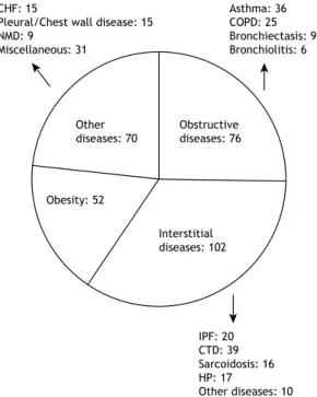
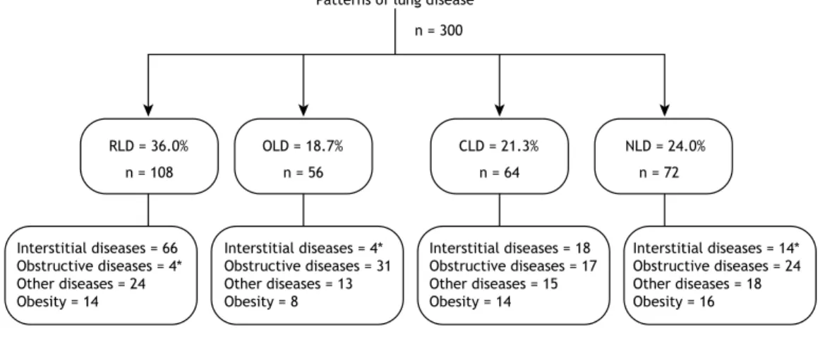
341 - Lung volumes and airway resistance in patients with a possible restrictive pattern on spirometry
Kenia Schultz, Luiz Carlos D’Aquino, Maria Raquel Soares, Andrea Gimenez, Carlos Alberto de Castro Pereira
348 - Prevalence of latent Mycobacterium tuberculosis infection in prisoners Pedro Daibert de Navarro, Isabela Neves de Almeida, Afrânio Lineu Kritski, Maria das Graças Ceccato, Mônica Maria Delgado Maciel, Wânia da Silva Carvalho, Silvana Spindola de Miranda
356 - Staphylococcal superantigen-speciic IgE antibodies: degree of sensitization and association with severity of asthma
José Elabras Filho, Fernanda Carvalho de Queiroz Mello, Omar Lupi,
ISSN 1806-3713
Published once every two months J Bras Pneumol. v.42, number 5, p. 307-398 September/October 2016
C
o
n
te
n
ts
362 - Gel pillow designed speciically for obstructive sleep apnea treatment with continuous positive airway pressure
Adriana Salvaggio, Anna Lo Bue, Serena Iacono Isidoro, Salvatore Romano, Oreste Marrone, Giuseppe Insalaco
367 - Effects of indacaterol versus tiotropium on exercise tolerance in patients with moderate COPD: a pilot randomized crossover study
Danilo Cortozi Berton, Álvaro Huber dos Santos, Ivo Bohn Jr., Rodrigo Quevedo de Lima, Vanderléia Breda, Paulo José Zimermann Teixeira
REVIEW ARTICLE
374 - Is there a rationale for pulmonary rehabilitation following successful chemotherapy for tuberculosis?
Marcela Muñoz-Torrico, Adrian Rendon, Rosella Centis, Lia D’Ambrosio, Zhenia Fuentes, Carlos Torres-Duque, Fernanda Mello, Margareth Dalcolmo, Rogelio Pérez-Padilla, Antonio Spanevello, Giovanni Battista Migliori
IMAGING IN PULMONARY MEDICINE
386 - Prominent bronchial vasculature, hemoptysis, and bilateral ground-glass opacities in a young woman with mitral stenosis
Fabian Aigner, Rudolf Speich, Macé Matthew Schuurmans
CASE REPORT
387 - Applications for a hybrid operating room in thoracic surgery: from
multidisciplinary procedures to image-guided video-assisted thoracoscopic surgery Ricardo Mingarini Terra, Juliano Ribeiro Andrade, Alessandro Wasum Mariani, Rodrigo Gobbo Garcia, Jose Ernesto Succi, Andrey Soares, Paulo Marcelo Zimmer
LETTER TO THE EDITOR
391 - An uncommon chest mass: oleothorax Bruno Hochhegger, Gláucia Zanetti, Edson Marchiori
392 - A rare case of hemorrhagic pneumonia due to Cladosporium cladosporioides Sérgio Grava, Francisco Antonio Dias Lopes, Rodrigo Silva Cavallazzi, Melyssa Fernanda Norman Negri Grassi, Terezinha Inez Estivalet Svidzinski
CORRESPONDENCE
395 - Cervical computed tomography in patients with obstructive sleep apnea: inluence of head elevation on the assessment of upper airway volume Shailendra Singh Rana, Om Prakash Kharbanda
Fábio José Fabrício de Barros Souza, Anne Rosso Evangelista, Juliana Veiga Silva, Grégory Vinícius Périco, Kristian Madeira
397 - Pulmonary rehabilitation in severe COPD with hyperinlation: some insights into exercise performance
Luiz Alberto Forgiarini Junior, Antonio Matias Esquinas Andre Luis Pereira de Albuquerque,
http://dx.doi.org/10.1590/S1806-37562016000400002
Diagnosing alpha-1 antitrypsin deiciency:
does it prevent or improve the course of
COPD?
Irma Godoy1
1. Disciplina de Pneumologia, Departamento de Clínica Médica, Faculdade de Medicina de Botucatu, Universidade Estadual Paulista, Botucatu (SP) Brasil. Alpha-1 antitrypsin (AAT) is a protein whose main
function is the inhibition of neutrophil elastase. The gene that encodes AAT is transmitted by simple autosomal codominant Mendelian inheritance through two alleles, one from each parent. The normal allele is designated Pi*M,
and the most common deicient alleles are the Pi*S and
Pi*Z alleles, which encode abnormal proteins that undergo polymerization in the liver. Therefore, 80-90% of the Z
proteins and 40-50% of the S proteins are retained within
the hepatocytes, grouped into polymers. The loss of the
anti-inlammatory and antiproteolytic activity, together with the pro-inlammatory effects of polymers, contribute to protein degradation and the exacerbation of inlammation,
resulting in an increased risk of developing COPD, with a predominance of emphysema, especially in smokers.
AAT deiciency (AATD) is a rare disease and, like most
such diseases, is underdiagnosed. The diagnosis is usually made late (at an average patient age of 45 years), and estimates suggest that 85% of patients with AATD have gone un diagnosed.(1) These indings indicate the low adherence to the recommendations of the World Health Organization, as well as to the guidelines of the American
Thoracic Society and the European Respiratory Society,
suggesting that patients with COPD or persistent airway obstruction should be tested for AATD.(2,3) The potential reasons for misdiagnosis include a lack of knowledge about the disease, about the tests necessary for the diagnosis, the lack of availability of such tests, and about
the algorithm required in order to conirm the diagnosis.
The prevalence of AATD varies depending on the population studied. Attempts to determine the prevalence of genetic predisposition in patient populations will inevitably overestimate the prevalence in the general population, whereas limiting screening to the healthy portion of the general population can underestimate
the prevalence. In Europe, the prevalence of the Pi*Z
mutation is highest in the northeastern region, where it ranges from 0.029 to 0.049.(4-6) In the United States, the prevalence is similar (0.019-0.030).(7,8) In Asia, the prevalence is extremely low (0.006).(1)
The data presented in the study conducted by Russo
et al.,(9) published in the current issue of the JBP, are unprecedented in that they show the prevalence of
AATD in 926 COPD patients in ive Brazilian states. The
diagnosis of AATD was based on an AAT cut-off point of
< 2.64 mg/dL in dried blood samples on ilter paper. For
patients in whom the AAT value was below the cut-off point, the serum concentration of AAT was determined.
For those with a serum AAT concentration < 113 mg/
dL, genetic sequencing was performed. In inconclusive cases, SERPINA gene sequencing was performed. Among
the patients evaluated by Russo et al.,(9) the AAT values
indicated the need for additional investigation in 9.2%, the serum AAT concentration was < 113 mg/dL in 2.6%, and the prevalence of the PI*Z allele was 0.8%, similar to that described in other countries.(9)
The study underscores the need to investigate AATD in patients with COPD, in accordance with the recommendations and guidelines mentioned above.(2,3) The alternative strategy is to prioritize investigation for
speciic risk groups, including patients with early-onset
emphysema or emphysema that is predominantly in the lower lobes, as well as family clusters of COPD patients
or irst-degree relatives of individuals diagnosed with
moderate or severe AATD.(10) It is also important to screen for AATD in individuals with unexplained liver disease, including newborns, children, and adults, as well
as in adults with necrotizing panniculitis. Screening for
AATD is recommended in adults with bronchiectasis of unclear etiology, adolescents with a persistent obstructive pattern, and adolescents with cytoplasmic-antineutrophil cytoplasmic antibody-positive vasculitis.(11)
Given that the main risk factor in individuals with AATD is smoking, which promotes the earlier emergence of COPD in smokers than in non-smokers,(12) early identiication of the disease allows preventive measures to be taken, the most important of which are avoiding smoking (active and passive) and exposure to environmental pollutants, both of which are determinants of the prognosis of COPD.
Early identiication of COPD allows lung function to be
monitored and the therapeutic decision to administer supplemental therapy while pulmonary function is still relatively preserved. The treatment of patients with COPD and AATD includes the usual therapy for the disease (smoking cessation, vaccination, use of bronchodilators, rehabilitation, and long-term home oxygen therapy when indicated).(10) The speciic treatment of AAT replacement
through the administration of concentrated puriied AAT
from human plasma is now available in Brazil. (11) However, that treatment is extremely expensive (approximately
US$100,000 per year); the indications for its use and
its eficacy are not well deined; and it is not recom
-mended by the US National Institute for Health and
Care Excellence,(13) although its use can be considered
in young patients with COPD, according to the Global Initiative for Chronic Obstructive Lung Disease (grade C recommendation).(14,15)
In brief, despite the recommendations of the World
Health Organization and the American Thoracic Society/ European Respiratory Society, many physicians and COPD
patients do not fully comprehend the risk of a rapid increase in airway obstruction associated with AATD. J Bras Pneumol. 2016;42(5):307-308
Diagnosing alpha-1 antitrypsin deiciency: does it prevent or improve the course of COPD?
With the availability of effective smoking cessation interventions, testing patients with COPD, especially those most at risk, to identify carriers of AATD is
important and justiiable. During the treatment of COPD,
efforts should be directed toward early detection of airway obstruction and avoiding exposure to risk factors, especially smoking, the most important intervention to reduce the progression of the disease.
REFERENCES
1. Greulich T, Vogelmeier CF. Alpha-1-antitrypsin deiciency: increasing awareness and improving diagnosis. Ther Adv Respir Dis. 2016;10(1):72-84. http://dx.doi.org/10.1177/1753465815602162 2. Alpha 1-antitrypsin deiciency: memorandum from a WHO meeting.
Bull World Health Organ. 1997;75(5):397-415.
3. American Thoracic Society; European Respiratory. American Thoracic Society/European Respiratory Society statement: standards for the diagnosis and management of individuals with alpha-1 antitrypsin deiciency. Am J Respir Crit Care Med. 2003;168(7):818-900. http:// dx.doi.org/10.1164/rccm.168.7.818
4. Dahl M, Tybjaerg-Hansen A, Lange P, Vestbo J, Nordestgaard BG. Change in lung function and morbidity from chronic obstructive pulmonary disease in alpha1-antitrypsin MZ heterozygotes: A longitudinal study of the general population. Ann Intern Med. 2002;136(4):270-9. http://dx.doi.org/10.7326/0003-4819-136-4-200202190-00006
5. Hutchison DC. Alpha 1-antitrypsin deiciency in Europe: geographical distribution of Pi types S and Z. Respir Med. 1998;92(3):367-77. http://dx.doi.org/10.1016/S0954-6111(98)90278-5
6. Sveger T. Liver disease in alpha1-antitrypsin deiciency detected by screening of 200,000 infants. N Engl J Med. 1976;294(24):1316-21. http://dx.doi.org/10.1056/NEJM197606102942404
7. Lieberman J, Winter B, Sastre A. Alpha 1-antitrypsin Pi-types in 965 COPD patients. Chest. 1986;89(3):370-3. http://dx.doi.org/10.1378/ chest.89.3.370
8. Morse JO, Lebowitz MD, Knudson RJ, Burrows B. Relation of protease inhibitor phenotypes to obstructive lung diseases in a community. N Engl J Med. 1977;296(21):1190-4. http://dx.doi.
org/10.1056/NEJM197705262962102
9. Russo R, Nascimento OA, Manzano B, Ivanaga IT, Fritscher L, Lundgren F, et al. Prevalence of deiciency of alpha-1 antitrypsin and allele frequency in patients with COPD in Brazil. J Bras Pneumol. 2016;42(5):311-16.
10. Ferrarotti I, Poplawska-Wisniewska B, Trevisan MT, Koepke J, Dresel M, Koczulla R, et al. How Can We Improve the Detection of Alpha1-Antitrypsin Deiciency? PLoS One. 2015;10(8):e0135316. http:// dx.doi.org/10.1371/journal.pone.0135316
11. Stoller JK, Aboussouan LS. A review of 1-antitrypsin deiciency. Am J Respir Crit Care Med. 2012;185(3):246-59. http://dx.doi. org/10.1164/rccm.201108-1428CI
12. Petrache I, Fijalkowska I, Zhen L, Medler TR, Brown E, Cruz P, et al. A novel antiapoptotic role for alpha1-antitrypsin in the prevention of pulmonary emphysema. Am J Respir Crit Care Med. 2006;173(11):1222-8. http://dx.doi.org/10.1164/rccm.200512-1842OC
13. National Institute for Health and Care Excellence (NICE) [homepage on the Internet]. London: NICE; c2016 [updated 2010 Jun; cited 2016 Jul 18]. Chronic obstructive pulmonary disease in over 16s: diagnosis and management. NICE guidelines [CG101]. Available from: https:// www.nice.org.uk/guidance/cg101/chapter/1-guidance
14. Global Initiative for Chronic Obstructive Lung Disease (GOLD) [homepage on the Internet] Bethesda: GOLD [cited 2016 Jan 11]. Available from: http://goldcopd.org/
15. Stoller JK. Alpha-1 antitrypsin deiciency: An underrecognized, treatable cause of COPD. Cleve Clin J Med. 2016;83(7):507-14.
http://dx.doi.org/10.1590/S1806-37562016000000223
Intracavitary nodule
Edson Marchiori1,2, Bruno Hochhegger3,4, Gláucia Zanetti2,5
1. Universidade Federal Fluminense, Niterói (RJ) Brasil. 2. Universidade Federal do Rio de Janeiro, Rio de Janeiro (RJ) Brasil. 3. Santa Casa de Misericórdia de Porto Alegre, Porto Alegre (RS) Brasil.
4. Universidade Federal de Ciências da Saúde de Porto Alegre, Porto Alegre (RS) Brasil. 5. Faculdade de Medicina de Petrópolis, Petrópolis (RJ) Brasil.
A 41-year-old male presented with complaints of anorexia and weight loss followed by cough with hemoptysis. The
inal diagnosis was aspergilloma in previous tuberculous
lesions.
The inding of a nodule in a lung cavity has important
diagnostic and therapeutic implications. Although aspergilloma is the most common cause of intracavitary nodules, a number of other conditions should be included in the differential diagnosis. These conditions primarily include neoplasms (particularly bronchial carcinoma), angioinvasive aspergillosis during the recovery phase,
Rasmussen aneurysm, and clots. Other, rarer, causes
include foreign bodies, thick pus, dehydrated caseous material, teratoma, and hydatidosis. The air crescent sign is commonly seen in patients with intracavitary nodules, regardless of the etiology. It corresponds to a collection
of air in the form of a crescent or half moon, located in the periphery of a nodule or mass of soft tissue density, separating the nodule from the cavity wall.
A useful imaging criterion for the differential diagnosis is a change in the position of the nodule in the cavity when patient position is changed, especially during examination in the supine and prone positions. It is extremely important to determine whether the central mass is free or attached to the cavity wall because, unlike
a fungus ball or a clot, cavitary lung cancer and Rasmussen aneurysm present as masses that are ixed to the cavity wall; that is, they do not move when patient position
is changed. Contrast enhancement of the nodule on CT scans can aid in differentiating between aspergilloma
and Rasmussen aneurysm. In cases of Rasmussen
aneurysm, which is a pulmonary artery pseudoaneurysm secondary to pulmonary tuberculosis, hemoptysis is a common initial manifestation and can be fatal when it is
massive. However, hemoptysis is also a common inding
in patients with aspergilloma.
A fungus ball or aspergilloma is the most common cause of intracavitary nodules, generally resulting from fungal colonization of pre-existing lung cavities. Although such cavities are most commonly due to tuberculosis, fungus balls can develop in cysts, bullae, and bronchiectasis. Colonization with Aspergillus spp. occurs in most cases, which is why the term “aspergilloma” is commonly used. However, the air crescent sign has been reported in association with other fungal infections and bacterial infections, including coccidioidomycosis, actinomycosis, nocardiosis, and candidiasis. In conclusion, although aspergilloma is the most common cause of intracavitary nodules, other conditions should be considered in the differential diagnosis, including intracavitary tumors and
Rasmussen aneurysm.
Figure 1. CT scan of the chest with lung window settings at the level of the main pulmonary artery showing volume loss in the right upper lobe, as well as bronchiectasis and a
cavitary lesion containing a nodular density. Note air interposed
between the nodule and the cavity wall (the air crescent sign)
RECOMMENDED READING
1. Fraser RS, Muller NL, Colman NC, Pare PD, editors. Fraser and Pare’s Diagnosis of Diseases of the Chest. 4th ed. Philadelphia: Saunders; 1999.
J Bras Pneumol. 2016;42(5):309-309
ISSN 1806-3713 © 2016 Sociedade Brasileira de Pneumologia e Tisiologia
http://dx.doi.org/10.1590/S1806-37562016000000296
Randomization: beyond tossing a coin
Juliana Carvalho Ferreira1,2, Cecilia Maria Patino1,3
1. Methods in Epidemiologic, Clinical and Operations Research–MECOR–program, American Thoracic Society/Asociación Latinoamericana del Tórax. Montevídeo, Uruguay. 2. Divisão de Pneumologia, Instituto do Coração – InCor – Hospital das Clínicas, Faculdade de Medicina, Universidade de São Paulo, São Paulo, Brasil.
3. Department of Preventive Medicine, Keck School of Medicine, University of Southern California, Los Angeles, CA, USA.
INTRODUCTION
Randomization is a research strategy used in order to
increase the validity of clinical trials evaluating the effect of interventions (e.g., drugs or exercise). It involves the random allocation of participants to either intervention or control groups and requires that participants have an equal chance of being allocated to either group. When properly implemented, randomization prevents selection bias and produces comparable study groups in terms of known
and unknown baseline risk factors. For randomization
to work, investigators and participants must be unable to predict to which group each of the participants will
be allocated—this is called allocation concealment; in
addition, investigators must be unable to change the allocation of any participant after randomization.
COMMONLY USED RANDOMIZATION STRATEGIES
Simple randomization is equivalent to tossing a coin: a
new participant has an equal chance of being assigned to intervention or control groups, independently of previous assignments. Instead of tossing a coin, however, a randomization list is generated by a computer and used to prepare sequentially numbered, sealed envelopes, or, preferably, that list is administered by a central telephone service or website. The advantages of simple randomization are that it is inexpensive and easy to implement. The disadvantages include the risk of producing imbalances in the number of participants in the groups, as well as in the distribution of baseline risk factors, in studies with
small sample sizes (N < 100; Figure 1).
In block randomization, the randomization list is a random sequence of blocks of participants instead of individual participants. The blocks have a pre-determined
size; for example, four participants in one block, with
six possible intervention and control sequences. This strategy ensures that intervention and control groups are
balanced in terms of the number of participants (Figure
1). To ensure allocation concealment using this method, random variation of block sizes should be used (four to eight participants per block).
Stratiied randomization is an alternative when balance
for key baseline risk factors is desired. Each new partic
-ipant is irst classiied into strata according to baseline
characteristics (e.g., age or disease severity), and each stratum has a separate randomization list. Thereafter,
once the participants are categorized into their stratum, they are randomized to either the intervention or the
control groups. Stratiication should be carried out using few relevant strata in order to work well. Stratiied and
block randomization strategies can be combined so that
patients are irst categorized into a stratum and then
randomized in blocks.
Adaptive randomization uses computer algorithms that take into consideration baseline risk factors and the allocation of previous participants to allocate the next participant. The advantage of this method is that it
accommodates more baseline risk factors than stratiication
and produces optimized group balance at the same time. However, it is more complex and requires a web-based randomization center available 24 h a day.
HOW TO CHOOSE
Simple randomization is easy to implement, is inex
-pensive, and can be a good option for large trials (N >
200). Block randomization is a good option when balance in the number of participants in each group is desired.
Stratiication is a good option to provide balance for
important covariates. Adaptive randomization methods may be a good option when the trial structure includes
statisticians and information technology support. For
all methods, adequate implementation is paramount to ensure allocation concealment and to prevent manipulation and selection bias.
Figure 1. A) Simple randomization of 12 participants (black
for intervention, white for control). This random sequence resulted in 7 subjects assigned to intervention and 5 to the
control group; B) Block randomization of 12 participants with
blocks of 4, resulting in 6 participants in each group.
A
B
REFERENCES
1. Kang M, Ragan BG, Park JH. Issues in outcomes research: an overview of randomization techniques for clinical trials. J Athlc Train. 2008;43(2):215-21. http://dx.doi.org/10.4085/1062-6050-43.2.215
2. Vickers AJ. How to randomize. J Soc Integr Oncol. 2006;4(4):194-8. http://dx.doi.org/10.2310/7200.2006.023
J Bras Pneumol. 2016;42(5):310-310
310
http://dx.doi.org/10.1590/S1806-37562015000000180
ABSTRACT
Objective: To determine the prevalence of alpha 1-antitrypsin (AAT) deiciency (AATD), as
well as allele frequency, in COPD patients in Brazil. Methods: This was a cross-sectional
study involving 926 COPD patients 40 years of age or older, from ive Brazilian states. All patients underwent determination of AAT levels in dried blood spot (DBS) samples by nephelometry. Those with DBS AAT levels ≤ 2.64 mg/dL underwent determination of serum AAT levels. Those with serum AAT levels of < 113 mg/dL underwent genotyping.
In case of conlicting results, SERPINA1 gene sequencing was performed. Results: Of
the 926 COPD patients studied, 85 had DBS AAT levels ≤ 2.64 mg/dL, and 24 (2.6% of the study sample) had serum AAT levels of < 113 mg/dL. Genotype distribution in this subset of 24 patients was as follows: PI*MS, in 3 (12.5%); PI*MZ, in 13 (54.2%); PI*SZ, in 1 (4.2%); PI*SS, in 1 (4.2%); and PI*ZZ, in 6 (25.0%). In the sample as a whole, the overall prevalence of AATD was 2.8% and the prevalence of the PI*ZZ genotype (severe
AATD) was 0.8% Conclusions: The prevalence of AATD in COPD patients in Brazil is
similar to that found in most countries and reinforces the recommendation that AAT levels be measured in all COPD patients.
Keywords: alpha 1-antitrypsin deiciency/epidemiology; pulmonary disease, chronic
obstructive/epidemiology; Alleles; alpha 1-antitrypsin.
Prevalence of alpha-1 antitrypsin deiciency
and allele frequency in patients with COPD
in Brazil
Rodrigo Russo1,2, Laura Russo Zillmer1, Oliver Augusto Nascimento1, Beatriz Manzano1, Ivan Teruaki Ivanaga1, Leandro Fritscher3,
Fernando Lundgren4, Marc Miravitlles5, Heicilainy Del Carlos Gondim6, Gildo Santos Junior7, Marcela Amorim Alves4, Maria Vera Oliveira8,
Altay Alves Lino de Souza9, Maria Penha Uchoa Sales10, José Roberto Jardim1
Correspondence to:
Rodrigo Russo. Departamento de Medicina, Universidade Federal de São João Del Rei, Praça Dom Helvécio, 74, Campus Dom Bosco (DCNAT), Sala 17, Fábrica, CEP 36301-160, São João Del Rei, MG, Brasil.
Tel.: 55 32 9931-5515 or 55 32 3051-0132. E-mail: rodrigo_russo@yahoo.com.br
Financial support: This study received inancial support from the Fundação de Amparo à Pesquisa do Estado de São Paulo (FAPESP, São Paulo Research Foundation).
INTRODUCTION
Alpha-1 antitrypsin (AAT) deiciency (AATD) is an autosomal codominant disorder
primarily affecting the lungs and the liver.(1,2) The incidence of AATD is 1 per
2,000-5,000 live births; analysis of a database of 4.4 billion people from 58 countries estimated that 116 million individuals have the MS or MZ phenotype and that 3.4
million have the SS, SZ, or ZZ phenotype.(3,4)
AAT is a glycoprotein consisting of a chain of 394 amino acids and three carbo-hydrate side chains, being considered the prototype of a superfamily of proteins known as serpins (serine protease inhibitors). Also known as protease inhibitor (PI), AAT is encoded by the SERPINA1 gene, located on the long arm of chromosome 14 (14q32.1), and inhibits neutrophil elastase, trypsin, and protease-3.(3,5,6)
Although smoking is a major cause of airlow obstruction, it is estimated that
only 15-30% of smokers develop COPD.(7-9) Despite the clear association between smoking and COPD, the effects of smoking vary across individuals.(10) Studies have shown that AATD can increase the impact of smoking on the lungs, resulting in an increased rate of decline in lung function and early emphysema in smokers. Mutant
S and Z alleles are the most commonly involved in severe AATD.(11,12)
The fact that the Brazilian population is racially diverse and includes immigrants
from European countries where the frequency of alleles involved in early lung changes
is high suggests that AATD is underdiagnosed in the country. Despite the estimated 5-7 million COPD patients in Brazil,(13) the prevalence of AATD in this population remains unknown, as does allele frequency. Therefore, the objective of the present study was to assess the prevalence of AATD, as well as allele frequency, in COPD
patients from ive Brazilian states.
1. Centro de Reabilitação Pulmonar, Escola Paulista de Medicina, Universidade Federal de São Paulo, São Paulo (SP) Brasil.
2. Departamento de Medicina, Universidade Federal de São João Del Rei, São João Del Rei (MG) Brasil.
3. Divisão de Pneumologia, Pontifícia Universidade Católica do Rio Grande do Sul, Porto Alegre (RS) Brasil. 4. Divisão de Pneumologia, Hospital Otávio
de Freitas, Recife (PE) Brasil.
5. Servicio de Neumología, Hospital
Universitari Vall d’Hebron, Centro de Investigación Biomédica en Red de Enfermedades Respiratorias – CIBERES – Barcelona, España.
6. Departamento de Pneumologia, Hospital Geral de Goiânia Alberto Rassi, Goiânia (GO) Brasil.
7. Departamento de Biologia Molecular, Associação Fundo de Incentivo à Pesquisa – AFIP – São Paulo (SP) Brasil.
8. Divisão de Pneumologia, Hospital do
Servidor Público Estadual de São Paulo – HSPE-SP – São Paulo (SP) Brasil.
9. Departamento de Psicobiologia, Universidade Federal de São Paulo, São Paulo (SP) Brasil.
10. Departamento de Pneumologia, Hospital de Messejana, Fortaleza (CE) Brasil.
Submitted: 5 September 2015.
Accepted: 9 May 2016.
Study carried out at the Centro de Reabilitação Pulmonar, Escola Paulista de Medicina, Universidade Federal de São Paulo, São Paulo (SP); in the Divisão de Pneumologia, Pontifícia Universidade Católica do Rio Grande do Sul, Porto Alegre (RS); in the Divisão de Pneumologia, Hospital Otávio de Freitas, Recife (PE); in the Departamento de Pneumologia, Hospital Geral de Goiânia Alberto Rassi, Goiânia (GO); in the Divisão de Pneumologia, Hospital do Servidor Público Estadual de São Paulo, São Paulo (SP); and in the Departamento de Pneumologia, Hospital de Messejana, Fortaleza (CE) Brasil. J Bras Pneumol. 2016;42(5):311-316
Prevalence of alpha-1 antitrypsin deiciency and allele frequency in patients with COPD in Brazil
METHODS
Study design
The present study was approved by the Research Ethics Committee of the Federal University of São Paulo
Hospital São Paulo (Protocol no. 0633/10), located in
the city of São Paulo, Brazil, as well as by the research
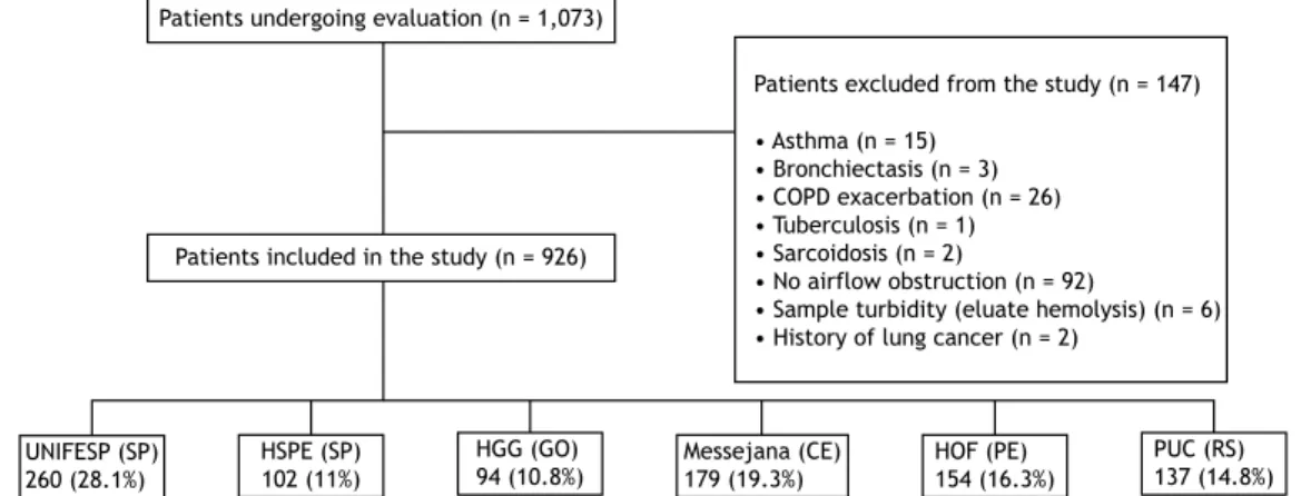
ethics committees of all participating centers. Between July of 2011 and August of 2012, 1,073 COPD patients followed at any of the six participating centers (two in northeastern Brazil, two in southeastern Brazil, one in southern Brazil, and one in central-western Brazil) were evaluated.
Patients
The inclusion criteria were as follows: being 40
years of age or older; having been diagnosed with
COPD (on the basis of clinical history and spirometry results, including a post-bronchodilator percent
predicted FEV1/FVC ratio—FEV1/FVC%—below the lower limit of normal); and having been stable for at
least four weeks.(14) The exclusion criteria were as follows: having been diagnosed with any other lung disease or systemic disease that can increase serum
AAT levels (including infections and inlammatory processes); having previously been diagnosed with AATD; being a relative of an index case of AATD; and having asthma (Figure 1).
The goal was to include 200 COPD patients from each participating center. At the end of the study period, no more patients were added to the study, regardless of whether or not the desired number of patients had been attained for each center.
Spirometry
The reference values for calculating percent predicted
FVC, percent predicted FEV1, and FEV1/FVC% were
based on the National Health and Nutrition Examination
Survey equations.(15) Spirometry was performed with
a portable spirometer (Easy One®; ndd Medical Tech
-nologies, Inc., Andover, MA, USA). At all participating
centers, the American Thoracic Society acceptability
and reproducibility criteria were used.(16)
Quantiication of AAT
The study was divided into three phases. In the irst
phase, all patients underwent determination of AAT
levels in dried blood spot (DBS) samples in order to
identify those with a possible diagnosis of AATD. In
the second phase, patients with DBS AAT levels ≤ 2.64
mg/dL (suspected AATD) underwent determination of serum AAT levels.(17) Finally, in the third phase, patients with serum AAT levels of < 113 mg/dL underwent
genotyping. In case of conlicting results between
serum AAT measurements and genotyping, genetic
sequencing was performed (Figure 2). To determine the sensitivity and speciicity of the eluate method,
Zillmer et al. used the bootstrap resampling method, comparing the AAT values measured in serum with
those measured in eluates from DBS samples in
order to determine a cut-off point for AAT values in
eluates; the value obtained was 2.02 mg/dL (97%
CI: 1.45-2.64). (17) All patients whose DBS AAT levels were below 2.64 mg/dL underwent measurement of serum AAT levels in order to prevent AATD from going undiagnosed.
Genotyping
Blood samples were collected with the use of ilter paper cards (Whatman 903, lot W101; Whatman/ GE Healthcare, Florham Park, NJ, USA). They were transported to the Federal University of São Paulo
Hospital São Paulo Central Laboratory, located in the
city of São Paulo, Brazil, under temperature-controlled conditions (i.e., at a constant temperature of −20°C),
in accordance with applicable International Air Transport
Association regulations. All cards were stored at −20°C for subsequent analysis (determination of DBS AAT
levels, genotyping, and SERPINA1 gene sequencing).
Serum and eluate samples were analyzed on a Siemens BNII system (Siemens Healthcare, Indianapolis, IN, USA) in July of 2012.
Patients excluded from the study (n = 147)
• Asthma (n = 15) • Bronchiectasis (n = 3) • COPD exacerbation (n = 26) • Tuberculosis (n = 1) • Sarcoidosis (n = 2)
• No airflow obstruction (n = 92)
• Sample turbidity (eluate hemolysis) (n = 6) • History of lung cancer (n = 2)
Patients included in the study (n = 926)
UNIFESP (SP) 260 (28.1%)
HSPE (SP) 102 (11%)
HGG (GO)
94 (10.8%) Messejana (CE)179 (19.3%)
HOF (PE) 154 (16.3%)
PUC (RS) 137 (14.8%) Patients undergoing evaluation (n = 1,073)
Figure 1. Flowchart of the patients included in the study and their distribution, by participating center. UNIFESP:
Universidade Federal de São Paulo; HSPE-SP: Hospital do Servidor Público Estadual de São Paulo; HGG: Hospital Geral de Goiânia Alberto Rassi; Messejana: Hospital de Messejana; HOF: Hospital Otávio de Freitas; PUC: Pontifícia Universidade Católica; SP: Brazilian state of São Paulo; GO: Brazilian state of Goiás; CE: Brazilian state of Ceará; PE: Brazilian state of Pernambuco; and RS: Brazilian state of Rio Grande do Sul.
Russo R, Zillmer LR, Nascimento OA, Manzano B, Ivanaga IT, Fritscher L, et al.
For DNA extraction, DBS samples were removed from the cards with a 6-mm paper punch, DNA being extracted with the QIAamp DNA Blood Mini Kit (QIAGEN,
Hilden, Germany), in accordance with the manufacturer
instructions. For identiication of S and Z alleles in exons 3 and 5, respectively, real-time PCR was used
with TaqMan® SNP Genotyping Assays (Thermo Fisher Scientiic Inc., Waltham, MA, USA). All patients with
AATD but without S and Z alleles underwent SERPINA1
gene sequencing (exons 2-5) in order to identify other polymorphisms described in the literature.
Statistical analysis
Continuous variables were expressed as mean ± standard deviation, whereas categorical variables were expressed as absolute numbers and proportions. Data were entered into an Oracle database and analyzed
with the Statistical Package for the Social Sciences, version 18.0 for Windows (SPSS Inc., Chicago, IL, USA).
RESULTS
Of the 1,073 patients who were being followed at any of the six participating centers during the study period, 926 met the eligibility criteria and
were therefore included in the study (Figure 1). The
demographic characteristics of the patients included in
the present study are shown in Table 1. There was no
statistically signiicant difference between males and
females regarding the prevalence of AATD, and two thirds of the participants were White. Although former smokers predominated (having accounted for 83.9% of the study sample), 36 (3.7%) of the patients had
never smoked; 410 patients (44.3% of the sample)
had a body mass index within normal limits, and 56 (6%) were underweight. As expected for patients with
COPD, FEV1/FVC% and percent predicted FEV1 were
low, characterizing obstructive lung disease. Of the 926 COPD patients included in the present
study, 85 had DBS AAT levels ≤ 2.64 mg/dL, therefore
being suspected of having AATD. Of those 85 patients, 2 died. Therefore, 83 patients underwent determination of serum AAT levels. Of those 83 patients, 24 had serum AAT levels < 113 mg/dL and therefore underwent genotyping. Genotype distribution was as follows:
PI*MS, in 3 (12.5%); PI*MZ, in 13 (54.2%); PI*SZ, in 1 (4.2%); PI*SS, in 1 (4.2%); and PI*ZZ, in 6
(25%). Although determination of serum AAT levels
was not performed in the 2 patients who had DBS AAT levels ≤ 2.64 mg/dL and died, genotyping and
SERPINA1 gene sequencing were performed from the
previously collected DBS samples, which had been stored at −20°C for subsequent analysis. Genotype
distribution was as follows: PI*M1I, in 1; and PI*ZZ,
Figure 2. Flowchart of alpha 1-antitrypsin (AAT) deiciency screening and genotype distribution. PI: protease inhibitor; and DBS: dried blood spot. *Although 2 patients died before undergoing determination of serum AAT levels, SERPINA1 gene
sequencing was performed with previously collected DBS samples. †Only 1 PI*MZ patient underwent gene sequencing,
because of discrepant results between determination of serum AAT levels and genotyping.
Patients included in the study (n = 926)
DBS AAT levels ≤ 2.64 mg/dL (n = 85; 9.2%) DBS AAT levels > 2.64 mg/dL (n = 841; 90.8%)
Serum AAT levels ≥ 113 mg/dL (n = 59; 71.1%)
PI*MS
(n = 3; 12.5%) (n = 13; 54.2%)PI*MZ (n = 1; 4.2%)PI*SS (n = 1; 4.2%)PI*SZ (n = 6; 25%)PI*ZZ
PI*M1Z† Genetic
sequencing
Serum AAT levels < 113 mg/dL (n = 24; 28.9%)
genotyping
Deaths (n = 2)* PI*M1I PI*ZZ
Prevalence of alpha-1 antitrypsin deiciency and allele frequency in patients with COPD in Brazil
in 1 (Figure 2). Given the discrepant results and the
deaths, genetic sequencing was performed, and the following genotypes were found: PI*M1Z; PI*M1I; and
PI*ZZ. These genotypes were included in a second analysis of allele frequency, and the results were as
follows: PI*M, 28.8%; PI*M1, 3.8%; PI*S, 11.5%;
PI*Z, 53.8%; and PI*I, 1.9%.
Table 2 shows the demographic characteristics of a
subset of 24 patients with DBS AAT levels ≤ 2.64 mg/
dL and serum AAT levels < 113 mg/dL. As expected, the patients with the PI*ZZ genotype were younger and had lower serum AAT levels than those with other genotypes (p < 0.001). However, there were no differences in gender, smoking history, spirometric
values, Medical Research Council scale scores, or COPD
Assessment Test scores across patient genotypes. Among the 926 COPD patients included in the present study, the overall prevalence of AATD was 2.8% and the
prevalence of the PI*ZZ genotype (severe AATD) was 0.8%. Analysis of allele frequency in the subgroup of patients with serum AAT levels < 113 mg/dL (including
the alleles found in the 2 patients who died, whose DBS AAT levels were ≤ 2.64 mg/dL) revealed frequencies of 53.8%, 11.5%, and 1.9% for the Z, S, and I alleles,
respectively (Table 3). Table 4 shows the genotype
prevalence among the ive regions of Brazil.
DISCUSSION
This is the irst study to show the prevalence of
AATD, as well as allele frequency, in a population of COPD patients in Brazil. The prevalence of AATD (i.e., 2.8%) and the Z allele (i.e., 0.8%) was found to be similar to that found in other countries.(18,19)
The decision to use the maximum conidence interval
of the cut-off point for AAT values in eluates(17) was due to the need to identify all individuals who were suspected of having AATD and should therefore undergo genotyping, thus minimizing the chances of not identifying patients with AATD. Although AATD is one of the most common genetic diseases, the prevalence of AATD is low. However, failure to identify any patient with AATD would have had an impact on
the inal results of the present study. Nevertheless, the use of the maximum conidence interval width
resulted in more patients being re-evaluated.(17) The cut-off point of 113 mg/dL was used in an attempt to Table 1. Demographic characteristics of the 926 COPD
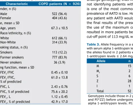
patients included in the present study.
Characteristic COPD patients (N = 926)
Gender, n (%) Male Female
522 (56.4) 404 (43.6) Age, mean ± SD
Age, years 67.3 ± 10.5
Race/ethnicity, n (%) White
Non-White
612 (66.1) 314 (33.9) Smoking status, n (%)
Smokers 113 (12.2)
Former smokers 777 (83.9)
Never smokers 36 (3.9)
Lung function, mean ± SD
FEV1/FVC 0.45 ± 0.10
FEV1/FVC, % of predicted
61.0 ± 13.8
FVC, L 2.43 ± 0.76
FVC, % of predicted 75.6 ± 20.2
FEV1, L 1.12 ± 0.45
FEV1, % of predicted 42.9 ± 17.0
Table 2. Demographic characteristics of a subset of 24 COPD patients with serum alpha 1-antitrypsin levels < 113 mg/dL, by genotype.a
Characteristic Genotype p*
PI*MS PI*MZ PI*SS PI*SZ PI*ZZ
Male gender, n (%) 2 (16.7) 7 (58.3) 1 (8.3) 1 (8.3) 1 (8.3) 0.07
Age, years 69.3 ± 9.4 69.0 ± 10.1 59.0 74.0 47.0 ± 2.3 < 0.001
Smoking history, pack-years 55.0 53.5 ± 41.1 40.0 12.6 19.1 ± 16.7 0.07
Post-BD FEV1, % of predicted 33.8 ± 8.3 41.1 ± 14.0 54.7 45.8 37.5 ± 19.9 0.63
FEV1/FVC 49.4 ± 5.8 57.4 ± 9.0 59.0 56.5 55.7 ± 12.6 0.92
Serum AAT, mg/dL 100 ± 13.5 93.7 ± 14.0 93.8 66.0 27.1 ± 4.8 < 0.001
MRC scale score 2.6 ± 1.1 2.7 ± 1.0 3.0 2.0 3.3 ± 1.6 0.27
CAT score, total 20.3 ± 6.4 16.6 ± 7.3 30 18 17.8 ± 6.3 0.42
Patients, n 3 13 1 1 6 N/A
PI: protease inhibitor; AAT: alpha 1-antitrypsin; MRC: Medical Research Council; BD: bronchodilator; and CAT:
COPD Assessment Test. aValues expressed as mean ± SD, except where otherwise indicated. *PI*ZZ vs. the
remaining genotypes.
Table 3. Allele frequency in a subset of 24 COPD patients with serum alpha 1-antitrypsin levels < 113 mg/dL, including the alleles found in 2 patients with dried blood spot alpha
1-antitrypsin levels ≤ 2.64 mg/dL, both of whom died.
Allele n %
M 15 28.8
M1 2 3.8
S 6 11.5
Z 28 53.8
I 1 1.9
Total 52 100
Genotypes include those in 2 patients who died (PI*M1I
and PI*ZZ) before undergoing determination of serum alpha 1-antitrypsin levels. PI: protease inhibitor.
Russo R, Zillmer LR, Nascimento OA, Manzano B, Ivanaga IT, Fritscher L, et al.
identify not only patients with severe AATD but also those with moderate AATD.(20-22)
Despite methodological differences and the fact that not all participants underwent genotyping, our results regarding the PI*Z allele are similar to those found in the literature.(11,18,23) The frequency of the mutant PI*Z allele and other AATD-associated alleles in the present study can be explained by the large number
of immigrants from Europe, primarily from countries
where the prevalence of AATD is high, such as Portugal and Italy.(23-26)
Although DBS analysis is particularly useful as an initial screening test for AATD, it is not suficient for a deinitive diagnosis. Clinical history, physical examination indings, and family history should be
taken into account when interpreting the results,
which should be conirmed by measuring serum
AAT levels in patients suspected of having AATD. If
the results of DBS analysis are conirmed by serum
ATT measurements, genotyping or phenotyping are
necessary for a deinitive diagnosis.
Health professionals providing care to patients with COPD should bear in mind that 2.8% of all COPD patients have some degree of AATD. Our study reinforces the knowledge that AATD is one of the most prevalent
genetic diseases. Further studies are warranted, given
that an AATD diagnosis can have a major impact on COPD prevention, especially in young smokers. In
addition, our inding that the prevalence of the PI*ZZ
genotype in the study population is 0.8% shows that severe AATD is present in patients with COPD in Brazil and reinforces the 1999 World Health Organization recommendation that “all COPD patients should be screened once for AATD using a quantitative test. Those with abnormal results on screening should undergo PI typing”.(27) Determination of AAT levels in COPD patients has also been recommended by the American Thoracic
Society/European Respiratory Society(11) and, more
recently, the Canadian Thoracic Society.(28) However,
these patients typically present at a younger age (< 45
years) with lower lobe emphysema. Family screening is useful for appropriate counseling. Few countries
are as racially diverse as is Brazil, which is populated by a large number of immigrants, including Asians,
Africans, Arabs, and, in particular, Europeans. The
Portuguese brought centuries of genetic admixture
among Europeans, including Celts, Romans, Germans,
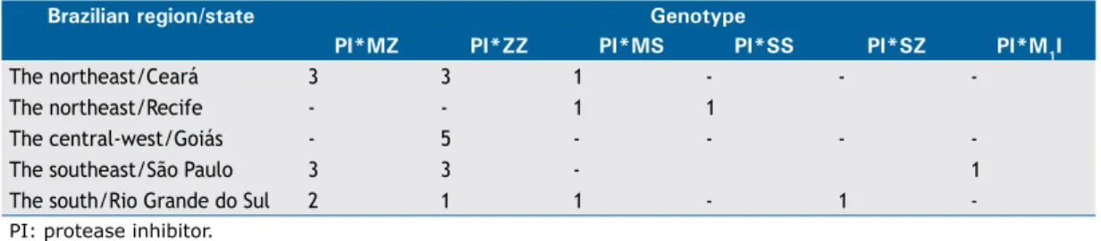
and Lusitanians. The differences in genotype prevalence
among the ive regions of Brazil might be due to the
different immigrant origins.
One limitation of the present study is that not all participants underwent genotyping. However, serum AAT levels were measured in all patients with the
use of the maximum conidence interval width, thus
preventing AATD from going undiagnosed.
The prevalence of AATD in COPD patients in Brazil was found to be similar to that found in most countries, despite the racial diversity of the Brazilian population. The actual prevalence of AATD in this population can be best determined by investigating neonates. Genetic studies aimed at determining the ancestry of this population are critical in order to establish a correlation between mutated alleles and the actual ancestry of the individuals.
ACKNOWLEDGMENTS
We would like to thank Siemens for the technical and scientiic support, which was crucial to the development
of the present study. We would also like to thank Grifols Brasil Ltda. for their support in creating the database.
Finally, we would like to thank the Associação Fundo
de Incentivo à Pesquisa (AFIP, Association for the Incentive Funding of Research) for their technical
support in performing the laboratory measurements. Table 4. Genotypes involved in alpha 1-antitrypsin deiciency, distributed by mutation of the SERPINA1 gene (genotype) and by participating center.
Brazilian region/state Genotype
PI*MZ PI*ZZ PI*MS PI*SS PI*SZ PI*M1I
The northeast/Ceará 3 3 1 - -
-The northeast/Recife - - 1 1
The central-west/Goiás - 5 - - -
-The southeast/São Paulo 3 3 - 1
The south/Rio Grande do Sul 2 1 1 - 1
-PI: protease inhibitor.
REFERENCES
1. Fagerhol MK, Laurell CB. The polymorphism of “prealbumins” and alpha-1-antitrypsin in human sera. Clin Chim Acta. 1967;16(2):199-203. http://dx.doi.org/10.1016/0009-8981(67)90181-7
2. Lai EC, Kao FT, Law ML, Woo SL. Assignment of the alpha 1-antitrypsin gene and a sequence-related gene to human chromosome 14 by molecular hybridization. Am J Hum Genet. 1983;35(3):385-92.
3. Stoller JK, Aboussouan LS. Alpha1-antitrypsin deiciency. Lancet. 2005;365(9478):2225-36. http://dx.doi.org/10.1016/S0140-6736(05)66781-5
4. de Serres FJ. Worldwide racial and ethnic distribution of alpha1-antitrypsin deiciency: summary of an analysis of published genetic epidemiologic surveys. Chest. 2002;122(5):1818-29. http://dx.doi. org/10.1378/chest.122.5.1818
5. Stockley RA. The pathogenesis of chronic obstructive lung diseases: implications for therapy. QJM. 1995;88(2):141-6.
6. Janoff A. Elastases and emphysema. Current assessment of the protease-antiprotease hypothesis. Am Rev Respir Dis. 1985;132(2):417-33.
Prevalence of alpha-1 antitrypsin deiciency and allele frequency in patients with COPD in Brazil
diagnosis and treatment of patients with COPD: a summary of the ATS/ERS position paper. Eur Respir J. 2004;23(6):932-46. http:// dx.doi.org/10.1183/09031936.04.00014304
8. Silverman EK, Chapman HA, Drazen JM, Weiss ST, Rosner B, Campbell EJ, et al. Genetic epidemiology of severe, early-onset chronic obstructive pulmonary disease. Risk to relatives for airlow obstruction and chronic bronchitis. Am J Respir Crit Care Med. 1998;157(6 Pt 1):1770-8. http://dx.doi.org/10.1164/ ajrccm.157.6.9706014
9. Lokke A, Lange P, Scharling H, Fabricius P, Vestbo J. Developing COPD: a 25 year follow up study of the general population. Thorax. 2006;61(11):935-9. http://dx.doi.org/10.1136/thx.2006.062802 10. Bascom R. Differential susceptibility to tobacco smoke: possible
mechanisms. Pharmacogenetics. 1991;1(2):102-6. http://dx.doi. org/10.1097/00008571-199111000-00008
11. American Thoracic Society; European Respiratory Society. American Thoracic Society/European Respiratory Society statement: standards for the diagnosis and management of individuals with alpha-1 antitrypsin deiciency. Am J Respir Crit Care Med. 2003;168(7):818-900. http://dx.doi.org/10.1164/rccm.168.7.818
12. de Serres FJ, Blanco I, Fernández-Bustillo E. Genetic epidemiology of alpha-1 antitrypsin deiciency in North America and Australia/New Zealand: Australia, Canada, New Zealand and the United States of America. Clin Genet. 2003;64(5):382-97. http://dx.doi.org/10.1034/ j.1399-0004.2003.00143.x
13. Menezes AM, Perez-Padilla R, Jardim JR, Muiño A, Lopez MV, Valdivia G, et al. Chronic obstructive pulmonary disease in ive Latin American cities (the PLATINO study): a prevalence study. Lancet. 2005;366(9500):1875-81. http://dx.doi.org/10.1016/S0140-6736(05)67632-5
14. Cazzola M, MacNee W, Martinez FJ, Rabe KF, Franciosi LG, Barnes PJ, et al. Outcomes for COPD pharmacological trials: from lung function to biomarkers. Eur Respir J. 2008;31(2):416-69. http://dx.doi. org/10.1183/09031936.00099306
15. Hankinson JL, Odencrantz JR, Fedan KB. Spirometric reference values from a sample of the general U.S. population. Am J Respir Crit Care Med. 1999;159(1):179-87. http://dx.doi.org/10.1164/ ajrccm.159.1.9712108
16. Standardization of Spirometry, 1994 Update. American Thoracic Society. Am J Respir Crit Care Med. 1995;152(3):1107-36. http:// dx.doi.org/10.1164/ajrccm.152.3.7663792
17. Zillmer LR, Russo R, Manzano BM, Ivanaga I, Nascimento OA, Souza AA, et al. Validation and development of an immunonephelometric assay for the determination of alpha-1 antitrypsin levels in dried blood spots from patients with COPD. J Bras Pneumol. 2013;39(5):547-54.
http://dx.doi.org/10.1590/S1806-37132013000500004
18. Lieberman J, Winter B, Sastre A. Alpha 1-antitrypsin Pi-types in 965 COPD patients. Chest. 1986;89(3):370-3. http://dx.doi.org/10.1378/ chest.89.3.370
19. de la Roza C, Costa X, Vidal R, Vilá S, Rodríguez-Frias F, Jardí R, et al. Screening program for alpha-1 antitrypsin deiciency in patients with chronic obstructive pulmonary disease, using dried blood spots on ilter paper [Article in Spanish]. Arch Bronconeumol. 2003;39(1):8-12. 20. Costa X, Jardi R, Rodriguez F, Miravitlles M, Cotrina M, Gonzalez C,
et al. Simple method for alpha1-antitrypsin deiciency screening by use of dried blood spot specimens. Eur Respir J. 2000;15(6):1111-5. 21. Miravitlles M, Herr C, Ferrarotti I, Jardi R, Rodriguez-Frias F, Luisetti M, et al. Laboratory testing of individuals with severe alpha1-antitrypsin deiciency in three European centres. Eur Respir J. 2010;35(5):960-8. http://dx.doi.org/10.1183/09031936.00069709 22. Ferrarotti I, Scabini R, Campo I, Ottaviani S, Zorzetto M, Gorrini M, et
al. Laboratory diagnosis of alpha1-antitrypsin deiciency. Transl Res. 2007;150(5):267-74. Erratum in: Transl Res. 2008;151(4):232. http:// dx.doi.org/10.1016/j.trsl.2007.08.001
23. Vidal R, Blanco I, Casas F, Jardí R, Miravitlles M; Committee on the National Registry of Individuals with Alpha-1 Antitrypsin Deiciency. Guidelines for the diagnosis and management of alpha-1 antitrypsin deiciency [Article in Spanish]. Arch Bronconeumol. 2006;42(12):645-59. http://dx.doi.org/10.1157/13095974
24. Blanco I, Fernández E, Bustillo EF. Alpha-1-antitrypsin PI phenotypes S and Z in Europe: an analysis of the published surveys. Clin Genet. 2001;60(1):31-41. http://dx.doi.org/10.1034/j.1399-0004.2001.600105.x
25. Sitkauskiene B, Serapinas D, Blanco I, Fernández-Bustillo E, Janciauskiene S, Sakalauskas R. Screening for alpha1-antitrypsin deiciency in Lithuanian patients with COPD. Respir Med. 2008;102(11):1654-8. http://dx.doi.org/10.1016/j.rmed.2008.07.003 26. Blanco I, de Serres FJ, Cárcaba V, Lara B, Fernández-Bustillo E.
Alpha-1 Antitrypsin Deiciency PI*Z and PI*S Gene Frequency Distribution Using on Maps of the World by an Inverse Distance Weighting (IDW) Multivariate Interpolation Method. Hepat Mon. 2012;12(10 HCC):e7434.
27. Alpha 1-antitrypsin deiciency: memorandum from a WHO meeting. Bull World Health Organ. 1997;75(5):397-415.
28. Marciniuk DD, Hernandez P, Balter M, Bourbeau J, Chapman KR, Ford GT, et al. Alpha-1 antitrypsin deiciency targeted testing and augmentation therapy: a Canadian Thoracic Society clinical practice guideline. Can Respir J. 2012;19(2):109-16. http://dx.doi. org/10.1155/2012/920918
http://dx.doi.org/10.1590/S1806-37562015000000069
ABSTRACT
Objective: Lung cancer is a global public health problem and is associated with high
mortality. Lung cancer could be largely avoided by reducing the prevalence of smoking. The objective of this study was to analyze the effects of social, behavioral, and clinical factors on the survival time of patients with non-small cell lung cancer treated at Cancer Hospital I of the José Alencar Gomes da Silva National Cancer Institute, located in the city
of Rio de Janeiro, Brazil, between 2000 and 2003. Methods: This was a retrospective
hospital cohort study involving 1,194 patients. The 60-month disease-speciic survival probabilities were calculated with the Kaplan-Meier method for three stage groups. The importance of the studied factors was assessed with a hierarchical theoretical model
after adjustment by Cox multiple regression. Results: The estimated 60-month
speciic-disease lethality rate was 86.0%. The 60-month speciic-disease-speciic survival probability ranged from 25.0% (stages I/II) to 2.5% (stage IV). The performance status, the intention to treat, and the initial treatment modality were the major prognostic factors identiied
in the study population. Conclusions: In this cohort of patients, the disease-speciic
survival probabilities were extremely low. We identiied no factors that could be modiied after the diagnosis in order to improve survival. Primary prevention, such as reducing the prevalence of smoking, is still the best method to reduce the number of people who will suffer the consequences of lung cancer.
Keywords: Lung neoplasms/epidemiology; Carcinoma, non-small-cell lung; Survival
analysis.
Factors associated with disease-speciic
survival of patients with non-small
cell lung cancer
Mirian Carvalho de Souza1, Oswaldo Gonçalves Cruz2, Ana Glória Godoi Vasconcelos3
Correspondence to:
Mirian Carvalho de Souza. Rua Marquês de Pombal, 125, 7º andar, Centro, CEP 20230-240, Rio de Janeiro, RJ, Brasil. Tel.: 55 21 3207-5667. E-mail: miriancs@inca.gov.br
Financial support: None.
INTRODUCTION
Lung cancer is the most common type of cancer
worldwide; it is estimated that, in 2012, there were 1.8
million new cases.(1) In Brazil in 2015, 27,000 new cases were estimated.(2)
Although lung cancer has various histological types,
the most widely used classiication system is that which
divides tumors into small cell carcinomas (15%) and non-small cell carcinomas (85%).(3)
The 60-month survival probability of patients with
non-small cell lung carcinoma is lower than 15% in Europe.(4)
A study conducted in the United States obtained estimates
ranging from 66% (stage Ia) to 4% (stage IV). (5) A study
involving patients from a university hospital in the city of
Rio de Janeiro, Brazil, found that the 60-month survival
probability was 6%, with it being 14% for the early stages and 5% for the advanced stages.(6)
Among the prognostic factors studied for lung cancer patients(7) are stage, performance status,(8) weight loss, gender, age, smoking, smoking history, quality of life, marital status, depression, and genetic mutations.(6,9-11)
Epidemiological studies have indicated that the effects
of socioeconomic factors on health outcomes are indirect, occurring through behavioral and clinical factors. In this context, it is important to establish the hierarchy
of these factors in determining the occurrence of lung cancer and in the survival probability of patients with this type of cancer.(12,13)
The objective of the present article was to analyze the importance of social, behavioral, and clinical factors on the survival time of patients with non-small cell lung cancer treated at Hospital do Câncer I do Instituto Nacional de CâncerJosé Alencar Gomes da Silva (HCI/INCA, Cancer
Hospital I of the José Alencar Gomes da Silva National Cancer Institute), located in the city of Rio de Janeiro,
Brazil, between 2000 and 2003.
METHODS
This was a retrospective observational hospital cohort study in which the object of interest was the time from diagnosis to death from lung cancer or metastasis.
The target population consisted of patients diagnosed with primary non-small cell lung carcinoma, between 2000 and 2003, who were registered in the Registro Hospitalar de Câncer (RHC, Hospital Cancer Registry) of HCI/INCA, which is a tertiary referral hospital for the treatment of cancer in the state of Rio de Janeiro, Brazil.
Eligible patients were deined as those from the state of Rio de Janeiro, where HCI/INCA is located, in whom diagnosis was conirmed by either anatomic pathological
1. Divisão de Pesquisa Populacional,
Coordenação de Pesquisa e Educação, Instituto Nacional de Câncer José Alencar Gomes da Silva – INCA – Rio de Janeiro (RJ) Brasil.
2. Programa de Computação Cientíica, Fundação Oswaldo Cruz – Fiocruz – Rio de Janeiro (RJ) Brasil.
3. Departamento de Métodos Quantitativos em Saúde, Escola Nacional de Saúde Pública Sérgio Arouca – ENSP – Fundação Oswaldo Cruz – Fiocruz – Rio de Janeiro (RJ) Brasil.
Submitted: 30 March 2015.
Accepted: 27 January 2016.
Study carried out at the Instituto Nacional de Câncer José Alencar Gomes da Silva – INCA – and at the Escola Nacional de Saúde Pública Sérgio Arouca – ENSP – Fundação Oswaldo Cruz – Fiocruz – Rio de Janeiro (RJ) Brasil.
J Bras Pneumol. 2016;42(5):317-325