DOI: 10.1590/1518-8345.0897.2689 www.eerp.usp.br/rlae
How to cite this article
Araújo DD, Almeida NG, Silva PMA, Ribeiro NS, Werli-Alvarenga A, Chianca TCM. Prediction of risk and incidence
of dry eye in critical patients. Rev. Latino-Am. Enfermagem. 2016;24:e2689. [Access ___ __ ____]; Available in:
____________________. DOI: http://dx.doi.org/10.1590/1518-8345.0897.2689. month day year
URL
Prediction of risk and incidence of dry eye in critical patients
1Diego Dias de Araújo
2Natália Gherardi Almeida
3Priscila Marinho Aleixo Silva
4Nayara Souza Ribeiro
4Andreza Werli-Alvarenga
5Tânia Couto Machado Chianca
6Objectives: to estimate the incidence of dry eye, to identify risk factors and to establish a risk
prediction model for its development in adult patients admitted to the intensive care unit of a
public hospital. Method: concurrent cohort, conducted between March and June, 2014, with 230
patients admitted to an intensive care unit. Data were analyzed by bivariate descriptive statistics,
with multivariate survival analysis and Cox regression. Results: 53% out of 230 patients have
developed dry eye, with onset mean time of 3.5 days. Independent variables that signiicantly
and concurrently impacted the time for dry eye to occur were: O2 in room air, blinking more than
ive times per minute (lower risk factors) and presence of vascular disease (higher risk factor). Conclusion: dry eye is a common inding in patients admitted to adults intensive care units, and
care for its prevention should be established.
Descriptors: Dry Eye Syndromes; Corneal Diseases; Intensive Care Units; Nursing; Nursing
Diagnosis.
1 Paper extracted from Master’s Thesis “Prediction of risk and incidence of dry eye in critically ill patients” presented to Escola de Enfermagem,
Universidade Federal de Minas Gerais, Belo Horizonte, MG, Brazil. Supported by Conselho Nacional de Desenvolvimento Cientíico e Tecnológico (CNPq), process # 479539/2012-0.
2 Doctoral Student, Escola de Enfermagem, Universidade Federal de Minas Gerais, Belo Horizonte, MG, Brazil. Professor, Universidade Estadual de Montes Claros, Montes Claros, MG, Brazil.
3 RN, Secretaria Municipal de Saúde, Prefeitura Municipal de Belo Horizonte, Belo Horizonte, Brazil.
4 Undergraduate Student in Nursing, Escola de Enfermagem, Universidade Federal de Minas Gerais, Belo Horizonte, MG, Brazil.
5 PhD, Adjunct Professor, Departamento de Enfermagem Básica, Escola de Enfermagem, Universidade Federal de Minas Gerais, Belo Horizonte, MG, Brazil.
Introduction
Patients in very critical conditions are normally
admitted to Intensive Care Units (ICUs). Most of the time
these patients are sedated, in a coma, with Mechanical
Ventilation (MV), taking several medications and with
compromised ocular protection mechanisms(1-8).
In ICUs, so far, little importance has been attributed
to the care of damage or injury related to the visual
perception of critical patients, occurring for several
causes, since its approach requires knowledge and
participation of a multidisciplinary team, and care for
reducing ocular problems(4,8-9). In addition, ICUs favor
assistance to systems considered vital (cardiovascular, respiratory and neurological).
The dysfunction of the tear ilm, known as dry
eye, is a multifactorial alteration of tears and the
ocular surface that results in symptoms of discomfort,
visual disturbances and instability of the tear ilm, with
potential damage to the ocular surface. The problem is
followed by an increase in the osmolarity of the tear
ilm, and ocular surface inlammation(10).
The nursing diagnosis of risk of dry eye is deined
as “risk of ocular discomfort and damage to the cornea
and conjunctiva due to the reduced amount or quality
of tears to moisten the eye”. The risk factors for the
problem in patients admitted to ICUs involve sedation,
environmental factors (air conditioning and low humidity) related to the treatment (side effects of pharmaceutical
agents such as diuretics, analgesics, sedatives and
neuromuscular blocking agents), mechanical ventilation
therapy, neurological lesions with sensory or motor loss,
and damage to the ocular surface(9).
The preventive approach to ocular care is of utmost
importance for patients admitted to ICUs. The absence
of speciic care for prevention of dry eye can negatively
impact the lives of patients, both during hospitalization
and after discharge from the ICU, for generating
discomfort and ocular damage that may limit daily
activities and compromise the quality of life.
Only a few studies support dry eye in patients
admitted to ICUs. We were able to ind one study(4) related to the problem in critical patients, however, it
was about dry eye prevention, lacking the identiication
of incidence and risk factors of the problem.
This research is justiied by the need for knowledge
about the dry eye problem, determination of its incidence
and risk factors in critical patients, and thus implements
practices based on scientiic evidence, for prevention and treatment of this involvement identiied in patients
admitted to ICUs. It should be noted that no studies that
speciically cover the problem were identiied, although
NANDA I(9) has approved the Nursing Diagnosis of risk of
dry eye in the 2012-2014 edition.
This study aims to estimate the incidence of dry
eye, to identify risk factors and to establish a risk
prediction model for its development in adult patients
admitted to the intensive care unit of a public hospital.
Methods
This is a concurrent cohort study, conducted in an
ICU for adult patients of a public teaching hospital in
Belo Horizonte, Minas Gerais. Currently, in this hospital,
30 intensive care beds intended for adults are available
to the community.
Sample size calculation was carried out using the
ininite population formula, by conservative criterion,
since the studied population was unknown. In the
estimation of the sample we considered the ininite population, conidence degree of 95%, margin of error
of 6.5%, and proportion of interest of 55.1% in the
incidence of lesions in the punctate cornea(8), resulting
in a minimum sample calculation of 225 patients.
The inclusion criteria were: to have 18 years or
more, to not present dry eye at the time of admission,
to remain hospitalized in intensive care for at least 24
hours, to consent to participate in the research or have
their participation authorized by the responsible person
through a free and informed consent form.
The target population of this research consisted
of 258 patients hospitalized in the ICU between March
and June, 2014. Eight out of 258 patients were excluded
because their relatives did not allow the participation in
the study, one for being underage, and the other 19 for
having dry eye diagnosis at the time of admission to the
unit. Therefore, after the application of the inclusion and
exclusion criteria, a total of 230 patients was sampled.
For data collection we used an evaluation tool for
admission, which included sociodemographic and clinical
information, and risk factors for the development of
dry eye. Twenty-four hours after the admission, the
patients were evaluated with the instrument of daily
developments, which included clinical data and risk
factors for the development of dry eye, identiied in the
literature(1-23).
The dependent variable was the time for the
occurrence of dry eye in patients admitted to adults ICU.
The independent variables, selected in literature(1-23),
were: age, gender, origin unit, Sepsis Related Organ
Failure Assessment (SOFA), Acute Physiology and Chronic Health Evaluation (APACHE II), Therapeutic Intervention Scoring System (TISS 28), type of patient, death,
postoperative time, days of hospitalization, referral to
score in Ramsay sedation scale, score in the Glasgow
Coma scale (GCS), tracheal intubation, tracheostomy (TQT), Mechanical Ventilation (MV), days with MV, MV type, Fraction of Inspired Oxygen (FiO2), End-expiratory Pressure (PEEP), orotracheal tube ixing, Noninvasive Ventilation (NIV), NIV time, Oxygen (O2) in room air, O2 by Nasal Catheter (NC), use of macronebulization,
oxygen low, blinking rate per minute, eyeball exposure,
oedema, conjunctival hemorrhage, severity of corneal
injury, medicines, oral diet allowed, feeding tube,
nutritional status, accumulated luid balance (FB), positioning (degree of elevation of the headboard), and
white blood cells.
Before data collection, the nurse researcher
was trained to evaluate the cornea by a nurse with
experience and training in corneal evaluation of the
critically ill patients. The training consisted in theoretical
explanation about corneal injury and practical training
of eye evaluation, in addition to reading articles and
texts on the subject. This nurse was considered the gold
standard for performing corneal evaluation due to the
experience with assistance, research and publication in
the area. We found Kappa coeficient of 0.84 between
the nurse researcher and the nurse considered expert,
i.e., an almost perfect agreement.
Data collection was carried out every day of the
week by the nurse researcher until the patient developed
an outcome, were discharged from ICU, transferred or
passed away. To evaluate the tear volume we used the
Schirmer I test, which consists in the installation of a
strip of Whatman no. 41 or 50, with 5 mm in width and
35 mm in length, with folded extremity (about 5 mm),
attached to the bottom of the lower eyelid bag in the
temporal part (the outer corner of the lower eyelid).
After 5 minutes, the tape was removed, measured,
and the extension of the moistened part was noted. For
corneal evaluation we installed a drop of luorescein in
each eye of the patient, and after 1 to 2 minutes, under
low light conditions, the cornea was examined with the
aid of an ophthalmoscope with cobalt blue light ilter and
magnifying glass, for best viewing of possible corneal
changes. Data were immediately noted in the data
collection instrument.
In the treatment of the data, we performed double
typing in Epi Info program, version 3.5.1, and after
verifying the consistency of the data they were exported
to the Statistical Package for Social Science (SPSS),
version 19.0.
In the analysis, we used simple frequencies, measures of central tendency (mean and median), and measures of variability (standard deviation). The incidence (global incidence and incidence rate) of dry
eye and risk factors were determined. For analysis of the
potential risk factors with the time to the occurrence of
dry eye in patients hospitalized in ICU, we used bivariate
analysis for the variables studied, from the survival
analysis. With that, we obtained the relation between
each independent variable and the outcome variable
(time before the occurrence of dry eye), being measured the strength of the association by the Hazard Ratio (HR), considering the conidence interval (CI) of 95%. To identify surveyed covariates that exerted inluence on
the time from the monitoring to the outcome, we used
the Cox regression model. Variables whose p value was
≤ 0.25 in the bivariate analysis were included in the
multivariate analysis model. We performed the global
adjustment of the model by the probability ratio test,
estimated the survival function, failure rate regarding
the time before the occurrence of dry eye, and risk
proportionality test.
The study is in accordance with Resolution 466/12,
which provides for research with human beings.
The project was referred to the Ethics and Research
Committee of the Federal University of Minas Gerais and
obtained a favorable opinion under the CAAE Protocol -
15616313.4.0000.5149.
Results
Among the 230 patients, 122 presented dry eye.
The global incidence of dry eye was, therefore, of 53%
in the period of the study.The incidence rate of dry eye
was of 0.184 cases/patient a day (5.51 cases/patient per month), ranging from 0.153 cases/patient a day (4.58 cases/patient per month) to 0.219 cases/patient a day (6.58 cases/patient per month), with 95% conidence.
Most (55.7%) were male, mean age of 59 years (SD ± 19.2), median of 62 years, with minimal
variability of 18 years and a maximum of 97 years. Of
the total patients (230), 36% were sedated. Intubation was used in 110 (48%); tracheostomy in 6 (2.6%), and mechanical ventilation in 114 (50%). Among the
patients studied, 8% passed away. The seriousness of
the medical condition of the patients was evaluated by
the instruments, SOFA, APACHE II and TISS 28, applied
in the irst 24 hours of the patient’s hospitalization in
ICU. On average, they had a 4.9 SOFA, 20 APACHE II
and 31 of 28 TISS. For admission to the ICU, vascular
diseases were the most frequent (27%).
More than half of patients blinked the eyes more
than ive times per minute (51.3%), and 49.2% had the eyeball exposed (lagophthalmos).
Among the patients, 53% showed positive Schirmer
I test, and 54.3% presence of corneal injury.Of these,
52% showed punctate-type injury and 6% corneal
30% showed punctiform epithelial erosions, involving
the lower third of the cornea of the left and right eyes.
On average, for admission, the patients presented
a moistened extension of the Whatman Strip of 14.6
mm and 12.9 mm in the left eye, in the Schirmer test
I, during the study.For the right eye, for admission, the
result was of 15 mm of moistened extension, and 13.1
mm during the study.
In the bivariate analysis, we obtained variables that
showed statistical signiicance (p≤0.25) over time until
the occurrence of dry eye. For multivariate analysis, 40
variables were eligible, of which 30 showed statistical
signiicance (p<0.05), presented in Table 1.
Variables Group
Dry eye
Hazard Ratio (HR)
(CI 95%) p value
Yes No
n % n %
Sedation No 51 41.8 96 88,9 2.10 (1.46 - 3.02) < 0.001
Yes 71 58.2 12 11.1
Intubation No 27 22.1 93 86.1 3.16 (2.06 - 4.85) < 0.001
Yes 95 77.9 15 13.9
Mechanical Ventilation No 24 19.7 92 85.2 3.40 (2.17 - 5.32) < 0.001
Yes 98 80.3 16 14.8
O2 on room air No 116 95.1 50 46.3 0.12 (0.05 - 0.29) < 0.001
Yes 6 4.9 58 53.7
O2 by nasal catheter No 103 84.4 46 42.6 0.27 (0.16 - 0.44) < 0.001
Yes 19 15.6 62 57.4
Oral diet allowed No 62 50.8 37 34.3 0.43 (0.30 - 0.63) < 0.001
Yes 60 49.2 71 65.7
Feeding tube Enteral feeding 45 75.0 11 15.5 3.01 (1.64 - 5.49) < 0.001
Blinking Up to ive times
per minute
101 82.8 11 10.2 0.16 (0.10 - 0.26) < 0.001
More than ive times per minute
21 17.2 97 89.8
Eyeball exposure No 62 50.8 104 96.3 2.43 (1.70 - 3.48) < 0.001
Yes 60 49.2 4 3.7
Oedema No 12 9.8 40 37.0 2.39 (1.31 - 4.34) 0.004
Yes 110 90.2 68 63.0
Oedema site Chemosis – left
eye
45 36.9 9 8.3 1.79 (1.24 - 2.59) 0.002
Chemosis – right eye
45 36.9 9 8.3 1.79 (1.24 - 2.59) 0.002
MMII 70 57.4 34 31.5 1.47 (1.03 - 2.11) 0.033
Anasarca 9 7.4 1 0.9 3.05 (1.53 - 6.09) 0.001
Anticoagulants Yes 82 67.2 84 77.8 0.64 (0.43 - 0.93) 0.022
Hypnotics/sedatives/anxiolytics Yes 71 58.2 20 18.5 1.58 (1.10 - 2.27) 0.013
Antihypertensives Yes 19 15.6 32 29.6 0.53 (0.32 - 0.87) 0.013
Analgesic Yes 22 18.0 43 39.8 0.62 (0.39 - 0.99) 0.048
SOFA*† Yes 6.8 3.7 4.0 3.5 1.10 (1.05 - 1.15) < 0.001
APACHE II*‡ Yes 25.4 7.5 19.0 9.7 1.03 (1.01 - 1.04) < 0.001
TISS 28*§ Yes 35.8 8.1 26.7 8.7 1.06 (1.03 - 1.08) < 0.001
Postoperative time* Yes 2.9 1.0 2.6 1.1 0.44 (0.31 - 0.63) < 0.001
Hospitalization Time* Yes 3.0 0.9 2.8 1.2 0.05 (0.02 - 0.13) < 0.001
Glasgow Coma Scale* Yes 8.9 4.2 14.2 1.3 0.84 (0.80 - 0.89) < 0.001
Mechanical Ventilation Time* Yes 3.2 1.2 2.1 1.0 0.64 (0.50 - 0.82) < 0.001
O2 low catheter* Yes 2.3 1.4 1.6 0.7 1.41 (1.01 - 1.97) 0.039
Anticoagulants* Yes 82 67.2 84 77.8 0.64 (0.43 - 0.93) 0.022
Hypnotics/sedatives/anxiolytics* Yes 71 58.2 20 18.5 1.58 (1.10 - 2.27) 0.013
Antihypertensives* Yes 19 15.6 32 29.6 0.53 (0.32 - 0.87) 0.013
Fluid balance/Daily developments (Positive)*
Yes 894.3 978.1 595.2 1022.0 1.00 (1.000003 -
1.0004)
0.046
Table 1 – Variables with association with the time until occurrence of dry eye. Belo Horizonte, MG, Brazil, 2014
*Variables in which n corresponds to the mean and % to the standard deviation †SOFA – Sepsis-related Organ Failure Assessment
‡APACHE II – Acute Physiology and Chronic Health disease Classiication System II
Risk prediction model (multivariate analysis) among the demographic and clinical factors identiied,
O2 in room air, blinking more than ive times per minute,
and presence of vascular disease impacted signiicantly
and concurrently until the occurrence of dry eye.
Patients who remained in O2 for room air presented
probability of occurrence of dry eye regarding the risk
group which was not in O2 by room air, at any time, 66%
lower (HR=0.34), ranging from 13% to 87%, with 95% conidence (p=0.025).
Patients who blinked their eyes more than ive
times per minute showed probability of occurrence of
dry eye, at any time, 75% lower (HR=0.25) in relation to those who blinked their eyes up to ive times per minute, ranging from 57% to 86%, with 95% conidence (p<0.001).
For patients who have medical diagnosis of vascular
disease when admitted to the ICU, the risk of dry eye,
at any time, was 1.56 (HR=1.56) higher compared to
those who did not have such medical diagnosis. The risk
ranged from 1.03 to 2.38 times, with 95% conidence (p=0.037).
Among the studied patients, we observed that dry
eye occurred only from the second day, considering that
between the third and the fourth days of hospitalization
50% of patients presented an outcome, according to the
estimated model. The mean time determined for the
occurrence of dry eye appearance was 3.5 days.
The risk of failure, determined to the development
of dry eye, was of 0.1 time until the second day, achieving
0.5 times on the third day, 1.1 time on the fourth day,
and 2.6 times in the sixth day of hospitalization.
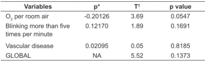
In Table 2 we observed that the values of the
Pearson correlation coeficient (p) are all close to zero.
In addition, we also observed that both the global test
and the tests for each variable do not show evidence for
rejecting the null hypothesis of proportional risks.
Discussion
In previous studies(11-15), conducted with patients in
outpatient units, dry eye prevalence ranged from 10.8%
to 57.1%. However, these studies were conducted
with a proile of patients different from the one of the
patients who participated in this study. The only study(4)
on dry eye in critical patients found in the literature is a
randomized clinical trial, comparing interventions for the
prevention of the problem. We highlight that such study
was conducted with a small sample (18 patients), and in
a reality different from Brazil’s.
As the reduction in the amount or the quality of
tears can be the beginning of major alterations in the
ocular surface, the prevention of dry eye becomes
essential for critically ill patients. This problem can
be identiied and prevented by the nurses, who have
nursing interventions to assist in the reduction of ocular
complications.
Studies(16-17) suggest that alterations in the ocular
surface are highly prevalent, especially in the early days
of hospitalization. It is estimated that the mean for the
development of corneal injury is between 24 hours and
8.9 days(1,6-8,18). In the study(4) that speciically addressed
dry eye, the mean for the emergence of the problem was
approximately three days, but this time was estimated
after the intervention implementation.
The emergence of dry eye is estimated at a relatively
small time interval. Thus, once the patient is admitted
to the ICU, ocular conditions should be evaluated, and
nursing interventions implemented to prevent possible
ocular complications that may impact negatively on the
lives of these patients during the hospitalization or after
discharge of the unit.
Among the variables that showed a signiicant association (p<0.05) with time until the occurrence of
dry eye and that predispose higher risk for developing
dry eye (HR≥ 1), authors(1-10) point out as possible risk factors: values obtained with APACHE II, TISS 28,
hospitalization time, intubation, Mechanical Ventilation
(MV), score in the Glasgow Coma scale, MV time, hypnotics/sedatives/anxiolytics, luid balance/daily evolution (positive), lagophthalmos, chemosis, and
anasarca.
Regarding the variables identiied as factors that
predispose lower risk of developing dry eye in critical
patients, by presenting HR<1, such variables may
be related to the better clinical pattern of patients,
since they would already be taking the allowed
diet, without intubation, breathing with the aid of
mechanical ventilation, with adequate blinking relex,
Variables p* T† p value
O2 per room air -0.20126 3.69 0.0547
Blinking more than ive times per minute
0.12170 1.89 0.1691
Vascular disease 0.02095 0.05 0.8185
GLOBAL NA 5.52 0.1373
Table 2 – Test of proportionality of risks. Belo Horizonte,
MG, Brazil, 2014
*Pearson correlation coeficient estimated between the Schoenfeld
standardized residues and the variable time response before the occurrence of the outcome
†Statistics of the test with Chi-square distribution, 3 degrees of freedom,
and may complain of pain. However, we highlight
that medications like anticoagulants, analgesics, and
antihypertensive drugs are commonly administered in
critical patients, hospitalized in ICUs and, although a
statistically signiicant association was found, further
studies are needed to evaluate whether there is a direct
causal relation between the use of such medicines and
the development of dry eye.
We also highlight that, in this study, lagophthalmos
(eyeball exposure) was identiied as the main risk
factor for changes in the ocular surface. The datum
is corroborated by other studies(17, 21-22), in which the
patients had lagophthalmos frequency ranging from
31% to 54%. The fact is also pointed out by a study(23)
in which this risk factor showed statistical signiicance (p=0.001), corroborating the results of this research.
Although the study(4) did not ind statistically signiicant
results which speciically addressed the dry eye, the
authors emphasize that lagophthalmos is one of the
most important predictive factors for the occurrence of
dry eye in critical patients.
Sedative drugs and mechanical ventilation were
also identiied as important risk factors for dry eye, and showed statistical signiicance (p=0.001). These data are conirmed by other studies(19,22-23). Both risk factors are important for the problem, despite the results of the
study(4), conducted speciically on dry eye, showing no
statistical signiicance.
Three covariates showed statistical signiicance (p<0.05) for the time until the occurrence of dry eye. Thus, patients who blinked their eyes more than ive times per minute (HR=0.25), and in room air (HR=0.34),
presented lower risk of dry eye. We can infer that the
patients of the group with lower risk had a clinical proile
less severe than patients that presented outcome.
Patients hospitalized with diagnosis of vascular
disease (HR=1.56) showed higher risk of dry eye, which
may be explained by the fact that these patients are
usually sedated, in a coma, breathing with the aid of
mechanical ventilation, using various medicines and
with compromised ocular protection mechanisms.
Thus, in a way, these patients were exposed to various
factors associated with ocular surface changes, and the
emergence of dry eye.
Regarding the function of failure, we found close
temporal relation between dry eye and hospitalization
in the ICU, i.e. the longer the hospitalization time, the
greater the risk of developing outcome in critically ill
patients.
The risk factors identiied in the study are related
to the medical condition and treatment of the patients.
Although the nursing staff cannot change these factors,
strategies for early identiication of risk of dry eye can
be adopted, as well as preventive measures to avoid
further ocular injury that can be implemented from the
ocular evaluation, using accurate tests such Schirmer I,
luorescein, and identiication of risk factors related to
the problem.
Some risk factors described in NANDA I(9) have
been validated in this study as: treatment side effects,
neurological lesions with sensory loss or relex motor (lagophthalmos, absence of spontaneous blinking relex due to reduced consciousness or other medical
conditions), and mechanical ventilation therapy.Certain
factors presented to this diagnosis, and constants in
the taxonomy were not validated for not presenting
statistically proven relevance, for instance, the case of
female patients with autoimmune diseases, aging, and
hormone use.
However, other factors in the diagnosis could not
be validated as well since they involve missing aspects
in patients of this sample such as lifestyle, history of
allergies, use of contact lenses, environmental factors,
and the place where they live. We suggest multicentric
research decrease in different populations, for comparing
different realities.
The model obtained was considered valid to describe
the relation between the time before the occurrence
of dry eye and associated risk factors, in addition to
anticipate which critically ill patients show risk of dry
eye. Furthermore, the analysis of proportional risks
tests shows that the model is appropriate, considering
that the proportional risks assumption was met, thus
determining the implementation of nursing care for its
prevention.
A limitation of this study was its performance with
a particular proile of patients, showing the need for
multicentric studies in different populations to establish
its external validity.
Conclusion
From the results, we can verify that dry eye in
patients admitted to adults ICUs is a common inding,
exposed to a set of internal and external risk factors that
can collaborate for the emergence of the problem.
After bivariate analysis and multivariate analysis
adjustment step, among the demographic and clinical
factors identiied, those that remained as better
predictors for the phenomenon studied were: O2 in
room air, blinking more than ive times per minute and
The early recognition of risk factors for dry eye
and, consequently, the adoption of preventive measures
will certainly reduce the probability of ocular surface
changes in critically ill patients.
It is recommended that the investigation of the risk
factors described in NANDA I and which could not be
validated according to the proile of the sample studied,
such as: lifestyle, history of allergies, contact lenses,
environmental factors and place where the patient lives.
In addition, we need studies that allow establishing
what is the best nursing care for the prevention of the
problem, particularly regarding critically ill patients.
We believe that this study may contribute to relect
on the relevance of the dry eye problem in critical and
non-critical patients, in addition to a greater awareness
and appreciation of the importance of eye care in
patients admitted to adults ICUs, being a fundamental
aspect for higher quality nursing care.
References
1. Desalu I, Akinsola F, Adekola O, Akinbami O, Kushimo
O, Adefuleositelu A. Ocular surface disorders in intensive
care unit patients in a Sub- Saharan teaching hospital.
Internet J Emerg Intensive Care Med. 2008;11(1):30-4.
2. Germano EM, Mello MJG, Sena DF, Correia JB, Amorim
MMR. Incidence and risk factors of corneal epithelial
defects in mechanically ventilated children. Crit Care
Med. 2009;37(3):1097-100.
3. Grixti A, Sadri M, Edgar J, Datta AV. Common ocular
surface disorders in patients in intensive care units. Ocul
Surface. 2012;10(1):26-42.
4. Güler EK, Eser I, Egrilmez S. Effectiveness of
polyethylene covers versus carbomer drops(Viscotears)
to prevent dry eye syndrome in the critically ill. J Clin
Nurs. 2011;20: 1916-22.
5. Imanaka H, Taenaka N, Nakamura J, Aoyama K,
Hosotani H. Ocular surface disorders in the critical ill.
Anesth Analg. 1997;85(2):343-6.
6. Joyce N. Eye Care for Intensive Care Patients:
A Systematic Review. Int J Evid Based Healthc.
2002;6(21):1-5.
7. Joyce N. Eye Care for Patients in the ICU. Int J Evid
Based Healthc. 2006;106(1):72A-72D.
8. Werli-Alvarenga A, Ercole FF, Botoni FA, Oliveira
JADMM, Chianca TCM. Corneal injuries: incidence and
risk factors in the Intensive Care Unit. Rev. Latino-Am.
Enfermagem. 2011;19(5):1088-95.
9. Herdman TH, editors. NANDA International nursing
diagnoses: deinitions and classiication, 2012-2014].
Porto Alegre: Artmed; 2013.
10. International Dry Eye Workshop (DEWS). The deinition and classiication of dry eye disease: report of the Deinition and Classiication Subcommittee of
the International Dry Eye Workshop. Ocul Surface.
2007;5(2):75-92.
11. Albietz JM. Prevalence of dry eye subtypes in clinical
optometry practice. Optom Vis Sci. 2000;77(7):357-63.
12. Farrell J, Grierson DJ, Patel S, Sturrock RDA.
Classiication for dry eyes following comparison of tear
thinning time with Schirmer tear test. Acta Ophthalmol.
1992;70(3):357-60.
13. Hikichiv T, Yoshida A, Fukui Y, Hamano T, Ri M,
Araki K, et al. Prevalence of dry eye in Japanese
eye centers. Graefe’s Arch Clin Exp Ophthalmol.
1995;233(9):555-8.
14. Toda I, Fujishima H, Tsubota K. Ocular fatigue is
the major symptom of dry eye. Acta Ophthalmologica.
1993;71(3):347-52.
15. Versura P, Cellini M, Torreggiani A, Profazio V,
Bernabini B, Caramazza R. Dryness symptoms, diagnostic
protocol and therapeutic management: a report on
1,200 patients. Ophthalmic Res. 2001;33(4):221-7.
16. Dawson D. Development of a new eye care guideline
for critically ill patients. Intensive Crit Care Nurs.
2005;21(2):119-22.
17. Mercieca F, Suresh P, Morton A, Tullo A. Ocular
surface disease in intensive care unit patients. Eye.
1990;13(2):231-6.
18. Oh EG, Lee WH, Yoo JS, Kim SS, Ko IS, Chu SH,
et al. Factors related to incidence of eye disorders in
Korean patients at intensive care units. J Clin Nurs.
2009;18(1):29-35.
19. Koroloff N, Boots R, Lipman J, Thomas P, Rickard
C, Coyer F. A randomised controlled study of the
eficacy of hypromellose and Lacri-Lube combination
versus polyethylene/Cling wrap to prevent corneal
epithelial breakdown in the semiconscious intensive care
patient. Intensive Care Med. 2004;30(6):1122-6.
20. Marshall AP, Elliott R, Rolls K, Schacht S, Boyle M. Eye
care in the critically ill: Clinical practice guideline. Austr
Crit Care. 2008;21(2):97-109.
21. Ezra DG, Chan MP, Solebo L, Malik AP, Crane E,
Coombes A, et al. Randomised trial comparing ocular
lubricants and polyacrylamide hydrogel dressings in
the prevention of exposure keratopathy in the critically
ill. Intensive Care Med. 2009;35(3):455-61.
22. Jammal H, Khader Y, Shihadeh W, Ababneh L, AlJizawi
Received: Apr. 8th 2015
Accepted: July 5th 2015
Copyright © 2016 Revista Latino-Americana de Enfermagem
This is an Open Access article distributed under the terms of the Creative Commons (CC BY).
This license lets others distribute, remix, tweak, and build upon your work, even commercially, as long as they credit you for the original creation. This is the most accommodating of licenses offered. Recommended for maximum dissemination and use of licensed materials.
Corresponding Author: Diego Dias de Araújo
Universidade Estadual de Montes Claros. Departamento de Enfermagem Campus Universitário Prof. Darcy Ribeiro
Av. Ruy Braga, s/n
Bairro: Vila Mauriceia , Prédio 6 (CCBS)
CEP: 39401-089, Montes Claros, MG, Brasil E-mail: [email protected]
23. Sivasankar S, Jasper S, Simon S, Jacob P, John
G, Raju R. Eye care in ICU. Indian J Crit Care Med.