Induced pluripotent stem cells reprogramming: Epigenetics
and applications in the regenerative medicine
KÁTIA MARIA SAMPAIO GOMES1*, ISMAEL CABRAL COSTA1, JENIFFER FARIASDOS SANTOS2, PAULO MAGNO MARTINS DOURADO3,
MARIA FERNANDA FORNI4, JULIO CESAR BATISTA FERREIRA1
1Department of Anatomy, Institute of Biomedical Sciences III, Universidade de São Paulo (ICB III/USP), São Paulo, SP, Brazil 2Department of Biochemistry, Universidade Federal de São Paulo (Unifesp), São Paulo, SP, Brazil
3Heart Institute, Faculdade de Medicina da Universidade de São Paulo (InCor/FMUSP), São Paulo, SP, Brazil 4Chemistry Institute, Universidade de São Paulo (IQ/USP), São Paulo, SP, Brazil
S
UMMARYStudy conducted at Universidade de
São Paulo (USP), São Paulo, SP, Brazil
Article received: 5/23/2016
Accepted for publication: 5/30/2016
*Correspondence:
Departamento de Anatomia, ICB III/USP Address: Av. Prof. Lineu Prestes, 2415,
Cidade Universitária São Paulo, SP – Brazil
Postal code: 05508-000 [email protected]
http://dx.doi.org/10.1590/1806-9282.63.02.180
Induced pluripotent stem cells (iPSCs) are somatic cells reprogrammed into an embryonic-like pluripotent state by the expression of speciic transcription factors. iPSC technology is expected to revolutionize regenerative medicine in the near future. Despite the fact that these cells have the capacity to self-renew, they present low eficiency of reprogramming. Recent studies have demonstrated that the previous somatic epigenetic signature is a limiting factor in iPSC performance. Indeed, the process of effective reprogramming involves a complete remodeling of the existing somatic epigenetic memory, followed by the establishment of a “new epigenetic signature” that complies with the new type of cell to be differentiated. Therefore, further investigations of epigenetic modiications associated with iPSC reprogramming are required in an attempt to improve their self-renew capacity and potency, as well as their application in regenerative medicine, with a new strategy to reduce the damage in degenerative diseases. Our review aimed to summarize the most recent indings on epigenetics and iPSC, focusing on DNA methylation, histone modiications and microRNAs, highlighting their potential in translating cell therapy into clinics.
Keywords: induced pluripotent stem cells, regenerative medicine, cell reprogramming, epigenetics, histones, microRNAs.
I
NTRODUCTIONHuman embryonic stem cells have great potential for self-renewal and the ability to differentiate into all tissues of the body (except embryonic attachments),1 forming an important source of material for regenerative medicine and cell therapy. However, the use of embryonic stem cells is limited by ethical and religious conlicts, as well as im-munological incompatibility.2 In order to reduce the dam-age caused by degenerative diseases, different strategies are being used in an attempt to optimize the use of em-bryonic stem cells.
The irst strategy used was that of somatic cell nucle-ar content (SCNT) for unfertilized and enucleated oo-cytes.3 However, the yield of this technique is still very low and the cells obtained can present phenotypic and gene expression abnormalities.4 Another strategy is the fusion of somatic cells with embryonic cells, reprogramming
their genome.5 However, although fusion-induced repro-gramming is very eficient (about 95%), the resulting hy-brid cells lack therapeutic potential due to their tetra-ploidy, as well as the presence of exogenous genes from the pluripotent cells used in the fusion.5 Therefore, a search for new strategies for the eficient use of cells with an embryonic proile is still needed.
medicine for Yamanaka in 2012, shared with John b. Gur-don.8 Since then, several surveys have been developed to explore this technology. In fact, several researchers were able to reprogram somatic cells (postmitotic) into iPSC using the aforementioned strategy.9-11
The advantage of using this method is that it allows the derivation of pluripotent cells from the donor, reduc-ing the risk of rejection by the immune system. In addition, this method provides a platform to study the molecular mechanisms of genetic and chronic diseases, minimizing the ethical, religious and political conlicts and opening up new perspectives for regenerative medicine. However, although cell identity can be modiied by the ectopic ex-pression of transcription factors, the eficiency of repro-gramming remains low (0.1 to 3%) and its cost is high.11,12
The low reprogramming eficiency of iPSC is associ-ated with residual epigenetic memory of the tissue from which they were derived, which complicates the repro-gramming process. Recent studies show that despite iPSC sharing common characteristics of pluripotency and self-renewal capacity, these cells still retain an epigenetic memory.13-15 In addition, there is evidence that the repro-gramming process involves complete remodeling of the existing somatic epigenetic memory, followed by the es-tablishment of a new “epigenetic signature” that conforms to the type of cell to be differentiated.16 Therefore, the epigenetic memory becomes a barrier in the process of cellular reprogramming. This fact highlights the need for new studies investigating the epigenetic changes associ-ated with cellular reprogramming in an attempt to im-prove the eficiency and effectiveness of the iPSC created, as well as their clinical application. As such, our review aimed to gather information about the epigenetic factors (DNA methylation, changes to histones and microRNAs) associated with iPSC reprogramming eficiency. In addi-tion, we have brought together the clinical studies using iPSC as cell therapy.
E
PIGENETICS AND IPSC
Waddington was the irst researcher to use the term epi-genetics in 1942 to explain how the genome interacts with the environment during the development process.17 There-fore, any reversible and inheritable change in the func-tional genome that does not alter the sequence of DNA nucleotides refers to epigenetics.18 Several pathologies are associated with epigenetic changes.19-22
The reprogramming eficiency of iPSC is also direct-ly related to epigenetic changes such as DNA methylation and the epigenetic memory of the source cells.23 The iPSC reprogramming process can be divided into three distinct
phases, called pre-iPSC, intermediate and full reprogram-ming. The reprogramming process is extremely slow, with low eficiency (0.1 to 3%) and high cost,11,12 and depends on suitable levels of gene expression in each phaseand speciic epigenetic changes.24 Djuric and Ellis compared the epigenetic changes that occur during the reprogram-ming process with a “seven headed dragon,” where a series of changes is necessary for efficient reprogramming, namely: 1) Endogenous reactivation of genes related to cell pluripotency, Nanog and Oct4; 2) Chromatin chang-es, such as trimethylation in H3K27 and changes in H3K4; 3) Hypomethylation of heterochromatin; 4) Reactivation of the inactive X chromosome; 5) Maintenance of DNA methylation marks; 6) Silencing the retrovirus that in-duces pluripotency; and, inally, 7) Two- or three-dimen-sional chromatin changes and location of nuclear sub-domains.24 Therefore, the control of epigenetic factors during reprogramming may improve the induction of iPSC and their eficiency.25
E
PIGENETIC CHANGESIN IPSC
REPROGRAMMING DNA methylation in iPSC reprogrammingDNA methylation is an epigenetic mechanism involved in many important cellular processes such as cell prolif-eration and differentiation, transcriptional repression, genomic imprinting, organization of chromatin and in-activation of the X chromosome.26 Thus, changes in the DNA methylation proile are associated with the appear-ance of many degenerative diseases.
The aforementioned studies demonstrate the impor-tance of DNA demethylation in cell reprogramming. Sev-eral studies have attempted to improve these and other epigenetic mechanisms in order to improve both the quality and eficiency of iPSC reprogramming. Another relevant topic for such improvement is the change in histones during the reprogramming process.
Changes to histones in iPSC reprogramming
Histones are basic proteins rich in lysine and may suffer several epigenetic changes. Most of these modiications happen in the N-terminal region of the histone, with the exception of ubiquitination, which occurs in the C-ter-minal region of H2A and H2B.37 Epigenetic modiications to histones may either promote or inhibit gene transcrip-tion by changing the level of chromatin folding.38,39
Taking into consideration the epigenetic changes in histones in iPSC, H3 is the histone researched the most, as it is directly related to genes expressed during embry-onic development, such as Oct3/4, Sox2 and Nanog. It has already been demonstrated that methylation of H3K27 is associated with the suppression of various genes, and that persistent trimethylation of the lysine 27 of histone 3 (H3K27me3) blocks reprogramming by repressing the chromatin region associated with the target genes of the stem cells. However, the methylation of H3K4 is associ-ated with the activation of different embryonic genes.24 In an attempt to improve the performance of iPSC and reach the ideal conditions for the induction of pluripo-tency, by reducing the “epigenetic memory” in somatic cells, different strategies that directly or indirectly affect the methylation/acetylation of H3 have been used.12 Sev-eral researchers have demonstrated that it is possible to perform the induction of pluripotency without the use of Yamanaka factors, using only chemical compounds/ molecules that interfere with the enzymes that control the chromatin structure.40
Recently, Rais et al. showed that the inhibition of Mbd3 – a subunit of the NuRD complex responsible for the deacetylation of histones, remodeling of nucleosomes and gene expression inhibition – is able to reactivate the genes expressed during embryonic development and im-prove the eficiency of iPSC reprogramming by almost 90%, both in human as well as mouse cells.41
Many other strategies have been tested in order to improve the iPSC reprogramming process, such as the use of small molecules like Forskolin (FSK),42 BIX-01294,43 valproic acid (VPA – HDAC histone deacetylase inhibitor)44 and vitamin C.45 Therefore, the induction of pluripo-tency of iPSC can only occur with the use of small
mol-ecules.46-48 The authors advocate the use of such because they are not immunogenic, with greater yield and easy production. Thus, the use of chromatin modulators can increase eficiency in the iPSC reprogramming process.49 To do so, it is necessary to use a small molecule that is able to demethylate the DNA in the promoter region and change speciic regions in histones.
MicroRNAs in iPSC reprogramming
MicroRNA or miRNA are important tools for regulating gene expression in post-transcriptional iPSC by promot-ing pluripotency to modulate the stability of messenger RNA (mRNA) and suppress the signs of differentiation during the self-renewal of embryonic stem cells.
MiRNAs also modulate the signaling cascades that are necessary for maintaining the pluripotent state.50 Wang et al. noted that the loss of function in the enzyme Dicer and DGCR8, proteins essential for the biogenesis of microRNAs in the embryonic stem cells of mice, pres-ent two differpres-ent phenotypes: 1) reduction in proliferation due to cell cycle arrest in G1; and 2) resistance to differ-entiation, which reveals a close relationship between mi-croRNAs, differentiation and the pluripotency of cells.51,52
MicroRNAs are small non-coding RNA molecules. They have 18-25 nucleotides (nt), and are derived from a larger precursor. The processing of microRNAs occurs as follows:
1. After DNA transcription by RNA polymerase II or III the primary microRNA is formed (pri-miRNA). This may be presented in the shape of a fork. The pri-miRNA irst undergoes processing by the enzyme ribonucle-ase (RNribonucle-ase) nuclear III-like DROSHA. The speciici-ty of the cleavage in this step is guided by the DGCR8 protein, which acts as a “molecular ruler,” position-ing the DROSHA at a distance of 11 nucleotides from the base of the pri-miRNA loop. After cleavage, a pre-miRNA (precursor pre-miRNA) is released, formed by about 60-70 nt.53
2. The pre-miRNA is actively transported to the cyto-plasm by exportin-5 (Exp5), when this protein is linked to its Ran-dependent GTP cofactor. In the cytoplasm, it undergoes another cleavage process, where it loses the loop and is reduced to a miRNA duplex approx-imately 18-25pb in length. This last stage of process-ing is conducted by DICER, an RNAse-III type en-zyme, aided by the TRBP protein.53
Only one of the microRNA duplex strands remains in the RISC complex (guide strand), with the other being degraded (passenger strand). The RISC com-plex is able to identify and bind to target messengers RNAs in region 3’UTR through complementarity of bases in the “SEED” region of the miRNA (nucleo-tides 2-8 from the 5’ end) in order to inhibit its trans-lation or promote its adenytrans-lation and degradation.53
MicroRNAs that promote the reprogramming of iPSC
• miR 290-295 (cluster): These constitute more than 70% of the entire population of microRNAs in the embryonic stem cells of mice. miR 291-3 p, miR-294 and miR-295 are part of this cluster and indirectly promote the transition of genes associated with en-try into the G1-S phase, blocking Cdkna (p21), a sup-pressor of the Cyclin E/Cdk2 complex, and regulator of the cell cycle. After its transfection into MEFs there is an increase of 0.01-0.05% to 0.1-0.3% in cell repro-gramming eficiency.54
• miR 302-367 and miR 371-373 (cluster): These miRNAs suppress the expression of MBD2 (methyl-CPG-bind-ing domain protein 2), which works like a demethyl-ase in cells, resulting in incredemethyl-ased expression of Nanog and conversion of completely reprogrammed iPSC. They are also able to reduce expression of the inhib-itors in the G1-S phase, as well as increasing the ki-netics of the mesenchymal-epithelial transition (MET) required for reprogramming.55,56 Only the use of miR-302a/b/c/d and miR-367 is able to reprogram adult cells, and with greater eficiency, when compared to the Yamanaka method.57 Data demonstrate that miR-302 in conjunction with Yamanaka factors inhibits NR2F2 (nuclear receptor subfamily 2, group F, mem-ber 2) and improves reprogramming eficiency through indirect positive regulation of Oct4.58
• miR-200b and -200c, miR-205: These promote MET via signaling of transforming growth factor β (TGF-β) and, in conjunction with the Yamanaka factors, exclude the need for signaling of bone morphogenetic protein (BMP) during the initial reprogramming phase.59 • miR-93, miR-106: They suppress the expression of
TGF-β and p21, leading to MET and increased pro-liferation.60
• miR-135b: This is highly expressed during the repro-gramming process, regulating the expression of TGF-β, IGFBP5 (insulin-like growth factor binding protein 5) and Wisp1 (inducible-signaling pathway protein 1), which are involved in the expression of extracellular matrix genes.61
MicroRNAs that are barriers to iPSC reprogramming
• miR Let-7 (cluster): This inhibits the Pou5f1/Oct4, Sox2, Nanog and Tcf3 targets – pluripotency factors – stabilizing a differentiated state. In addition, this miRNA inhibits the translation of CDK4, repressing the transition of the G1-S-phase. MiR Let-7 forms a negative feedback circuit, providing a molecular mech-anism that facilitates the decision between self-re-newal and differentiation of cells.50,62
• miR-34a, miR-34b/c: miR-34a represses the expres-sion of Nanog, Sox2 and c-Myc. Taken in conjunction, miR-34a and miR-34b/c target p53 (tumor suppres-sor gene), holding an essential role in the containment of somatic reprogramming.63
These studies show that miRNAs can be important tools in the mediation of iPSC reprogramming without the need for the ectopic expression of pluripotency induction factors, including OSKM factors. The tables below pres-ent a summary of differpres-ent approaches and their effects on iPSC reprogramming. Table 1 is related to changes in DNA methylation, Table 2 is related to modiications in histones and Table 3 is related to the use of miRNAs. In addition, Figure 1 presents these changes in summa-rized form.
C
LINICAL APPLICATION OF IPSC
SThe main discussion about the use of iPSC in regenerative medicine is related to their ability to transform into can-cer cells. Incomplete reprogramming of iPSC may be as-sociated with the emergence of various mutations.
In addition to the impact on iPSC reprogramming, OSKM factors are associated with the development of tumors. Oct4 is highly expressed in the cervical cancer cell line. Deletion of Sox2 is associated with regression of melanomas. The Klf4 and c-Myc factors regulate genes involved in cell growth and proliferation.64 However, over the last 10 years following the discovery of iPSC, this technology has undergone several changes, such as the use of episomal plasmids that are not integrated into the genome, diminishing the carcinogenic potential of these cells.65 Currently, several methods are used to develop iPSC lines, including the use of plasmids, transposons, adenovirus, Sendai virus, miRNA and chemical com-pounds, minimizing mutagenic factors. Given these ad-vances, iPSC have been used in pre-clinical tests and clinical trials.
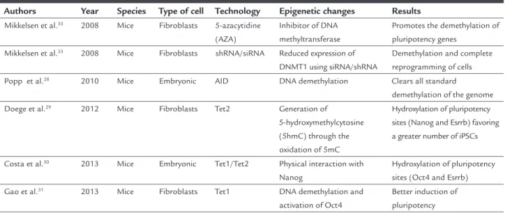
pa-TABLE 1 Epigenetic DNA changes.
Authors Year Species Type of cell Technology Epigenetic changes Results Mikkelsen et al.33 2008 Mice Fibroblasts 5-azacytidine
(AZA)
Inhibitor of DNA methyltransferase
Promotes the demethylation of pluripotency genes
Mikkelsen et al.33 2008 Mice Fibroblasts shRNA/siRNA Reduced expression of
DNMT1 using siRNA/shRNA
Demethylation and complete reprogramming of cells Popp et al.28 2010 Mice Embryonic AID DNA demethylation Clears all standard
demethylation of the genome
Doege et al.29 2012 Mice Fibroblasts Tet2 Generation of
5-hydroxymethylcytosine (5hmC) through the oxidation of 5mC
Hydroxylation of pluripotency sites (Nanog and Esrrb) favoring a greater number of iPSCs
Costa et al.30 2013 Mice Embryonic Tet1/Tet2 Physical interaction with
Nanog
Hydroxylation of pluripotency sites (Oct4 and Esrrb) Gao et al.31 2013 Mice Fibroblasts Tet1 DNA demethylation and
activation of Oct4
Better induction of pluripotency
iPSCs: induced pluripotent stem cells.
TABLE 2 Epigenetic changes in histones.
Authors Year Species Type of cell Technology Epigenetic changes Results
Huangfu et al.44 2008 Mice Fibroblasts VPA/TSA/SAHA Inhibitor of histone
deacetylation
Prevents histone deacetylation, increasing reprogramming eficiency by more than 100X compared to OSKM
Shi et al.43 2008 Mice Fibroblasts BIX-01294 G9a methyltransferase
inhibitor
Reduces histone H3K9 methylation, facilitating transcription. Improves reprogramming eficiency compared to Oct4 and Klf4
Esteban et al.35 2010 Humans Fibroblasts Vitamin C Nanog promoter, Oct4/
Histone demethylation
Decreases senescence during reprogramming
Liang et al.46 2010 Mice Fibroblasts Sodium butyrate Histone deacetylase
(HDAC) inhibitor
Facilitates reprogramming mediated by c-Myc
Liang et al.48 2012 Mice Fibroblasts Lentivirus increasing
the expression of Kdm2b
Activation of genes in the initial phase of reprogramming
Decreased H3K36me2 levels, contributing to reprogramming
Onder et al.47 2012 Mice Fibroblasts epz004777 DOT1L inhibitor
(Histone methyltransferase speciic for H3K79)
With the reduction of DOT1L, a reduction in the methylation of H3K79 occurs, improving the reprogramming by increasing transcription factors such as Nanog and Lin28
Chen et al.45 2013 Mice Fibroblasts Vitamin C Increases Kdm3 and
Kdm4 – Lysine-speciic demethylase
Reduces H3K9me2 and H3K9me3, important early in the reprogramming of iPSCs
Hou et al.42 2013 Mice Fibroblasts 3-deazaneplanocin
A (Dznep)
Histone methyltransferase inhibitor
Better reprogramming occurs with inhibition of histone methyltransferase by preventing methylation in the arginine and lysine residues of histones
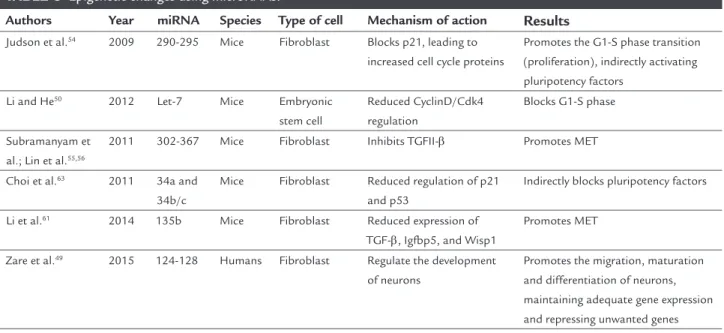
TABLE 3 Epigenetic changes using microRNAs.
Authors Year miRNA Species Type of cell Mechanism of action Results
Judson et al.54 2009 290-295 Mice Fibroblast Blocks p21, leading to
increased cell cycle proteins
Promotes the G1-S phase transition (proliferation), indirectly activating pluripotency factors
Li and He50 2012 Let-7 Mice Embryonic
stem cell
Reduced CyclinD/Cdk4 regulation
Blocks G1-S phase
Subramanyam et al.; Lin et al.55,56
2011 302-367 Mice Fibroblast Inhibits TGFII-β Promotes MET
Choi et al.63 2011 34a and
34b/c
Mice Fibroblast Reduced regulation of p21 and p53
Indirectly blocks pluripotency factors
Li et al.61 2014 135b Mice Fibroblast Reduced expression of
TGF-β, Igfbp5, and Wisp1
Promotes MET
Zare et al.49 2015 124-128 Humans Fibroblast Regulate the development
of neurons
Promotes the migration, maturation and differentiation of neurons, maintaining adequate gene expression and repressing unwanted genes
MET: mesenchymal-epithelial transition.
thology affects around 700,000 people in Japan, and is the most common form of blindness in people aged over 60, causing progressive loss of the retinal pigment epi-thelial monolayer. The transplant lasted around two hours and, according to the researchers, the patient did not suffer adverse effects, and there were an improvement in the morphology of the macula and neovascularization.66
This clinical trial was carried out by the group of Pro-fessor Takahashi, co-author of the manuscripts that won Professor Yamanaka the Nobel Prize in Medicine in 2012. However, in March 2015, clinical testing intended to treat six patients was suspended due to regulatory changes in regenerative medicine in Japan.67
Other clinical trials are currently underway to test the effectiveness of cellular therapy with iPSC in the treat-ment of AMD, Parkinson’s disease, spinal cord injury, diabetes and myocardial infarction.68 Preliminary results have not yet been presented.
Maintenance of the epigenetic factors of the iPSC after reprogramming has also been used to understand the molecular pathways involved in the development of diseases, development of new drugs and personalized medicine.69 The irst study conducted of this kind used a model of neuropathic disease. The authors repro-grammed ibroblasts from patients with Riley-Day syn-drome and monitored in vitro splicing of the IKBKAP (mutation associated with the disorder). Furthermore, the researchers also evaluated candidate drugs for rever-sal of the splicing. The study of iPSC gains relevance in this case, due to the inability of accessing the tissues
affected by Riley-Day syndrome.70 Other studies have been developed along this line,69 and are promising in the context of drug development. Figure 2 summarizes the use of iPSC.
C
ONCLUSIONG
O
M
ES
K
M
S
ET
AL
.
R
EV
A
SS
O
C
M
ED
B
R
AS
2
0
1
7
; 6
3
(2
):1
8
0
-1
8
9
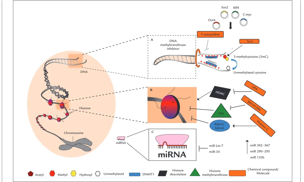
FIGURE 1 Epigenetic factors and iPSC reprogramming eficiency. The ectopic expression of Yamanaka factors, Oct4, Sox2, Klf4 and c-Myc (OSKM) are able to lead to DNA demethylation and reprogramming of somatic cells. A. The use of Tet1 (DNA hydroxylase) and of 5-azacytidine (inhibitor of the enzyme DNA methyltransferase) improves the reprogramming eficiency of iPSC cells. B. The use of valproic acid (VPA), BIX-01294 and vitamin C favors reprogramming through inhibition of histone deacetylase (HDAC), histone methyltransferase (G9a) and activation of the deme-thylases Kdm3/Kdm4, respectively. C. miRNAs 302-367, 290-295, 135b are able to increase reprogramming eficiency by promoting the progression of the cell cycle. miR Let-7 inhibits the cell cycle, and miR-34 inhibits the translation of p53, decreasing reprogramming eficiency.
Acetyl
Sox2 Klf4
C-myc Oct4
Tet1
miRNA DNA
Histone
Chromosome
Methyl Hydroxyl Unmethylated
5-azacytidine
miR Let-7
miR-34
miR 302∼367
miR 290∼295
miR 135b Kdm3/
Kdm4
HDAC
VPA
BIX -01294
Vit amin C
5-methylcytosine (5mC)
Unmethylated cytosine
DNMT1
DNMT1
DNMT1
A
B
C
Histone
deacetylase methyltransferaseHistone DNA
methyltransferase inhibitor
miRNA
Chemical compound/ Molecule –
the mechanisms of the aforementioned changes, optimiz-ing their application in regenerative medicine.
R
ESUMOCélulas-tronco de pluripotência induzida: papel da epi-genética na reprogramação e sua aplicabilidade clínica
As células-tronco de pluripotência induzida (CTPI) ou do inglês induced pluripotent stem cells (iPSCs)são células so-máticas reprogramadas para o estado embrionário por meio da expressão de fatores ectópicos de transcrição especíicos, tornando-as um alvo promissor para a medi-cina regenerativa. Apesar das CTPI compartilharem ca-racterísticas embrionárias, como pluripotência e capaci-dade de autorrenovação, elas possuem uma baixa eiciência de reprogramação, sendo a memória epigené-tica uma das principais barreiras nesse processo. A epige-nética é caracterizada por alterações reversíveis e herdáveis no genoma funcional que não alteram a sequência de nucleotídeos do DNA. Dentre as diferentes modiicações epigenéticas, destacam-se metilação de DNA, alterações em histonas e microRNA. Atualmente, sabe-se que o pro-cesso de reprogramação efetivo das CTPI envolve um completo remodelamento da memória epigenética somá-tica existente, seguido pelo estabelecimento de uma
“as-sinatura epigenética” que esteja de acordo com o novo tipo de célula a ser diferenciada. Modiicações epigenéti-cas personalizadas são capazes de melhorar o rendimen-to e a efetividade das CTPI geradas, abrindo uma nova perspectiva para a terapia celular. Nesta revisão reunimos as principais informações sobre os fatores epigenéticos que afetam a reprogramação das CTPI, bem como seus benefícios na aplicação da terapia celular.
Palavras-chave: células-tronco de pluripotência induzida, medicina regenerativa, reprogramação celular, epigenética, histonas, microRNA.
R
EFERENCES1. Biehl JK, Russell B. Introduction to stem cell therapy. J Cardiovasc Nurs. 2014; 24(2):98-103; quiz 104-5.
2. Lo B, Parham L. Ethical issues in stem cell research. Endocr Rev. 2009; 30(3):204-13.
3. Gurdon JB, Elsdale TR, Fischberg M. Sexually mature individuals of Xenopus laevis from the transplantation of single somatic nuclei. Nature. 1958; 182(4627):64-5.
4. Tachibana M, Amato P, Sparman M, Gutierrez NM, Tippner-Hedges R, Ma H, et al. Human embryonic stem cells derived by somatic cell nuclear transfer. Cell. 2014; 153(6):1228-38.
5. Cowan C a, Atienza J, Melton DA, Eggan K. Nuclear reprogramming of somatic cells after fusion with human embryonic stem cells. Science. 2005; 309(5739):1369-73.
6. Takahashi K, Yamanaka S. Induction of pluripotent stem cells from mouse embryonic and adult ibroblast cultures by deined factors. Cell. 2006; 126(4):663-76.
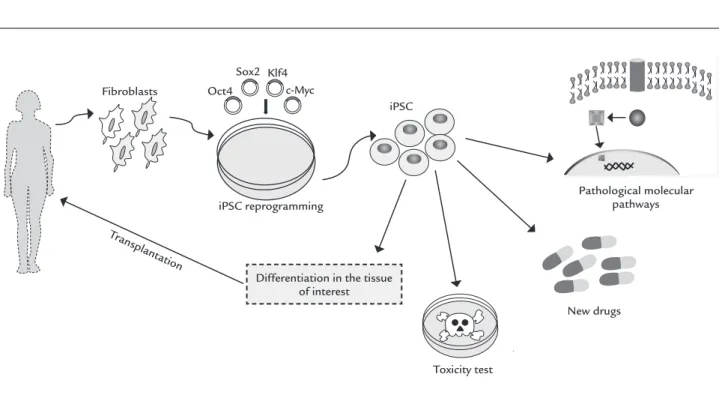
FIGURE 2 Somatic cells are reprogrammed into induced pluripotent stem cells (iPSCs). These cells are differentiated in the tissue of interest and transplanted in an attempt to reduce the damage caused by degenerative diseases. In addition, iPSCs are also being used in pre-clinical and clinical tests.
Fibroblasts
iPSC Oct4
Sox2 Klf4 c-Myc
iPSC reprogramming
Toxicity test
New drugs
Pathological molecular pathways
Differentiation in the tissue of interest
7. Patel M, Yang S. Advances in reprogramming somatic cells to induced pluripotent stem cells. Stem Cell Rev. 2010; 6(3):367-80.
8. Johnson MH, Cohen J. Reprogramming rewarded: the 2012 Nobel Prize for Physiology or Medicine awarded to John Gurdon and Shinya Yamanaka. Reprod Biomed Online. 2012; 25(6):549-50.
9. Lowry WE, Plath K. The many ways to make an iPS cell. Nat Biotechnol. 2008; 26(11):1246-8.
10. Yu J, Vodyanik MA, Smuga-Otto K, Antosiewicz-Bourget J, Frane JL, Tian S, et al. Induced pluripotent stem cell lines derived from human somatic cells. Science. 2007; 318(5858):1917-20.
11. Takahashi K, Yamanaka S. Induced pluripotent stem cells in medicine and biology. Development. 2013; 140(12):2457-61.
12. Apostolou E, Hochedlinger K. Chromatin dynamics during cellular reprogramming. Nature. 2013; 502(7472):462-71.
13. Chin MH, Mason MJ, Xie W, Volinia S, Singer M, Peterson C, et al. Induced pluripotent stem cells and embryonic stem cells are distinguished by gene expression signatures. Cell Stem Cell. 2009; 5(1):111-23.
14. Kim K, Doi A, Wen B, Ng K, Zhao R, Cahan P, et al. Epigenetic memory in induced pluripotent stem cells. Nature. 2010; 467(7313):285-90. 15. Polo JJM, Liu S, Figueroa MME, Kulalert W, Eminli S, Tan KY, et al. Cell
type of origin inluences the molecular and functional properties of mouse induced pluripotent stem cells. Nat Biotechnol. 2010; 28(8):848-55. 16. Nashun B, Hill PWS, Hajkova P. Reprogramming of cell fate: epigenetic
memory and the erasure of memories past. EMBO J. 2015; 34(10):1296-308. 17. Waddington CH. The epigenotype. 1942. Int J Epidemiol. 2012; 41(1):10-3. 18. Haig D. The (dual) origin of epigenetics. Cold Spring Harb Symp Quant
Biol. 2004; 69:67-70.
19. Kim SY, Morales CR, Gillette TG, Hill JA. Epigenetic regulation in heart failure. Curr Opin Cardiol. 2016; 31(3):255-65.
20. Abdolmaleky HM, Zhou J-R, Thiagalingam S. An update on the epigenetics of psychotic diseases and autism. Epigenomics. 2015; 7(3):427-49. 21. Faroogi AA, Tang JY, Li RN, Ismail M, Chang YT, Shu CW, et al. Epigenetic
mechanisms in cancer: push and pull between kneaded erasers and fate writers. Int J Nanomedicine. 2015; 10:3183-91.
22. Coppedè F. The potential of epigenetic therapies in neurodegenerative diseases. Front Genet. 2014; 5:220.
23. Gładych M, Andrzejewska A, Oleksiewicz U, Estécio MRH. Epigenetic mechanisms of induced pluripotency. Contemp Oncol (Pozn). 2015; 19(1A):A30-8.
24. Djuric U, Ellis J. Epigenetics of induced pluripotency, the seven-headed dragon. Stem Cell Res Ther. 2010; 1(1):3.
25. Liang G, Zhang Y. Embryonic stem cell and induced pluripotent stem cell: an epigenetic perspective. Cell Res. 2013; 23(1):49-69.
26. Hackett JA, Surani MA. DNA methylation dynamics during the mammalian life cycle. Philos Trans R Soc Lond B Biol Sci. 2013; 368(1609):20110328. 27. Nishino K, Toyoda M, Yamazaki-Inoue M, Fukawatase Y, Chikazawa E,
Sakaguchi H, et al. DNA methylation dynamics in human induced pluripotent stem cells over time. PLoS Genet. 2011; 7(5):5-8.
28. Popp C, Dean W, Feng S, Cokus SJ, Andrews S, Pellegrini M, et al. Genome-wide erasure of DNA methylation in mouse primordial germ cells is affected by AID deiciency. Nature. 2010; 463(7284):1101-5.
29. Doege CA, Inoue K, Yamashita T, Rhee DB, Travis S, Fujita R, et al. Early--stage epigenetic modiication during somatic cell reprogramming by Parp1
and Tet2. Nature. 2012; 488(7413):652-5.
30. Costa Y, Ding J, Theunissen TW, Faiola F, Hore TA, Shliaha PV, et al. NANOG--dependent function of TET1 and TET2 in establishment of pluripotency.
Na-ture. 2013; 495(7441):370-4.
31. Gao Y, Chen J, Li K, Wu T, Huang B, Liu W, et al. Replacement of Oct4 by Tet1 during iPSC induction reveals an important role of DNA methylation and hydroxymethylation in reprogramming. Cell Stem Cell. 2013; 12(4):453-69. 32. Watanabe A, Yamada Y, Yamanaka S. Epigenetic regulation in pluripotent stem cells: a key to breaking the epigenetic barrier. Phil Trans R Soc. 2013; 368:(1609):20120292.
33. Mikkelsen TS, Hanna J, Zhang X, Ku M, Wernig M, Schorderet P, et al. Dissecting direct reprogramming through integrative genomic analysis. Nature. 2008; 454(7200):49-55.
34. Wang T, Chen K, Zeng X, Yang J, Wu Y, Shi X, et al. The histone demethylases Jhdm1a/1b enhance somatic cell reprogramming in a vitamin-C-dependent manner. Cell Stem Cell. 2011; 9(6):575-87.
35. Esteban MA, Wang T, Qin B, Yang J, Qin D, Cai J, et al. Vitamin C enhances the generation of mouse and human induced pluripotent stem cells. Cell Stem Cell. 2010; 6(1):71-9.
36. Bagci H, Fisher AG. DNA demethylation in pluripotency and reprogramming: the role of Tet proteins and cell division. Cell Stem Cell. 2013; 13(3):265-9. 37. Sadakierska-Chudy A, Filip M. A comprehensive view of the epigenetic landscape. Part II: Histone post-translational modiication, nucleosome level, and chromatin regulation by ncRNAs. Neurotox Res. 2014; 27(2):172-97. 38. Eissenberg JC, Shilatifard A. Histone H3 lysine 4 (H3K4) methylation in
development and differentiation. Dev Biol. 2010; 339(2):240-9. 39. Becker JS, Nicetto D, Zaret KS. H3K9me3-dependent heterochromatin:
barrier to cell fate changes. Trends Genet. 2016; 32(1):29-41.
40. Lin T, Wu S. Reprogramming with small molecules instead of exogenous transcription factors. Stem Cells Int. 2015; 2015:794632.
41. Rais Y, Zviran A, Geula S, Gafni O, Chomsky E, Viukov S, et al. Deterministic direct reprogramming of somatic cells to pluripotency. Nature. 2013; 502(7469):65-70.
42. Hou P, Li Y, Zhang X, Liu C, Guan J, Li H, et al. Pluripotent stem cells induced from mouse somatic cells by small-molecule compounds. Science. 2013; 341(6146):651-4.
43. Shi Y, Desponts C, Do JT, Hahm HS, Schöler HR, Ding S. Induction of pluripotent stem cells from mouse embryonic ibroblasts by Oct4 and Klf4 with small-molecule compounds. Cell Stem Cell. 2008; 3(5):568-74. 44. Huangfu D, Maehr R, Guo W, Eijkelenboom A, Snitow M, Chen AE, et al.
Induction of pluripotent stem cells by deined factors is greatly improved by small-molecule compounds. Nat Biotechnol. 2008; 26(7):795-7. 45. Chen J, Liu H, Liu J, Qi J, Wei B, Yang J, et al. H3K9 methylation is a barrier
during somatic cell reprogramming into iPSCs. Nat Genet. 2013; 45(1):34-42. 46. Liang G, Taranova O, Xia K, Zhang Y. Butyrate promotes induced pluripotent
stem cell generation. J Biol Chem. 2010; 285(33):25516-21.
47. Onder TT, Kara N, Cherry A, Sinha AU, Zhu N, Bernt KM, et al. Chromatin--modifying enzymes as modulators of reprogramming. Nature. 2012;
483(7391):598-602.
48. Liang G, He J, Zhang Y. Kdm2b promotes induced pluripotent stem cell generation by facilitating gene activation early in reprogramming. Nat Cell Biol. 2012; 14(5):457-66.
49. Zare M, Soleimani M, Akbarzadeh A, Bakhshandeh B, Aghaee-Bakhtiari SH, Zarghami N. A novel protocol to differentiate induced pluripotent stem cells by neuronal microRNAs to provide a suitable cellular model. Chem Biol Drug Des. 2015; 86(2):232-8.
50. Li MA, He L. microRNAs as novel regulators of stem cell pluripotency and somatic cell reprogramming. Bioessays. 2012; 34(8):670-80.
51. Wang Y, Medvid R, Melton C, Jaenisch R, Blelloch R. DGCR8 is essential for microRNA biogenesis and silencing of embryonic stem cell self-renewal. Nat Genet. 2007; 39(3):380-5.
52. Wang Y, Baskerville S, Shenoy A, Babiarz JE, Baehner L, Blelloch R. Embryonic stem cell-speciic microRNAs regulate the G1-S transition and promote rapid proliferation. Nat Genet. 2008; 40(12):1478-83.
53. He L, Hannon GJ. MicroRNAs: small RNAs with a big role in gene regulation. Nat Rev Genet. 2004; 5(7):522-31.
54. Judson RL, Babiarz JE, Venere M, Blelloch R. Embryonic stem cell-speciic microRNAs promote induced pluripotency. Nat Biotechnol. 2009; 27(5):459-61. 55. Subramanyam D, Lamouille S, Judson RL, Liu JY, Bucay N, Derynck R, et al. Multiple targets of miR-302 and miR-372 promote reprogramming of human ibroblasts to induced pluripotent stem cells. Nat Biotechnol. 2011; 29(5):443-8.
56. Lin SL, Chang DC, Lin CH, Ying SY, Leu D, Wu DTS. Regulation of somatic cell reprogramming through inducible mir-302 expression. Nucleic Acids Res. 2011; 39(3):1054-65.
57. Anokye-Danso F, Trivedi CM, Juhr D, Gupta M, Cui Z, Tian Y, et al. Highly eficient miRNA-mediated reprogramming of mouse and human somatic cells to pluripotency. Cell Stem Cell. 2011; 8(4):376-88.
58. Hu S, Wilson KD, Ghosh Z, Han L, Wang Y, Lan F, et al. MicroRNA-302 increases reprogramming eficiency via repression of NR2F2. Stem Cells. 2013; 31(2):259-68.
59. Samavarchi-Tehrani P, Golipour A, David L, Sung HK, Beyer TA, Datti A, et al. Functional genomics reveals a BMP-driven mesenchymal-to-epithelial transition in the initiation of somatic cell reprogramming. Cell Stem Cell. 2010; 7(1):64-77.
60. Li Z, Yang C, Nakashima K, Rana TM. Small RNA-mediated regulation of iPS cell generation. EMBO J. 2011; 30(5):823-34.
62. Zhong X, Li N, Liang S, Huang Q, Coukos G, Zhang L. Identiication of microRNAs regulating reprogramming factor LIN28 in embryonic stem cells and cancer cells. J Biol Chem. 2010; 285(53):41961-71.
63. Choi YJ, Lin C, Ho JJ, He X, Okada N, Bu P, et al. miR-34 miRNAs provide a barrier for somatic cell reprogramming. Nat Cell Biol. 2011; 13(11):1353-60. 64. Curry EL, Moad M, Robson CN, Heer R. Using induced pluripotent stem cells as a tool for modelling carcinogenesis. World J Stem Cells. 2015; 7(2):461-9. 65. Takahashi K, Yamanaka S. A decade of transcription factor-mediated reprogramming to pluripotency. Nat Rev Mol Cell Biol. 2016; 17(3):183-93. 66. First iPS cell transplant patient makes progress one year on. The Japan Times [internet]. 2015 [cited 23 May 2016]. Available from: http://www. japantimes.co.jp/news/2015/10/02/national/science-health/irst-ips-cell-transplant-patient-makes-progress-one-year#.WJxP59JVikq
67. Garber K. RIKEN suspends irst clinical trial involving induced pluripotent stem cells. Nat Biotechnol. 2015; 33(9):890-1.
68. Trounson A, DeWitt ND. Pluripotent stem cells progressing to the clinic. Nat Rev Mol Cell Biol. 2016; 17(3):194-200.
69. Avior Y, Sagi I, Benvenisty N. Pluripotent stem cells in disease modelling and drug discovery. Nat Rev Mol Cell Biol. 2016; 17(3):170-82.
70. Lee G, Papapetrou EP, Kim H, Chambers SM, Tomishima MJ, Fasano CA, et al. Modelling pathogenesis and treatment of familial dysautonomia using patient-speciic iPSCs. Nature. 2009; 461(7262):402-6.