glmS
Kwangsoo Kim1, Jae Ho Jeong1, Daejin Lim1, Yeongjin Hong1, Misun Yun2, Jung-Joon Min2, Sahng-June Kwak3, Hyon E. Choy1*
1Department of Microbiology, Chonnam National University Medical School, Dong-gu, Gwangju, Republic of Korea,2Department of Nuclear Medicine, Chonnam National University Medical School, Dong-gu, Gwangju, Republic of Korea,3Department of Biochemistry, Dankook University Medical College, Chungnam, Cheonan, Anseo, Korea
Abstract
During the last decade, an increasing number of papers have described the use of various genera of bacteria, includingE. coliandS. typhimurium, in the treatment of cancer. This is primarily due to the facts that not only are these bacteria capable of accumulating in the tumor mass, but they can also be engineered to deliver specific therapeutic proteins directly to the tumor site. However, a major obstacle exists in that bacteria because the plasmid carrying the therapeutic gene is not needed for bacterial survival, these plasmids are often lost from the bacteria. Here, we report the development of a balanced-lethal host-vector system based on deletion of theglmSgene in E. coliandS. typhimurium. This system takes advantage of the phenotype of the GlmS2mutant, which undergoes lysis in animal systems that lack the nutrients required for proliferation of the mutant bacteria, D-glucosamine (GlcN) or N-acetyl-D-glucosamine (GlcNAc), components necessary for peptidoglycan synthesis. We demonstrate that plasmids carrying aglmSgene (GlmS+p) complemented the phenotype of the GlmS2 mutant, and thatGlmS+p was maintained faithfully both
in vitro and in an animal system in the absence of selection pressure. This was further verified by bioluminescent signals fromGlmS+pLux carried in bacteria that accumulated in grafted tumor tissue in a mouse model. The signal was up to several hundred-fold stronger than that from the control plasmid, pLux, due to faithful maintenance of the plasmid. We believe this system will allow to package a therapeutic gene onto an expression plasmid for bacterial delivery to the tumor site without subsequent loss of plasmid expression as well as to quantify bioluminescent bacteria using in vivoimaging by providing a direct correlation between photon flux and bacterial number.
Citation:Kim K, Jeong JH, Lim D, Hong Y, Yun M, et al. (2013) A Novel Balanced-Lethal Host-Vector System Based onglmS. PLoS ONE 8(3): e60511. doi:10.1371/ journal.pone.0060511
Editor:Hiroshi Shiku, Mie University Graduate School of Medicine, Japan ReceivedJuly 3, 2012;AcceptedFebruary 28, 2013;PublishedMarch 28, 2013
Copyright:ß2013 Kim et al. This is an open-access article distributed under the terms of the Creative Commons Attribution License, which permits unrestricted use, distribution, and reproduction in any medium, provided the original author and source are credited.
Funding:This work was supported by the Intelligent Synthetic Biology Center of the Global Frontier Project funded by the Ministry of Education, Science, and Technology (MEST) (2011- 0031958). JJM was supported by the National Research Foundation of Korea (NRF) (No. 2012-0006072). YH was supported by the Pioneer Research Center Program ‘‘Bacteriobot’’ through NFR funded by the Ministry of Education, Science, and Technology (MEST) (2012- 0001031). The funders had no role in study design, data collection and analysis, decision to publish, or preparation of the manuscript.
Competing Interests:The authors have declared that no competing interests exist. * E-mail: [email protected]
Introduction
Cancer therapy using bacteria takes advantage of multiple types of bacteria, includingSalmonella,Escherichia,Clostridium,Proteusand
Streptococcus, due to their tendency to accumulate preferentially in tumor tissue, although the mechanism that mediates this remains unknown [1]. Another obvious advantage of the use of bacteria as antitumor agents is that they can be engineered to deliver a specific protein of interest directly to the tumor mass. This feature makes bacterial therapy attractive compared to other therapies, including chemotherapy and radiation therapies, which are often toxic to normal cells.
Our laboratory has been developing bacterial therapy usingE. coli and the non-virulent strain Salmonella typhimurium, which is defective in ppGpp synthesis, [2,3] to deliver therapeutic/bio-imaging proteins specifically to tumor masses in murine models [4–6]. In general, the expression of cargo proteins in these bacterial strains is achieved by introducing recombinant plasmids carrying genes encoding the desired proteins. Therefore, plasmid stability is the most critical parameter for the successful delivery of
cargo proteins into the tumor mass. However, the use of antibiotic resistance genes as a selective determinant for plasmid mainte-nance is impracticalin vivo. This problem was first addressed by the construction of a balanced-lethal system in which theasdgene of
St. mutanswas introduced in a plasmid that complements an asd
mutation in the chromosome of theSalmonellastrain [7]. Theasd
gene encodes aspartate-semialdehyde dehydrogenase, an enzyme required for the synthesis of diaminopimeic acid (DAP), an essential component of the cell wall peptidoglycan of Gram-negative bacteria [8]. In the absence of DAP,asdmutants quickly undergo lysis. Since DAP is not present in mammalian tissues, this balanced-lethal system ensures that all surviving asd mutant
Salmonellaecarry the recombinant Asd+plasmid [7].
[10,11] of these amino sugars are encoded by five genes of thenag
regulon [12,13] [14]. In the absence of these amino sugars, bacteria must synthesize glucosamine-6-phosphate (GlnC-6-P) from fructose-6- phosphate and glutamine via the enzyme glucosamine-6-phosphate synthase (L-glutamine: D-fructose-6-phosphateamidotransferase; EC 2.6.1.16), which is encoded by the geneglmS. GlcN-6-P is then converted to GlcN-1-P by GlmM and subsequently acetylated to yield UDP-GlcNAc by GlmU. UDP-GlcNAc is the primary cytoplasmic intermediate in the synthesis of lipid A and peptidoglycan [15]. Mutant bacteria defective in the synthesis of these intermediates are strictly dependent on the presence of exogenous GlcN or GlcNAc. Elimination of these compounds from culture conditions causes a rapid loss of viability and cell lysis [16–18]. Thus, we constructed a balanced-lethal system in which the glmS gene is present in a plasmid that complements glmSmutation in the chromosome of theE. coliandS.typhimurium. We demonstrate that GlmS2mutant bacteria failed to accumulate in tumor tissue in a murine model and that GlmS2 mutant bacteria carrying the GlmS+
plasmid proliferated as well as wild-type bacteria. We also show that only 1/1000,1/10,000 wild-type bacteria maintained the GlmS+
plasmid, while all GlmS2 mutant bacteria retained the GlmS+ plasmid, thus confirming its function in a balanced-lethal host system.
Materials and Methods
Bacterial strains and constructions
The bacterial strains used in this study are summarized in Table 1. All E. coli strains were derived from the MG1655 background. The GlmS2 mutant strain (IBPC750) was kindly provided by J. Plumbridge (France). TheglmS:tetRwas transferred to test strains by P1 phage [19]. The streptomycin resistant strain (CH1436) was obtained from colonies grown on LB plates containing streptomycin (10mg/ml). The GlmS2 mutant S.
typhimurium (SKS1001) was constructed from SCH2005 (14028s)
by the method developed by Datsenko and Wanner [20]. ADNA
fragment carrying akanR
cassette in place of theglmSopen reading frame was generated by PCR amplification using the primer pairs pKDS.tglmS59 (TTA CTC AAC CGT AAC CGA TTT TGC CAG GTT ACG CGG CTG GTC AAC GTC GGT GCC TTG ATT GTG TAG GCT GGA GCT GCT TCG AA) and pKD
S.t
glmS39(ATG TGT GGA ATT GTT GGC GCG ATC GCG CTT CGT GAT GTA GCT GAA TCC TTC TTG AAG GTC ATA TGA ATA TCC TCC TTC GTT CC). The DppGpp/ GlmS2 mutant Salmonella (SKS1002) was constructed by trans-duction using p22 phage.
Plasmids
The luminescence-expressing plasmid (pLux) was previously decribed [5]. Briefly,pLuxcontaining theluxoperon (luxCDABE) of
Photobacterium leiognathi was inserted into the pUC19 plasmid
backbone using anXbaI restriction enzyme site [21].The original
pLuxcontains 9.5 kb of theluxoperon and approximately 700 bp of unknown sequence upstream. The unknown sequence was replaced by thelacpromoter sequence as follows. Theluxoperon without the upstream sequence was PCR-amplified using two primers: lux1 (59-GGGAATTCTATACCGAAACTACATAC-39) and lux2 (59-GGGTCTAGAGCACTTAATGCCGCTACT-39). This 8.8 kb DNA fragment was digested withXbaI andEcoRI and ligated into the same site in pUC19, generating the construct pDNlux. TheE. coli lacpromoter sequence was obtained using the forward primer 59 -GGGAATTCCATGGTCATAGCTGTTTC-39 and the reverse primer 59
-GTGAGCTCGGGATCCTCTA-GAGTCGA-39. This 100 bp fragment was cloned into the pGEM-T Easy vector (Promega, Madison, WI, USA), digested withEcoRI, and ligated into the same site in the pDNlux vector, generating pLacP::Lux (pLux).
The expression vector forglmSwas constructed as follows. The
glmS gene was amplified from E. coli genomic DNA using the forward primer 59 -GGAAGCTTATGTGTGGAATTGTT-GGCG-39 and the reverse primer 59 -GGTCTAGATTACT-CAACCGTAACAGATTTTG-39. This 1.8 kb fragment was digested with HindIII and XbaI and ligated into the same site in the pUC19 vector, generatingE.cGlmS+p.
To construct a plasmid containing both thelux operon cassette andglmS, theglmSgene fromE. coli(MG1655) was amplified by PCR using the forward primer 59- AAGTCGACATGTGTG-GAATTGTTGGCG-39 and the reverse primer 59- GGGTCG-ACTTACTCAACCGTAACAGATTTTG-39.This 1.8 kb frag-ment was digested with Sal I and ligated into the same site in thepLuxvector, generatingE.cGlmS+pLux
. To construct E.cGlmS+
pGFP, the GFP gene was amplified by PCR using thepEGFP plasmid as a template. The primers used were 59- GGCCCGGGGTGAGTTAGCTCACTCATTAG (for-ward) and 59- AAACCCGGGGAA TTCTAGAGTCGCCGC (reverse). This 1.1 kb fragment was digested withSmaIand ligated into the same site in theE.cGlmS+
pvector, generatingE.cGlmS+
pGFP. TheglmSgene ofS. typhimurium(S.tglmS) was amplified with the forward primer 59- GGGCTAGAATGTGTGGAATTGTTGGC -39 and the reverse primer 59- GGGAAGCTTTTACTC-TACGGTAACCGATTTC -39 using S. typhimurium (14028s) genomic DNA as a template. Using XbaI and HindIII restriction sites, the PCR product was inserted into the pBAD24 vector, resulting inScGlmS+
p.
To construct a plasmid containing both the lux cassette and
S.t
glmS+p, theglmSgene from the genomic DNA of 14028s was amplified by PCR. This 1.8 kb fragment was digested with SalI
and ligated into the same site in thepLuxvector, generatingS.tglmS+
pLux.
Bacterial growth conditions
Bacteria were grown in Luria–Bertani (LB) medium (Difco Laboratories, USA) containing 1% NaCl or M9 minimal medium supplemented with 0.2% glucose, 1mg/ml thiamine, and 1mg/ml calcium pantothenate. For solid support medium, 1.5%-bacto agar was included. All media were supplemented with antibiotics as follows: ampicillin at 100mg/ml, tetracycline 15mg/ml and streptomycin 10mg/ml. When needed, N-acetyl-D-glucosamine (GlcNAc) was added (100mg/ml).
Cell culture
HeLa, 4T-1, and CT26 cell lines were grown in high-glucose Dulbecco’s modified Eagle medium (DMEM), and ASPC1 in RPMI1640 medium, both containing 10% FBS and 1% penicillin-streptomycin.
Salmonella invasion assay
Bacterial invasion assays were performed as described previ-ously. Overnight cultures ofS. typhimurium(14028S) were grown at 37uC in Luria-broth (LB) medium.S. typhimuriumwas inoculated into fresh cultures and grown for 4 hrs at 37uC, and then resuspended at the appropriate dilution in cell culture medium for infection of cell monolayers at an MOI 1:10 for 30 min. Cells (16105) were seeded in 24-well plates and grown in DMEM with 10% FBS at 37uC in a 5% CO2 incubator. Infected cells were washed three times with PBS (pH 7.4). DMEM containing gentamicin (10 mg/ml; Sigma Chemical) was added, and the
mixtures were incubated for 30 min. Intracellular bacteria were harvested by extraction with lysis buffer (0.05% Triton X-100 in PBS, pH 7.4) in triplicate for colony counting on brain–heart infusion agar plates supplemented with GlcNAc.
Protein preparation and Western blot analysis
Protein samples were boiled for 5 min and separated by SDS-PAGE. The separated proteins were then transferred electropho-retically to a nitrocellulose membrane. The membrane was blocked with 5% skim milk and probed with a mouse anti-GFP antibody (1:5000; Sigma-Aldrich, UK) at 4uC overnight. The membrane was then incubated with anti- mouse IgG antibody linked to horseradish peroxidase (Sigma-Aldrich, UK) for 1 hr and bound proteins were visualized by ECL (Amersham Biosciences). The bacterial spent media and bacterial pellets were prepared as follows: the pellets were lysed by sonication in phosphate-buffer saline with lysis buffer (10 mM lysozyme, 10% SDS). The spent media were filtered (0.22mm pore filter) and the proteins were precipitated with 10% trichloroacetic acid (1 hr, 4uC).
Measurement of plasmid stability
Overnight cultures were subcultured in fresh media (1/1000) without ampicillin every 12 hours for 4 days. Samples were taken every 24 hrs and serially diluted in sterile 0.9% NaCl, and appropriate volumes were spread in triplicate on LB agar plate containing GlcNAc with or without ampicillin. The number of colonies was used to calculate the concentration of total viable cells and the percentage of plasmid-carrying bacteria.
b-galactosidase assay
b-galactosidase assay was performed as described by Miller [22] using bacterial pellets that were permeabilized with Koch’s lysis solution [23] or filtered spent media. Formation of ONP was determined as A420/min/ml.
Animal model
Five- to six-week-old male BALB/c (for 4T-1 and CT26) and BALB/c athymic nu-/nu- mice (for ASPC1) (20–30 g body weight) were purchased from the Samtako Company, Korea. All animal care, experiments, and euthanasia were performed in accordance with protocols approved by the Chonnam National University Animal Research Committee. Animals were anesthe-tized with isoflurane (2%) during imaging or a mixture of ketamine (200 mg kg21) and xylasine (10 mg kg21) for surgery. Mice carrying subcutaneous tumors were generated as follows:in vitro
cultured tumor cells were harvested, suspended in 100ml PBS and
injected subcutaneously into the right thigh of each mouse: 16106 cells for 4T-1 and CT26, and 16107 cells for ASPC1. Tumor volumes (mm3) were estimated using the formula (L6H6W)/2 where L is the length, W is the width, and H is the height of the tumor in millimeters [21].
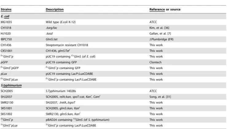
Table 1.Bacterial strains and plasmids used in this study.
Strains Description Reference or source
E. coli
MG1655 Wild type (E.coliK-12) ATCC
CH1018 Darg/lac Kim, et al. [36]
HJ1020 Dasd Gallan, et al. [7]
IBPC750 GlmS::tet J.Plumbridge(FR)
CH1436 Streptomycin resistant CH1018 This work
CKS1001 CH1436,glmS:Tetr This work
E.c
GlmS+
p pUC19 containingE.c
GlmS(ofE. coli) This work
pGFP pUC19 containing GFP Clontech
E.c
GlmS+
pGFP E.cGlmS+
pcontaining GFP This work
pLux pUC19 containing LacP::LuxCDABE This work
E.c
GlmS+
pLux E.c
GlmS+
pcontaining LacP::LuxCDABE This work
S.typhimurium
SCH2005 S.Typhimurium14028s ATCC
SHJ2037 SCH2005,relA::kan, spoT::cat, Kanr
, Camr Song, et al. [31]
SMR2130 SHJ2037,DrelA,DspoT This work
SKS1001 SCH2005,glmS::kan, Kanr
This work
SKS1002 SMR2130,glmS::kan, Kanr
This work
S.t
GlmS+
p pBAD24 containingS.t
GlmS (ofS. typhimurium) This work
S.t
GlmS+
pLux S.t
GlmS+
pcontaining LacP::LuxCDABE This work
Injection of bacteria into animals
BioluminescentE. coli orS. typhimurium (16108) suspended in 100ml PBS were injected intravenously into tumor-bearing mice through the lateral tail vein using a l cc insulin syringe [21].
Optical bioluminescence imaging
To image bacterial bioluminescence, anesthetized animals were placed in the light-tight chamber of the IVIS100 (Caliper, Hopkinton, MA, USA) equipped with a cooled charged couple detector (CCD) camera. Photons emitted from luciferase-express-ing bacteria were collected and integrated over one-minute periods. Pseudocolor images representing photon counts were overlaid on photographs of the mice using Living Image software v. 2.25 (Caliper, Hopkinton, MA). A region of interest (ROI) was selected manually based on signal intensity. The area of the ROI was kept constant, and the intensity was recorded as the maximum number of photons (photons s21cm22sr21) within a ROI [21].
Preparation of tissue extracts
Spleen and liver extracts of mice were prepared by adding organ homogenates directly to M9 media. The bone marrow extract was prepared from cancellous bone and marrow cavity of pig femur. The tissue sample was lysed using French pressure (1,000psi) and homogenizer, and the extract was taken after centrifugation (5,000 rpm, 5 min, Eppendorf) and filtration (0.45mm).
Statistical analysis
Statistical analysis was performed using the SPSS 18.0 statistical package (SPSS Inc., Chicago, IL, USA). A two-tailed Student’s
t-test was used to determine the statistical significance of tumor growth between the control and treatment groups. AP value of
,0.05 was considered statistically significant.
Results
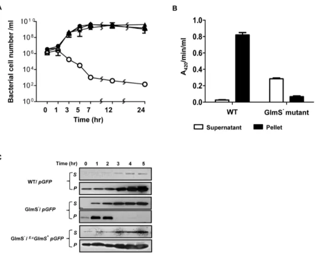
Characterization of E. coli GlmS+p vector in vitro GlmS2E. colirequires exogenous GlcN/GlcNAc for its survival [24]. GlmS2 mutant bacteria was generated and its phenotype was assessed by culturing the mutant strain (CKS1001) in minimal media (M9) (Fig. 1A). Bacteria grown in the presence of supplemental GlcNAc were subcultured into minimal media and samples taken at the indicated times were plated on LB agar plates supplemented with GlcNAc for viable cell counting (Colony Forming Units, CFU). On LB plates, GlmS2 mutant bacteria replicated for a few rounds and then underwent lysis, indicating that an insufficient amount of nutrients for survival of GlmS2 mutants was present in LB (see below). The assay revealed that the viability of the GlmS2mutant reduced drastically from 106CFU to 102CFU over 24 hrs. When GlcNAc was present in the culture media, however, the CFUs of the mutant increased in size, as did those of the wild type. These phenotypes were similar to those previously observed in GlmM2 mutant bacteria [25]. Subse-quently, the fate of GlmS2 mutant bacteria in the absence of supplemental GlcNAc was further examined by determining whether or not bacterial death was due to cell lysis. A GlmS2 mutant carrying a plasmid, in which DNA fragment of
hdeABp::lacZYA[26] was cloned, was used to determine the degree of bacterial lysis in the absence of GlcNAc (Fig. 1B). Samples were taken 5 hr after subculturing in minimal media and used forb -galactosidase assay of the supernatant (without any additional processing) and in the bacterial pellets (after treatment with cell lysis solution) [23]. A420/min/ml (product formation) instead of A420/min/ml/A600 (specific enzyme activity) was determined,
since the A600 value (cell mass) of the GlmS2 mutant would be meaningless due to the significant decrease in CFU (over 104-fold) over the period of 5 hrs. The sum of A420/min/ml in the supernatant and pellet was 0.85 for wild-type bacteria and 0.35 for GlmS2mutant bacteria. However, the A420/min/ml determined for the GlmS2 mutant was predominantly in the supernatant (.80%), while that for the wild type was exclusively in the pellet, suggesting that GlmS2mutant bacteria undergo lysis under these culture conditions. To further verify lysis of the GlmS2mutant in the absence of supplemental GlcNAc, wild type and GlmS2 mutant E. coli were transformed with pGFP and cultured in minimal media as in Figure 1A. Supernatant and pellet samples were taken at the indicated times and analyzed by Western blot analysis for GFP using a GFP-specific antibody (Fig. 1C). Samples from the culture of GlmS2mutant bacteria (CKS1001) carrying
pGFPcontained GFP in the supernatant beginning at 1 hr and increasing thereafter, whereas GFP in the pellet was detected only up to the 2 hr time point. Samples from the culture of wild type bacteria (CH1018) carryingpGFPshowed GFP exclusively in the pellet for the duration of the experiment.
Based on the above observations, a balanced-lethal host-vector system was constructed in which theglmSgene was incorporated into a plasmid that would complement the chromosomal glmS
mutation. A 1.8 Kb DNA fragment carrying theglmSofE. coliwas obtained by PCR amplification and placed under the control of thelacpromoter in a pUC19 plasmid (see Materials and Methods). The GlmS+
plasmid (E.cGlmS+
p) was used to transform GlmS2 mutant bacteria, which were then tested for complementation of the GlmS2mutant phenotype (Fig. 1A). GlmS2mutant bacteria carryingE.cGlmS+
pwere cultured in minimal media and assessed for CFU at the indicated times. The mutant carrying E.cGlmS+
p
survived as well as wild-type bacteria in the absence of supplemental GlcNAc. We also examined the complementation of GlmS2 mutant byE.cGlmS+
pGFP(Fig. 1C). Samples from the culture of GlmS2 mutant bacteria carrying E.cGlmS+pGFP
was analyzed and shown that GFP exclusively in the pellet, similar to wild-type bacteria carrying pGFP. Subsequently, wild-type and GlmS2 mutant bacteria were transformed with E.cGlmS+
p, and maintenance of the plasmid in the absence of antibiotics was determined (Fig. 2). Bacteria were grown in minimal media and subcultured (1:1000) every 12 hrs. Samples were taken on the indicated days to assess plasmid maintenance by plating the bacteria on plates containing ampicillin. In the wild-type background, the plasmid was lost rapidly; 92% by day 2 and over 99% by day 3. In the GlmS2mutant background, no loss of the plasmid was observed for the duration of the experiment (4 days). Clearly, this result demonstrated the feasibility of using glmS
mutant bacteria in a balanced-lethal system to maintain plasmid expression in the absence of antibiotics.
Characterization of the E. coli GlmS+p vector in a mouse
model
A prerequisite for the use of a balanced-lethal system based on
streptomycin-resistant mutant was generated and the allele was moved to the test strains. This was necessary to correct for contamination or bacteria pre-existing in the mice. Chromosom-ally-acquired streptomycin resistance is mainly due to mutations in the gene encoding the ribosomal protein S12, rpsL [27]. The tumor tissues were sampled on the indicated days, homogenized and spread on LB plates supplemented with GlcNAc and containing streptomycin (Fig. 3). At day 1, approximately equal numbers of bacteria were observed for both wild-type and GlmS2 mutant bacteria (,16106). While the number of wild-type bacteria increased to approximately 109CFU by day 5, the number of GlmS2 mutant bacteria decreased to approximately 56103CFU by day 7. In this study, Asd2mutant bacteria were also enumerated. The number of Asd2mutant bacteria at day 1 was similar to that of wild-type bacteria, but this value decreased to approximately 56104CFU by day 7. Taken together, these findings demonstrate that animal systems lack sufficient amounts of the nutrients required for the proliferation of GlmS2 mutant bacteria, similar to Asd2mutant bacteria. GlmS2mutant bacteria carrying E.cGlmS+p proliferated as well as wild-type bacteria,
Figure 1. Phenotype of GlmS2mutantE. coli.(A) Growth of GlmS2mutantE. coliunder various media conditions. GlmS2 mutantE. coli (CKS1001, open circles) and GlmS2mutantE. colicarryingE.cGlmS+p
(closed circles) grown overnight in LB supplemented with 0.2% GlcNAc were diluted 50-fold in minimal media or media supplemented with 0.2% GlcNAc (open triangles) and grown for 24 hrs. Wild-type parentalE. coli(CH1436, closed triangles) were grown in the same way in minimal media. Samples were taken at the indicated times for CFU determination on supplemented LB plates. (B) GlmS2mutant (CKS1001) or parental wild-typeE. coli(CH1436) carryingwhdeABp:lacZYAgrown overnight in LB supplemented with 0.2% GlcNAc were diluted 50-fold in minimal media and grown for 5 hrs.b-galactosidase activity (A420/min/ml) in the supernatants and lysed pellets was determined. (C) GlmS2mutant bacteria (CKS1001) carryingE.cGlmS+pGFP
orpGFPgrown overnight in LB supplemented with 0.2% GlcNAc were diluted 50-fold in minimal media and cultured for the indicated times. Wild-type parentalE. coli(CH1436) carryingpGFPwas grown the same way in minimal media. Samples were taken at the indicated times, the supernatant (s) and pellet (p) fractions were isolated, and the fractions were separated by 12% SDS-PAGE for determination of GFP by Western blotting.
doi:10.1371/journal.pone.0060511.g001
Figure 2. Plasmid maintenance inE. coliusing theglmSsystem. GlmS2 mutant bacteria (CKS1001) or the parental strain (CH1436) carryingE.c
GlmS+
pwere subcultured (1/1000) in minimal media every 12 hrs. The fraction of bacteria carryingE.cGlmS+pat the indicated time
was determined on GlcNAc-supplemented LB plates containing ampicillin (50 mg/ml).
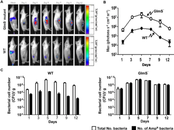
demonstrating that the glmS gene on the plasmid was able to complement the chromosomal mutation in anin vivomouse model. Our laboratory previously reported a quantitative and nonin-vasive imaging technique that enables the monitoring of bacterial migration in living subjects [5]. In this technique, bioluminescent bacteria are generated by transforming bacteria with an expression plasmid (pLux) that contains theluxCDABEoperon [5,21]. Using this method, a mouse tumor model carrying grafted CT26 was injected with wild-type or GlmS2bacteria carrying E.cGlmS+pLux via the tail vein. Expression of theluxgene was monitored using a cooled charge coupled device camera (Fig. 4A). Within 30 min of bacterial injection, bioluminescent signals were detected in the spleens and livers of the mice. At day 1, the signals from both types of bacteria had diminished in the liver but were detected exclusively in the tumor region. It should be noted that the signals from the GlmS2 mutant were significantly stronger than those from wild-type bacteria. Photon fluxes in tumor tissues were measured at the indicated days after the injection (Fig. 4B). The photon flux from GlmS2 mutant bacteria was 10- to 100-fold stronger than that from the wild-type E. coli. This was further confirmed by counting the number of CFU carrying the
E.c
GlmS+pLux (Fig. 4C). Tumor tissues were sampled on the indicated day, homogenized and spread on LB plates containing streptomycin and supplemented with GlcNAc for enumeration of the total number of bacteria, and on plates containing ampicillin and GlcNAc to assess the number of bacteria carrying the plasmid (AmpR). For both types of bacteria, the total number of bacteria increased from 107at day 1 to 56109 at day 5, and decreased gradually thereafter. However, the AmpR wild-type bacteria decreased by approximately 50-fold by day 1, and approximately 1000-fold by day 5, and thereafter, while the number of GlmS2 mutant bacteria carrying the plasmid did not decrease. This suggested that the balanced-lethal system using glmS is effective within an animal system.
Characterization of the Salmonella GlmS+p vector In addition toE. coli, Salmonella spp. shown to be localized to transplanted tumors in animals has also been extensively developed to carry anti-tumoral cargo proteins [6]. Thus, we attempted to establish a balanced-lethal system inSalmonellausing the glmS gene. First, the glmS gene on the chromosome of S. typhimuriumwas disrupted using thelRed system [28]. A 1.8 Kb DNA fragment carryingglmSof S. typhimuriumwas placed under
control of thePBADpromoter in the pBAD24 plasmid to construct
the Salmonella GlmS+
plasmid (S.tGlmS+
p). The phenotype of GlmS2 mutantSalmonella was examined by culturing the mutant strain (SKS1001) in minimal media, as described previously for the
E. colimutant (Fig. 5). The assay revealed that the viability of the GlmS2 mutant was reduced from 56104CFU to 56102CFU over 12 hrs. In the presence of GlcNAc, however, the CFU of the mutant increased, similar to wild-type, from 56104to 26109. In addition, GlmS2mutantSalmonellacarrying thes.tGlmS+plasmid multiplied to a degree similar to that of the wild-type control in the absence of GlcNAc. Interestingly,E.cGlmS+
failed to complement GlmS2mutantS. typhimurium, even though the GlmS proteins of the two species are virtually identical: only 8 out of 609 amino acids differ (see NCBI sequence accession number: NP_418185 and YP_005240128 for E. coli and S. typhimurium glmS genes, respectively).
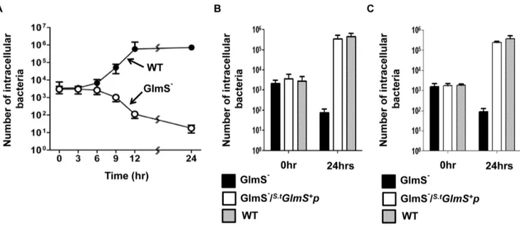
S. typhimuriumis capable of invading and replicating in animal cells [29]. Thus, the characteristics of GlmS2 mutant S. typhimuriumwas examined in cultured HeLa cells and peritoneal macrophages extracted from BALB/c mice. First, a time course assay was performed, in which intracellular bacteria were enumerated at 3 hr intervals. Wild-type and GlmS2 mutant Salmonellae grown in the presence of GlcNAc were mixed with HeLa cells and intracellular bacteria were counted in the presence of gentamycin (10mg/ml)[30] (Fig. 6A and B). The number of bacteria that invaded HeLa cells were approximately equal for both wild-type and GlmS2 mutant bacteria (3,46103CFU at T = 0). The number of wild-typeSalmonellaestarted to increase at 6 hrs post-infection and eventually reached 106CFU at 24 hrs post-infection. Conversely, the number of the GlmS2 mutant
Salmonellaestarted to decline at 6 hrs post-infection and eventually reached less than 10 CFU at 24 hrs post-infection. This result again confirmed that the nutrients necessary for cell wall and membrane synthesis in GlmS2 mutant Salmonellae are not sufficiently present within animal cells. The decline in the number of intracellular GlmS2mutant bacteria was most likely due to the effects of failed peptidoglycan synthesis, since onset of the decrease coincided with the time at which the number of wild-type bacteria began to increase (T = 6 hr). Subsequently, invasion and intracel-lular multiplication of GlmS2mutantSalmonellaetransformed with
S.t
GlmS+
p was tested (Fig. 6B). Enumeration of intracellular bacteria at 0 and 24 hrs indicated that the complemented GlmS2 mutant bacteria invaded HeLa cells and multiplied intracellularly as effectively as wild-type bacteria. The same findings were obtained in peritoneal macrophages (Fig. 6C).
The fidelity of the balanced-lethal system usingglmSwas tested
in vitro using wild-type and GlmS2 mutant Salmonellae carrying
S.t
GlmS+
p(Fig. 7). In the wild-type background, theS.tGlmS+
pwere lost rapidly in the absence of antibiotics. In the GlmS2 mutant background, over 99% ofS.tGlmS+
pwas lost at day 6 when cultured in media supplemented with GlcNAc, but strictly maintained when cultured in media lacking GlcNAc. Lastly, theSalmonella glmS
balanced-lethal system was tested in an animal model with wild-type and GlmS2 mutant Salmonellae carrying S.tGlmS+
p. Since S. typhimuriumis highly virulent in rodents, an attenuated strain ofS. typhimuriumdefective inDppGpp synthesis [2,3] was used (ppGpp synthesized by relA and spoT is required for virulence of S. Typhimurium[31]). The ppGpp-null mutant (relA::kan,spot::cat) and the ppGpp-null mutant carrying the glmS mutation, which are both resistant to kanamycin and chloramphenicol, were trans-formed with ampicillin-resistants.tglmS+p. Mice carrying grafted CT26 (mouse colon cancer) cells were constructed as described previously. After 14 days,DppGpp or DppGpp/GlmS2 Salmo-nellae carrying GlmS+
pLux (36107CFU) were injected
intrave-Figure 3. Targeting and proliferation of various mutantE. coli
strains in CT26 tumor-bearing mice. GlmS2 mutant bacteria (CKS1001), GlmS2 mutant bacteria carrying E.cGlmS+p
, Asd2 mutant bacteria (HJ1019), and the parental wild-type E. coli(CH1436) were injected into CT26 tumor-bearing mice through the tail vein (16108CFU), and the number of bacteria in the tumor tissues were
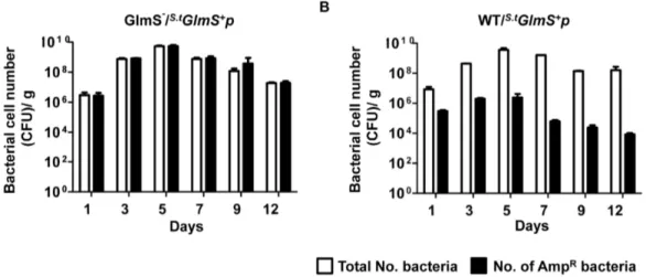
nously into each mouse via the tail vein. Tumor tissues were sampled on the indicated days, homogenized and spread on LB plates supplemented with GlcNAc containing kanamycin and chloramphenicol and/or ampicillin. Both the total number of bacteria (KanR CatR) and the number of bacteria carrying the plasmid (AmpR) were counted (Fig. 8). The total number of both types of bacteria increased from 107at day 1 to 56109at day 5 and decreased gradually thereafter. However, the number of AmpRparental bacteria was 50-fold less at day 1, 5000-fold less at day 5 and more than 10,000-fold less at day 12. Conversely, the number of AmpR GlmS2 mutant bacteria was the same as the total number of bacteria. This was further verified using two other tumor models: BALB/c mice carrying 4T-1 (mouse breast cancer) and nude mice carrying ASPC-1 (human pancreatic cancer). The numbers of bacteria carrying the plasmid were counted at day 7 in the homogenized tumor tissue (Table 2). Similarly as with CT26-bearing mice, virtually all GlmS2 mutant Salmonella carried the plasmid, but only,0.1% wild typeSalmonellacarried the plasmid
irrespective of types of tumor models. These data demonstrated that theSalmonella glmSbalanced-lethal host-vector system ensured maintenance of the plasmid in the absence of a selective determinant in animals.
Figure 5. Phenotype of GlmS2mutantS. typhimurium.(A) Growth of GlmS2mutantSalmonellaeunder various media conditions. GlmS2 mutant Salmonella (SKS1001,open circles) and the GlmS2 mutant Salmonella carrying S.t
GlmS+p (closed circles) grown overnight in LB
supplemented with 0.2% GlcNAc were diluted 50-fold in minimal media or media supplemented with 0.2% GlcNAc (open triangles) and grown for 24 hrs. Wild-type parentalSalmonella (SCH2005, closed triangles) were grown in the same way in minimal media. Samples were taken at the indicated times for CFU determination on supplemented LB plates. doi:10.1371/journal.pone.0060511.g005
Figure 4. Maintenance ofpLuxinE. coliproliferating in tumor tissue.(A) GlmS2mutant bacteria (CKS1001) and parental wild typeE. coli (CH1436) carryingE.cGlmS+pLux
were injected into CT26 tumor-bearing mice through the tail vein (16108CFU). Bioluminescent signals frompLux
were monitored at the indicated times using anin vivoimaging system. (B) The photon intensity of the tumor region was plotted as a function of time for GlmS2 mutant and wild-typeE. coli. The region of interest (ROI) was selected manually over the tumor region and the area was kept constant. Photon intensity was recorded as the maximum intensity (photons s21cm22sr21) within the ROI. Data represent the means and SEM of three independent experiments. (C) Tumor tissues were sampled on the indicated days. The total number of bacteria and the number of bacteria carryingE.cGlmS+pLux
Discussion
The system based on the asd gene is the most acclaimed balanced-lethal host system [7]. Here, we have demonstrated that, as for the Asd2mutant, animal tissues lack the nutrients required for survival of GlmS2mutant bacteria (Fig. 3). Since the level of DAP is insufficient in mammalian tissue, the balanced-lethal system of asdsystem coerces multiplying Asd2 mutantSalmonella
within the animal to carry the recombinant Asd+plasmid [8] [13]. In this study, we presented multiple lines of evidence demonstrat-ing that GlmS2 mutant E. coli and S. typhimurium undergo lysis unless GlcNAc is supplied exogenously and/or the bacteria are complemented by an E.cGlmS+p orS.t
GlmS+pvector, respectively. For the successful application of a balanced-lethal host system
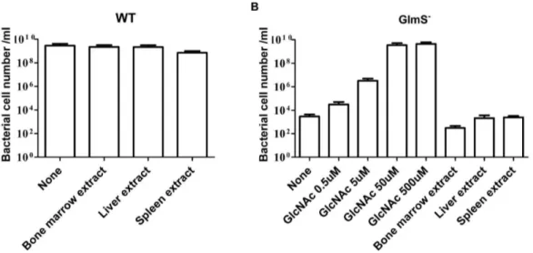
based onglmS, would be insufficient supply of those intermediates fot synthesis of GlcNAc in animal tissues. In mammals, GlcNAc is a component of glycoproteins, proteoglycans, glycosaminoglycans (GAGs) and other connective tissue building blocks [32]. Despite being a building block of biomacromolecules, GlcNAc seldom exists in free form [33]. We determined the growth of GlmS2 mutantE. coliin the presence of bone marrow, spleen, and liver extract, found the supplementations did not support the growth of the mutants (Fig. 9). GlmS2 mutant E. coli required ,50 mM
GlcNAc for normal growth. Thus, the necessary intermediates for the synthesis of GlcNAc are present at insufficient levels in these animal tissues to sustain the proliferation of GlmS2 mutant bacteria. This is consistent with the recent determination, median
Figure 7. Plasmid maintenance inS. typhimuriumusing theglmSsystemin vitro.(A) GlmS2mutant (SKS1001) salmonellae carryings.tGlmS+p
were subcultured (1/1000) in minimal media (closed circles) or media supplemented with 0.2% GlcNAc (open circles) every 12 hrs. The fraction of bacteria carryings.tGlmS+p
was determined on the indicated days by the plating method. (B) The same was done with wild-typeSalmonellae (SCH2005) in the absence (open circles) or presence (closed circles) of ampicillin.
doi:10.1371/journal.pone.0060511.g007
Figure 6. Intracellular growth of GlmS2 mutantS. typhimurium. (A) 5
6105CFU of GlmS2 mutant (SKS1001) and wild-type (SCH2005)
Salmonellae grown in LB supplemented with 0.2% GlcNAc was mixed with HeLa cells. Gentamycin-resistant intracellular Salmonellae were enumerated by determining the number of CFU at the indicated times. (B) The intracellular GlmS2mutant bacteria, GlmS2mutant bacteria carrying
S.t
GlmS+p, and wild-type bacteria were enumerated at 0 and 24 hrs in HeLa cells (B) and peritoneal macrophages (C).
endogenous glucosamine concentrations in plasma and synovial fluid in human were 0.29mM and 0.21mM, respectively [34].
Lytic cell death of GlmS2in the absence of GlcNAc supply was demonstrated to be due to leakage of cellular contents (Figs. 2 and 4). It should be noted that bacteria apparently fail to detect the absence of the building blocks necessary for membrane synthesis and continue to expand until they undergo lysis. This is in contrast to the situation that occurs in the absence of sufficient supply of amino acids or nucleotides, under which conditions bacteria cease proliferation through the accumulation of DppGpp [27]. The balanced-lethal host-vector system takes advantage of this phenomenon to ensure that the bacteria maintain the plasmid underin vivoconditions in which there is no selective pressure.
The observation that up to 99.99% of wild-type bacteria abandoned the GlmS+plasmid within 3–5 days afteri.v. injection was remarkable (Figs. 4 and 8). It indicates that maintenance of a plasmid that is not needed for survival imposes a great stress on bacteria, especiallyin vivo, where bacteria must struggle to ensure they acquire the nutrients necessary for survival while escaping the immunological assault of the host animal. This observation underscored the capacity of a balanced-host lethal system to maintain plasmids carrying genes for therapeutic proteins. Bioluminescent signals from the GlmS2 mutant were up to several hundred-fold stronger than those from wild-type bacteria
(Fig. 4). This suggests that bacterial therapies utilizing S. typhimuriumcarrying a plasmid containing an anti-tumoral protein gene [4,6] would be significantly more effective in aglmS-based balanced-lethal system.
While in vivoimaging of bioluminescent bacteria is a powerful tool that allows us to visualize the process of bacterial-tumor targeting, quantify bacterial growth noninvasively in target tissues, and monitor bacterial migration in real time, [5] it requires direct correlation between the photon flux and the number of bacteria. The plasmid (pLux) that contains the luxCDABE operon from
Photobacterium leiognathiemployed in this study is appropriate for this since it does not require an exogenous source of substrate to produce bioluminescence [5,35]. This allows direct measurement of photon flux from bacteria in deep tissues without the inconvenience of light scattering and attenuation through body tissues due to the lack of an excitation source. In the absence of selection pressure, however, the loss of plasmid carryingluxoperon would be an obstacle. ThepLuxexpression vector loaded with the
GlmS+
p/DglmS balanced-lethal system offers a solution that allows for the direct quantification of bacteria within living animals by determination of bioluminescence via IVIS imaging.
Table 2.Maintenance ofS.t
GlmS+pinS. typhimuriumproliferating in tumor tissues.
Designation Origin GlmS WT
KmR, CmR AmpR/KmR, CmR KmR, CmR AmpR/KmR, CmR
ASPC-1 Pancreatic adenocarcinoma 1.96108 2.46108 3.06108 3.06105 64.4 64.81 63.29 62.69 4T-1 Murine mammary carcinoma 6.66108 4.76108 4.46108 1.46105
61.28 62.16 62.82 64.4
SMR2130(GlmS2) and
SKS1002(WT) strains carryingS.t
GlmS+
pLux(36107CFU), were injected intravenously into mouse bearing 4T-1 (mouse breast cancer) or ASPC-1
(human pancreatic cancer). Tumor tissue were sampled at 7 days after the injection, homogenized, spread on LB plates containing kanamycin and chloramphenicol and/or amphicilin, and enumerated total number of bacteria (KmRCmR) and those carrying plasmid(AmpR).
doi:10.1371/journal.pone.0060511.t002
Figure 8. Maintenance ofs.tGlmS+
pinS. typhimuriumproliferating in tumor tissue.(A) GlmS2mutant (SKS1002, A) and parentalSalmonellae (SHJ2037, B) carryings.tGlmS+p
were injected into CT26 tumor-bearing mice through the tail vein (36107CFU). Tumor tissues were sampled on the
indicated days, homogenized, and spread on GlcNAc-supplemented LB plates containing kanamycin and chloramphenicol for the enumeration of total number of bacteria and ampicillin for the determination of plasmid-carrying bacteria.
Author Contributions
Helped to revise the manuscript: SJK. Conceived and designed the experiments: YH JJM HEC. Performed the experiments: KSK JHJ DL MY. Analyzed the data: HEC. Contributed reagents/materials/analysis tools: HEC JJM. Wrote the paper: HEC.
References
1. Forbes NS (2010) Engineering the perfect (bacterial) cancer therapy. Nat Rev Cancer 10: 785–794.
2. Na HS, Kim HJ, Lee HC, Hong Y, Rhee JH, et al. (2006) Immune response induced by Salmonella typhimurium defective in ppGpp synthesis. Vaccine 24: 2027–2034.
3. Song M, Kim HJ, Ryu S, Yoon H, Yun J, et al. (2010) ppGpp-mediated stationary phase induction of the genes encoded by horizontally acquired pathogenicity islands and cob/pdu locus in Salmonella enterica serovar Typhimurium. J Microbiol 48: 89–95.
4. Jiang SN, Phan TX, Nam TK, Nguyen VH, Kim HS, et al. (2010) Inhibition of tumor growth and metastasis by a combination of Escherichia coli-mediated cytolytic therapy and radiotherapy. Mol Ther 18: 635–642.
5. Min JJ, Kim HJ, Park JH, Moon S, Jeong JH, et al. (2008) Noninvasive real-time imaging of tumors and metastases using tumor-targeting light-emitting Escherichia coli. Mol Imaging Biol 10: 54–61.
6. Nguyen VH, Kim HS, Ha JM, Hong Y, Choy HE, et al. (2010) Genetically engineered Salmonella typhimurium as an imageable therapeutic probe for cancer. Cancer Res 70: 18–23.
7. Galan JE, Nakayama K, Curtiss R, 3rd (1990) Cloning and characterization of the asd gene of Salmonella typhimurium: use in stable maintenance of recombinant plasmids in Salmonella vaccine strains. Gene 94: 29–35. 8. Schleifer KH, Kandler O (1972) Peptidoglycan types of bacterial cell walls and
their taxonomic implications. Bacteriol Rev 36: 407–477.
9. Jones-Mortimer MC, Kornberg HL (1980) Amino-sugar transport systems of Escherichia coli K12. J Gen Microbiol 117: 369–376.
10. Holmes RP, Russell RR (1972) Mutations affecting amino sugar metabolism in Escherichia coli K-12. J Bacteriol 111: 290–291.
11. White RJ (1968) Control of amino sugar metabolism in Escherichia coli and isolation of mutants unable to degrade amino sugars. Biochem J 106: 847–858. 12. Bachmann BJ (1983) Linkage Map of Escherichia coli K-12, Edition 7.
Microbiol Rev 47: 454.
13. Mengin-Lecreulx D, Flouret B, van Heijenoort J (1983) Pool levels of UDP N-acetylglucosamine and UDP N-N-acetylglucosamine-enolpyruvate in Escherichia coli and correlation with peptidoglycan synthesis. J Bacteriol 154: 1284–1290. 14. Plumbridge JA (1989) Sequence of the nagBACD operon in Escherichia coli
K12 and pattern of transcription within the nag regulon. Mol Microbiol 3: 505– 515.
15. Plumbridge J (1995) Co-ordinated regulation of amino sugar biosynthesis and degradation: the NagC repressor acts as both an activator and a repressor for the transcription of the glmUS operon and requires two separated NagC binding sites. EMBO J 14: 3958–3965.
16. Plumbridge JA, Cochet O, Souza JM, Altamirano MM, Calcagno ML, et al. (1993) Coordinated regulation of amino sugar-synthesizing and -degrading enzymes in Escherichia coli K-12. J Bacteriol 175: 4951–4956.
17. Sarvas M (1971) Mutant of Escherichia coli K-12 defective in D-glucosamine biosynthesis. J Bacteriol 105: 467–471.
18. Wu HC, Wu TC (1971) Isolation and characterization of a glucosamine-requiring mutant of Escherichia coli K-12 defective in glucosamine-6-phosphate synthetase. J Bacteriol 105: 455–466.
19. Garrett S, Taylor RK, Silhavy TJ, Berman ML (1985) Isolation and characterization of delta ompB strains of Escherichia coli by a general method based on gene fusions. J Bacteriol 162: 840–844.
20. Kaiser P, Rothwell L, Galyov EE, Barrow PA, Burnside J, et al. (2000) Differential cytokine expression in avian cells in response to invasion by Salmonella typhimurium, Salmonella enteritidis and Salmonella gallinarum. Microbiology 146 Pt 12: 3217–3226.
21. Min JJ, Nguyen VH, Kim HJ, Hong Y, Choy HE (2008) Quantitative bioluminescence imaging of tumor-targeting bacteria in living animals. Nat Protoc 3: 629–636.
22. Zubay G, Morse DE, Schrenk WJ, Miller JH (1972) Detection and isolation of the repressor protein for the tryptophan operon of Escherichia coli. Proc Natl Acad Sci U S A 69: 1100–1103.
23. Putnam SL, Koch AL (1975) Complications in the simplest cellular enzyme assay: lysis of Escherichia coli for the assay of beta-galactosidase. Anal Biochem 63: 350–360.
24. Wu G, Sun Y, Qu W, Huang Y, Lu L, et al. (2011) Application of GFAT as a novel selection marker to mediate gene expression. PLoS One 6: e17082. 25. Mengin-Lecreulx D, van Heijenoort J (1993) Identification of the glmU gene
encoding N-acetylglucosamine-1-phosphate uridyltransferase in Escherichia coli. J Bacteriol 175: 6150–6157.
26. Shin M, Song M, Rhee JH, Hong Y, Kim YJ, et al. (2005) DNA looping-mediated repression by histone-like protein H-NS: specific requirement of Esigma70 as a cofactor for looping. Genes Dev 19: 2388–2398.
27. Wittmann HG, Apirion D (1975) Analysis of ribosomal proteins in streptomycin resistant and dependent mutants isolated from streptomycin independent Escherichia coli strains. Mol Gen Genet 141: 331–341.
28. Datsenko KA, Wanner BL (2000) One-step inactivation of chromosomal genes in Escherichia coli K-12 using PCR products. Proc Natl Acad Sci U S A 97: 6640–6645.
29. Galan JE (2001) Salmonella interactions with host cells: type III secretion at work. Annu Rev Cell Dev Biol 17: 53–86.
30. Mengin-Lecreulx D, Flouret B, van Heijenoort J (1982) Cytoplasmic steps of peptidoglycan synthesis in Escherichia coli. J Bacteriol 151: 1109–1117.
Figure 9. Growth of WT (CH1018, A) and GlmS2 (CKS1001, B) mutant E. coli in the minimal media with supplementations.
31. Song M, Kim HJ, Kim EY, Shin M, Lee HC, et al. (2004) ppGpp-dependent stationary phase induction of genes on Salmonella pathogenicity island 1. J Biol Chem 279: 34183–34190.
32. Chen JK, Shen CR, Liu CL (2010) N-acetylglucosamine: production and applications. Mar Drugs 8: 2493–2516.
33. El Sayed H. El Ashry1 aMREA (2007) Synthesis and Biological Relevance of N-Acetylglucosamine-containing Oligosaccharides. Pure Appl Chem 12, 2229– 2242.
34. Persiani S, Rotini R, Trisolino G, Rovati LC, Locatelli M, et al. (2007) Synovial and plasma glucosamine concentrations in osteoarthritic patients following oral
crystalline glucosamine sulphate at therapeutic dose. Osteoarthritis Cartilage 15: 764–772.
35. Jeong JH, Song M, Park SI, Cho KO, Rhee JH, et al. (2008) Salmonella enterica serovar gallinarum requires ppGpp for internalization and survival in animal cells. J Bacteriol 190: 6340–6350.