Association of Cytotoxic T-Lymphocyte
Antigen 4 (
CTLA4
) and Thyroglobulin (
TG
)
Genetic Variants with Autoimmune
Hypothyroidism
Hinal Patel1, Mohmmad Shoab Mansuri2, Mala Singh2, Rasheedunnisa Begum2*, Minal Shastri3, Ambikanandan Misra1*
1Pharmacy Department, Faculty of Technology & Engineering, The Maharaja Sayajirao University of Baroda, Vadodara, Gujarat, India,2Biochemistry Department, Faculty of Science, The Maharaja Sayajirao University of Baroda, Vadodara, Gujarat, India,3Medicine Department, Faculty of Medicine, Sir Sayajirao Gaekwad Hospital, The Maharaja Sayajirao University of Baroda, Vadodara, Gujarat, India
*misraan@hotmail.com(AM);rasheedunnisab@yahoo.co.in(RB)
Abstract
Autoimmune hypothyroidism is known to be caused by immune responses related to the thy-roid gland and its immunological feature includes presence of autoimmune antibodies. There-fore the aim was to analyze presence of anti-TPO antibodies in hypothyroidism patients in Gujarat. Cytotoxic T-Lymphocyte Antigen 4 (CTLA4) is one of the susceptibility genes for vari-ous autoimmune diseases. Hence, exon1 +49A/G and 3’UTR CT60A/G single nucleotide polymorphisms (SNPs) inCTLA4and its mRNA expression levels were investigated in auto-immune hypothyroidism patients. Thyroglobulin (TG) is known to be associated with autoim-mune thyroid disorders and thus exon 33 (E33) SNP inTGwas investigated. We analyzed the presence of anti-TPO antibodies in the plasma samples of 84 hypothyroidism patients and 62 controls by ELISA. PCR-RFLP technique was used for genotyping of polymorphisms. sCTLA4and flCTLA4mRNA expression levels were assessed by real time PCR. 59.52% of hypothyroid patients had anti-TPO antibodies in their circulation. The genotype and allele fre-quencies differed significantly for +49A/G(p= 0.0004 for +49AG,p= 0.0019 for +49GG & p= 0.0004 for allele), CT60 (p =0.0110 for CT60AG,p =0.0005 for CT60GG &p<0.0001 for allele) andTGE33 (p= 0.0003 for E33TCp<0.0001 for E33CC&p<0.0001 for allele) SNPs between patients and controls. Patients had significantly decreased mRNA levels of both sCTLA4(p= 0.0017) and flCTLA4(p<0.0001) compared to controls. +49A/G and CT60 poly-morphisms ofCTLA4were in moderate linkage disequilibrium. Logistic regression analysis indicated significant association of CT49A/G, CT60A/G andTGexon 33 polymorphisms with susceptibility to autoimmune hypothyroidism when adjusted for age and gender. Our results suggest +49A/G and CT60 polymorphism ofCTLA4and E33 polymorphism ofTGmay be genetic risk factors for autoimmune hypothyroidism susceptibility and down regulation of both forms ofCTLA4advocates the crucial role ofCTLA4in pathogenesis of autoimmune hypothyroidism.
OPEN ACCESS
Citation:Patel H, Mansuri MS, Singh M, Begum R, Shastri M, Misra A (2016) Association of Cytotoxic T-Lymphocyte Antigen 4 (CTLA4) and Thyroglobulin (TG) Genetic Variants with Autoimmune Hypothyroidism. PLoS ONE 11(3): e0149441. doi:10.1371/journal.pone.0149441
Editor:Ming Yang, Beijing University of Chemical Technology, CHINA
Received:September 12, 2015
Accepted:January 31, 2016
Published:March 10, 2016
Copyright:© 2016 Patel et al. This is an open access article distributed under the terms of the Creative Commons Attribution License, which permits unrestricted use, distribution, and reproduction in any medium, provided the original author and source are credited.
Data Availability Statement:All relevant data are within the paper and its Supporting Information files.
Funding:The authors have no support or funding to report.
Introduction
Hypothyroidism is an endocrine disorder characterized by decreased activity of thyroid gland leading to insufficient production of thyroid hormones. Subclinical or asymptomatic hypothy-roidism is characterized by elevated thyrotropin level and normal serum thyroid hormones level. Whereas, there remains elevated thyrotropin but decreased thyroid hormones serum lev-els in case of overt or clinical hypothyroidism [1] [2].
In India, hypothyroidism used to usually be categorized under the iodine deficient disorders and represented based on total goiter rate. Government of India has adopted the universal salt iodization program and since then there has been a decline in goiter prevalence in various
parts of the country [3–7]. As per World health organization (WHO) assessment report India
has undergone transition from iodine deficient state to iodine sufficient state [8–10]. A large, cross-sectional, comprehensive study recently carried out in adult population across the coun-try, indicates about 10.9% prevalence of hypothyroidism [11]; whereas, the prevalence of hypo-thyroidism in the developed countries is about 4–5% [12,13]. This indicates even though most of the regions of India have been made iodine sufficient there is still high prevalence of hypo-thyroidism. Hence, underlying pathogenesis may involve a complex interplay of genetic, envi-ronmental and endogenous factors and not only iodine deficiency. Clinical investigation of patients in India does not include evaluation of thyroid autoantibodies and hence iodine defi-ciency is believed to be the sole candidate for hypothyroidism pathogenesis which may not be the case.
Autoimmune hypothyroidism is characterized by gradual destruction of the thyroid gland due to loss of thyroid cells, leading to thyroid hormone deficiency. The immunological features of this disorder include the presence of anti-thyroidperoxidase (anti-TPO) antibodies and, less commonly, anti-thyroglobulin (anti-TG) antibodies, abnormalities in the circulating T cell population and a goiter with lymphocytic infiltration [14,15]. To date, significant progress has been made in identifying and characterizing genes involved in the disease pathogenesis. As both environmental and genetic factors appear to play a role in disease susceptibility [16], the precise mechanism for the pathogenesis of this disorder is not fully understood. The cytotoxic
T-lymphocyte antigen 4 (CTLA4) and thyroglobulin (TG) genes have been considered to be
major genetic factors involved in the development of autoimmune hypothyroidism.
TheCTLA4gene on human chromosome 2q33 is one of the candidate genetic markers for
autoimmune diseases, encodes a cell surface molecule that is expressed on the surface of acti-vated T lymphocytes and has the most remarkable function of down regulation of the immune response [17].CTLA4binds to the ligands, B7-1 and B7-2, as CD28 but with a 20–50-fold higher affinity. The interaction betweenCTLA4and B7 plays an essential role in regulation of self-tolerance, and hence susceptibility to autoimmune diseases [18]. TheCTLA4gene
pro-duces two differentCTLA4protein isoforms: full lengthCTLA4(flCTLA4) and solubleCTLA4
(sCTLA4). The flCTLA4serves as a transmembrane receptor on activated T cells to inhibit cell
proliferation. The role of sCTLA4is not yet known but it has been suggested that sCTLA4can block the B7-CD28 interaction by acting as functional receptor for B7 antigens and thus can interfere with the co-stimulation signal and inhibit T-cell proliferation [19].
Several polymorphic sites in theCTLA4gene such as promoter -318 C/T[20], exon 1 +49 A/
G [21,22], microsatellite (AT)n repeat in the 3’-untranslated region (UTR) of exon [23] and
three single nucleotide polymorphisms (SNPs) in the 6.1-kb 3’non-coding region such as
development of various autoimmune diseases, the exon 1 +49 A/G polymorphism inCTLA4 exon 1 has been reported to be involved in the development of autoimmune diseases including Graves’disease [28] and Hashimoto thyroiditis [27]. The CT60 in the 3’UTR region ofCTLA4 gene is also the most promising locus for the autoimmune thyroid diseases [29]. The meta-analysis study shows consistent associations of Graves’disease and Hashimoto thyroiditis with CT60 [30] and clarifies the important role of theCTLA4locus in determining the risk of auto-immune thyroid diseases.
TheTGgene on 8q24 locus has been strongly linked with autoimmune thyroid diseases
(AITD). Previous studies have demonstrated that an exon 10–12 SNP cluster and an exon 33
(E33) SNP are significantly associated with autoimmune thyroid diseases because amino acid substitution that occurs due to this polymorphism predisposes to autoimmune thyroid diseases [31].
Hypothyroidism diagnosis is limited to determination of thyroid hormone and thyrotropin serum levels and evaluation of autoimmune antibodies in patients is not currently practiced in India. We therefore investigated the presence of anti-TPO antibodies in patients with
hypothy-roidism and also analyzed the frequencies ofCTLA43’UTR CT60, exon 1 +49 A/G andTG
E33 polymorphisms andCTLA4expression in autoimmune hypothyroidism patients and
con-trols from Gujarat as indicators of thyroid disorder susceptibility.
Subjects and Methods
Subjects
The study plan was approved by‘Human Scientific and Ethics Review Committee for Human
Research’, Faculty of Medicine, The Maharaja Sayajirao University of Baroda, Vadodara,
Guja-rat, India. The importance of the study was explained to all participants and written consent was obtained from all patients and controls. The study group included 84 hypothyroidism patients comprised of 78 females and 6 males who referred to S.S.G. Hospital, Vadodara. The diagnosis of hypothyroidism was based on thyroid profile analysis (serum T3, T4and TSH
lev-els) and patients had no other associated autoimmune diseases. A total of 62 ethnically and sex-matched unaffected individuals were included as controls in this study. The control group comprised 55 females and 7 males (S1 Table). None of the healthy individuals had any evidence of hypothyroidism and any other diseases. The study plan was approved by the Institutional ethics committee for human research (IECHR), Faculty of Medicine, The Maharaja Sayajirao University of Baroda, Vadodara, Gujarat, India.
Estimation of anti-Thyroid Peroxidase (anti-TPO) antibodies levels
In the present study, plasma from hypothyroidism patients was examined to find the levels of anti-TPO antibodies compared to controls. Plasma samples of 84 hypothyroidism patients and 62 controls were analyzed for the presence of anti-TPO antibodies by ELISA. 5 ml venousblood was collected from the patients and healthy subjects in K3EDTA coated vacutainers (BD,
Franklin Lakes, NJ 07417, USA) and plasma was extracted. Presence of anti-TPO antibodies
were assessed by ELISA method as per manufacturer’s protocol (Genway Biotech, Inc. San
Diego, CA). Absorbance of all wells was measured at 450nm using 620nm as reference wavelength.
Determination of s
CTLA4
, fl
CTLA4
and
GAPDH
mRNA expression
RNA extraction and cDNA synthesis. Total RNA from whole blood was isolated and
protocol. RNA integrity was verified by agarose gel electrophoresis/ ethidium bromide staining
and O.D. 260/280 absorbance ratio>1.95. RNA was treated with DNase I (Ambion inc. Texas,
USA) before cDNA synthesis to avoid DNA contamination. One microgram of total RNA was used to prepare cDNA. cDNA synthesis was performed using the Verso cDNA Synthesis Kit (Thermo scientific, Lithuania, EU) according to the manufacturer’s instructions using Master-cycler Gradient PCR (Eppendorf, Germany).
Real-time PCR. The levels of full length, solubleCTLA4andGAPDHtranscripts were measured by real-time PCR using gene specific primers (S2 Table) (Eurofins, Bangalore, India). Expression of GAPDH gene was used as a reference. Real-time PCR was performed in
duplicates in 20μl volume using LightCycler1480 SYBR Green I Master (Roche Diagnostics
GmbH, Mannheim, Germany) following the manufacturer’s instructions and carried out in the
Light Cycler 480 Real-Time PCR (Roche Diagnostics GmbH, Mannheim, Germany). The ther-mal cycling conditions included an initial activation step at 95°C for 10 min, followed by 45 cycles of denaturation, annealing and amplification. The fluorescent data collection was per-formed during the extension step. At the end of the amplification phase a melt curve analysis was carried out on the product formed. The value of Cp was determined by the first cycle num-ber at which fluorescence was greater than the set threshold value.
Genotyping of
CTLA4
gene exon 1 +49A/G and 3
’
UTR CT60A/G
polymorphisms and
TG
gene exon 33 polymorphism
Genomic DNA was extracted from whole blood using‘QIAamp DNA Blood Kit’(QIAGEN
Inc., Valencia, CA 91355, USA) according to the manufacturer’s instructions. Polymerase
chain reaction–restriction fragment length polymorphism (PCR-RFLP) was used to genotype
exon 1 +49A/G and 3’UTR CT60A/G polymorphisms ofCTLA4gene and E33 polymorphism
ofTGgene and amplification was performed using Mastercycler Gradient PCR (Eppendorf,
Germany). according to the protocol: 95°C for 10 minutes followed by 30 cycles of 95°C for 30
seconds, primer (S2 Table) dependent annealing for 30 seconds, and 72°C for 30 seconds. The
amplified products were checked by electrophoresis on a 2.0% agarose gel stained with ethid-ium bromide.
Restriction enzymes (New England Biolabs, Beverly, MA) used for digesting amplicons of
exon 1 +49A/G and 3’UTR CT60A/G ofCTLA4gene and E33 polymorphism ofTGgene are
given inS2 Table. 15μL of the amplified products were digested for 16 hours at 37°C with 5 U
of the corresponding restriction enzyme. The digestion products with 100/50 base pair DNA ladder (Bioron, Ludwigshafen am Rhein, Germany) were resolved in 3.5% agarose gels stained with ethidium bromide and visualized under UV transilluminator.
Statistical analysis
Evaluation of the Hardy-Weinberg equilibrium (HWE) was performed for the polymorphisms in patients and controls by comparing the observed and expected frequencies of the genotypes using chi-square analysis. The distribution of the genotypes and allele frequencies ofCTLA4
exon 1 +49A/G and 3’UTR CT60A/G andTGE33 polymorphisms for patients and control
subjects were compared using the chi-square test using Prism 5 software (Graphpad software Inc; San Diego CA, USA, 2007). Logistic regression analysis was applied to evaluate whether
CTLA4exon 1 +49A/G and 3’UTR CT60A/G andTGE33 polymorphisms predict the
suscep-tibility to autoimmune hypothyroidism when adjusted for age and gender by using by SPSS sta-tistics 23.0 (IBM SPSS Inc., Chicago, IL). Haplotype analysis was carried out usinghttp://
analysis.bio-x.cn/myAnalysis.php.[32] The linkage disequilibrium (LD) coefficients D’= D/
the Haploview programe version 4.1.[33] Differences were considered as statistically significant if thep-value was less than 0.025 due to Bonferroni’s correction for multiple testing of +49A/G
and CT60A/G SNPs inCTLA4whereas, differences were considered to be statistically
signifi-cant if thep-value was less than 0.05 forTGSNP. Odds ratio (OR) with respective confidence interval (95% CI) for disease susceptibility was also calculated. Relative expression of both
flCTLA4and sCTLA4in patient and control groups was plotted and analyzed by
nonparamet-ric unpaired t-test using Prism 5 software (Graphpad software Inc; San Diego CA, USA, 2007). The statistical power of detection of the association with the disease at the 0.05 level of
signifi-cance was determined by using the GPower software [34].
Results
Anti-TPO antibody levels in hypothyroidism patients and controls
It was found that 59.52% (n = 50) of hypothyroidism patients (n = 84) had anti-TPO antibodies in their blood circulation suggesting that autoimmunity may play an important role in the pathogenesis of the disease moreover, these autoimmune hypothyroidism patients had signifi-cantly increased anti-TPO antibody levels as compared to controls (p<0.0001).
The expression of fl
CTLA4
and s
CTLA4
transcripts
Comparison of the findings showed significantly decreased expression of both full length and
solubleCTLA4in autoimmune hypothyroidism patients than in controls after normalization
withGAPDHexpression (p<0.0001 andp= 0.01 respectively) (Fig 1A). The 2-ΔΔCpanalysis
showed 0.166 and 0.342 fold decrease in the expression of flCTLA4and sCTLA4mRNA
expression, respectively, in patients as compared to controls (Fig 1B).
Genotype-phenotype correlations for fl
CTLA4
and s
CTLA4
in
autoimmune hypothyroidism patients and controls
Analysis of the mRNA expression of flCTLA4and sCTLA4based on the +49A/G and CT60A/
G genotypes of 45 autoimmune hypothyroidism patients and 60 controls was performed. The
expression levels of flCTLA4and sCTLA4for AA genotypes of exon 1 +49A/G polymorphism
did not differ significantly in autoimmune hypothyroidism patients as compared to controls (p= 0.0667 andp= 0.6260 respectively) (Fig 2A & 2B). However, the expression levels of both
flCTLA4and sCTLA4were decreased significantly for AG (p= 0.0141 andp= 0.0358
respec-tively) and GG (p= 0.0007 andp= 0.0102 respectively) genotype of exon 1 +49A/G
polymor-phism in autoimmune hypothyroidism patients as compared to controls (Fig 2A & 2B). The expression levels of flCTLA4and sCTLA4for AA (p= 0.5817 andp= 0.7099 respectively) and
AG (p= 0.2379 andp= 0.7478 respectively) genotypes of CT60 polymorphism did not differ
significantly in autoimmune hypothyroidism patients as compared to controls (Fig 2C & 2D).
However, the expression levels of both flCTLA4and sCTLA4were decreased significantly for
GG genotype of CT60 polymorphism in autoimmune hypothyroidism patients as compared to controls (p<0.0001 andp= 0.0001 respectively) (Fig 2C & 2D).
The expression levels of flCTLA4were found to be significantly decreased and associated
with GG and AG haplotypes in patients and controls (p= 0.0091 andp= 0.0429 respectively).
Other two haplotypes, such as, AA and GA did not differ with respect to flCTLA4expression
levels in patients and controls (p= 0.1271 &p= 0.3191). However, sCTLA4expression levels were found to be significantly decreased and associated with GG haplotypes in patients and
respect to sCTLA4expression levels in patients and controls (p= 0.8129 &p= 0.1948 & p= 0.2925respectively).
Ratio of s
CTLA4
and fl
CTLA4
mRNA expression in autoimmune
hypothyroidism patients and controls
The expression level of sCTLA4and flCTLA4was also analyzed as ratio of sCTLA4: flCTLA4in autoimmune hypothyroidism patients and controls. There was no significant difference in the
ratio of sCTLA4to flCTLA4mRNA expression between patients and controls (p= 0.1360)
(Fig 3A).
None of AA, AG and GG genotypes of exon 1 +49A/G polymorphism show any significant
difference for the ratio of sCTLA4and flCTLA4mRNA expression in patients compared to
controls (p= 0.9724,p= 0.2378 andp= 0.3405). Similarly, the ratio of sCTLA4and flCTLA4 mRNA expression was not found to significantly differ for AA, AG and GG genotypes of CT60
polymorphism in patients compared to control (p= 0.6213,p= 0.4425 andp= 0.2940
respec-tively) (Fig 4B).
Moreover, ratio of sCTLA4and flCTLA4mRNA expression did not differ significantly for
the haplotypes AA, AG and GA in patients and controls (p= 0.159,p= 0.068 andp= 0.966
respectively) (Fig 3B). But patients with GG haplotype showed significant increase in the ratio
ofsCTLA4toflCTLA4mRNA expression compared to controls (p= 0.0045).
Analysis of association between
CTLA4
gene exon 1 +49A/G
polymorphism and susceptibility to autoimmune hypothyroidism
PCR-RFLP for +49A/G polymorphism yielded a 271 bp undigested product corresponding to G allele and 249 bp and 22 bp digested products corresponding to A allele. The three genotypes Fig 1. Relative gene expression of flCTLA4and sCTLA4in controls and autoimmune hypothyroidism patients: (A)Expression of flCTLA4and sCTLA4mRNA in 45 autoimmune hypothyroidism patients and 60 controls as suggested by MeanΔCp. Autoimmune hypothyroidism patients showed significantly reduced mRNA levels of flCTLA4(p<0.0001) and sCTLA4(p= 0.001) as compared to controls.(B)Expression fold change of flCTLA4and sCTLA4in 45 autoimmune hypothyroidism patients and 60 controls showed approximately 0.166 and 0.342 fold decrease as determined by 2-ΔΔCpmethod
respectively.
Fig 2. Genotype—phenotype correlation of exon 1 +49A/G and 3’UTR CT60A/G polymorphisms of flCTLA4and sCTLA4in controls and
autoimmune hypothyroidism patients: (A)Relative mRNA expression of flCTLA4with respect to +49A/G genotypes in 45 patients and 60 controls. None of the three genotypes AA (p= 0.0667), AG (p= 0.0141) and GG (p= 0.0007) in patients showed significant difference for flCTLA4expression as compared to controls a suggested by MeanΔCp. [NS= non-significant](B)Relative mRNA expression of sCTLA4with respect to +49A/G genotypes in 45 patients and 60 controls. None of the three genotypes AA (p= 0.6260), AG (p= 0.0358) and GG (p= 0.0102) in patients showed significant difference for flCTLA4
expression as compared to controls a suggested by MeanΔCp. [NS= non-significant](C)Relative mRNA expression of flCTLA4with respect to CT60A/G genotypes in 45 patients and 60 controls. GG genotype showed significant decrease in the levels of flCTLA4mRNA (p<0.0001) in patients as compared to AA and AG genotype (p= 0.5817 &p= 0.2379 respectively) as suggested by MeanΔCp. [NS= non-significant](D)Relative mRNA expression of sCTLA4
with respect to CT60A/G genotypes in 45 patients and 60 controls. GG genotype showed significant decrease in levels of sCTLA4mRNA (p= 0.0001) in patients as compared to AA and AG genotype (p= 0.7099 &p= 0.7478 respectively) as suggested by MeanΔCp. [NS= non-significant].
Fig 3. Ratio of sCTLA4and flCTLA4mRNA expression in controls and autoimmune hypothyroidism patients: (A)sCTLA4/flCTLA4mRNA ratio was measured in 45 autoimmune hypothyroidism patients and 60 controls which was not found altered (p= 0.1360). [NS= non-significant](B)sCTLA4/flCTLA4
mRNA ratio was analyzed with respect to (+49A/G: CT60A/G) haplotypes in patients and controls. AA, AG and GA haplotypes did not show significant difference in the sCTLA4/flCTLA4mRNA ratio (p= 0.4165,p= 0.2964 andp= 0.9481), however, GG haplotype showed significant increase in the sCTLA4/ flCTLA4mRNA ratio in patients compared to controls (p= 0.0045 respectively). [NS= non-significant].
doi:10.1371/journal.pone.0149441.g003
Fig 4. Ratio of sCTLA4and flCTLA4mRNA expression with respect to +49A/G and CT60A/G genotypes in controls and patients: (A)sCTLA4/ flCTLA4mRNA ratio was analyzed with respect to +49A/G genotypes in patients and controls. None of AA, AG and GG genotypes showed significant difference in the sCTLA4/flCTLA4mRNA ratio in patients compared to controls (p= 0.9724,p= 0.2378 andp= 0.3405 respectively). [NS= non-significant] (B)sCTLA4/flCTLA4mRNA ratio was analyzed with respect to CT60A/G genotypes in patients and controls. None of AA, AG and GG genotypes showed significant difference in the sCTLA4/flCTLA4mRNA ratio in patients compared to controls (p= 0.6213,p= 0.4425 andp= 0.2940 respectively). [NS= non-significant].
identified by 3.5% agarose gel electrophoresis were: AA homozygous, AG heterozygous and
GG homozygous for +49A/G polymorphism ofCTLA4gene (S1A Fig).
AG and GG genotypes of Exon 1 +49A/G polymorphism ofCTLA4gene when compared
with AA genotype between patients and control by chi-square test-2x2 contingency table showed to increase susceptibility to autoimmune hypothyroidism (p= 0.0004 and odds ratio 5.333; p= 0.0019 and odds ratio 5.779 respectively) (Table 1). Furthermore logistic regression analysis showed that, when adjusted for age and gender, AG and GG genotype of Exon 1 +49A/G
poly-morphism ofCTLA4gene increase the risk of autoimmune hypothyroidism by 4.319 fold (95%
CI: 1.408 to 13.249,p= 0.011) and 4.309 fold (95% CI: 1.102 to 16.855,p= 0.036) respectively
(Table 2). From logistic regression, age and gender of the patients were not found to be associated
with susceptibility to autoimmune hypothyroidism as age and gender matched controls were selected in this study (Table 2). However our data (S1 Table) suggest that autoimmune hypothy-roidism is common in females than in males which is in accordance with previous prevalence studies on autoimmune thyroid disorders [35]. The mean age of onset is 38 years for females whereas for males it cannot be concluded because of relatively small sample size (S1 Table).
Furthermore, there was significantly high frequency of the G allele in patients with
autoim-mune hypothyroidism compared with controls (55% vs. 31% respectively;χ2= 12.58,
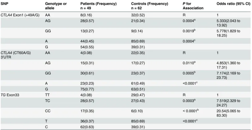
p= 0.0004) (Table 1). Both patient and control populations were found to be in Hardy-Wein-berg equilibrium for this polymorphism (p= 0.2783 andp= 0.0913 respectively) (Table 1). Table 1. Association studies forCTLA4gene exon 1 +49A/G and 3’UTR CT60A/G polymorphisms andTGgene exon 33 polymorphism in autoim-mune hypothyroidism patients.
SNP Genotype or
allele
Patients (Frequency) n = 49
Controls (Frequency) n = 62
Pfor Association
Odds ratio (95% CI)
CTLA4Exon1 (+49A/G) AA 8(0.16) 32(0.52) R 1
AG 28(0.57) 21(0.34) 0.0004a 5.333(2.043 to
13.92)
GG 13(0.27) 9(0.14) 0.0019b 5.778(1.829 to
18.25)
A 44(0.45) 85(0.69) 0.0004c
G 54(0.55) 39(0.31)
CTLA4(CT60A/G) 3’UTR
AA 4(0.08) 22(0.35) R 1
AG 15(0.31) 17(0.27) 0.0110a 4.853(1.360 to
17.31)
GG 30(0.61) 23(0.37) 0.0005b 7.174(2.169 to
23.73)
A 23(0.23) 61(0.49) <0.0001c
G 75(0.77) 63(0.51)
TGExon33 TT 4(0.08) 29(0.47) R 1
TC 28(0.57) 27(0.43) 0.0003a 7.519(2.329 to
24.27)
CC 17(0.35) 6(0.10) <0.0001b 20.54(5.065 to
83.30)
T 36(0.37) 85(0.69) <0.0001c
C 62(0.63) 39(0.31)
‘n’represents number of Patients/ Controls, CI refers to Confidence Interval, R refers to Reference Group
aandbrepresents Patients vs. Controls (genotype) using chi-squared test with 2 × 2 contingency table for AA vs AG and AA vs GG of +49A/G and CT60,
and respectively for TT vs CT and TT vs CC ofTGexon33.
crepresents Patients vs. Controls (allele) using chi-squared test with 2 × 2 contingency table.
This study has 83.01% statistical power for the effect size 0.5 to detect association of +49A/G polymorphism ofCTLA4at p<0.05 in patients and control population.
Analysis of association between 3
’
UTR
CTLA4
gene CT60A/G
polymorphism and susceptibility to autoimmune hypothyroidism
The genotyping of CT60A/G polymorphism revealed a 216 bp undigested product correspond-ing to G allele and 174 bp and 42 bp digested products correspondcorrespond-ing to A allele by PCR-RFLP method. The three genotypes identified by 3.5% agarose gel electrophoresis were: AA
homozy-gous, AG heterozygous and GG homozygous for CT60A/G polymorphism ofCTLA4gene
(S1B Fig).
AG and GG genotypes of 3’UTR CT60 polymorphism ofCTLA4gene when compared with
AA genotype between patients and control by chi-square test-2x2 contingency table showed to increase susceptibility to autoimmune hypothyroidism (p= 0.0110 and odds ratio 4.853;p= 0.0005 and odds ratio 7.174 respectively) (Table 1). Furthermore logistic regression analysis showed that,
when adjusted for age and gender, AG and GG genotype of 3’UTR CT60 polymorphism ofCTLA4
gene increase the risk of autoimmune hypothyroidism by 7.096 fold (95% CI: 1.616 to 31.171, p= 0.009) and 5.855 fold (95% CI: 1.467 to 23.368,p= 0.012) respectively (Table 2).
Furthermore, there was significantly high frequency of the G allele in patients with autoim-mune hypothyroidism compared with controls (77% vs. 51% respectively;χ2= 15.40,p<0.0001)
(Table 1). Patient population was found to be in Hardy-Weinberg equilibrium for this
polymor-phism however Control population deviated from the equilibrium (p= 0.3008 andp= 0.0004
respectively) (Table 1). This study has 83.01% statistical power for the effect size 0.5 to detect association of +49A/G polymorphism ofCTLA4at p<0.05 in patients and control population.
Analysis of association between
TG
gene E33 polymorphism and
susceptibility to autoimmune hypothyroidism
The genotyping ofTGgene Exon 33 polymorphism revealed a 375 bp undigested product
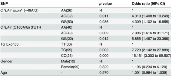
cor-responding to T allele and 208 bp and 167 bp digested products corcor-responding to C allele by Table 2. Association ofCTLA4exon1 +49A/G, 3’UTR CT60A/G andTGexon33 with autoimmune hypothyroidism when adjusted for age and gender using logistic regression.
SNP pvalue Odds ratio (95% CI)
CTLA4Exon1 (+49A/G) AA(26) R 1
AG(32) 0.011 4.319 (1.408 to 13.249)
GG(53) 0.036 4.309 (1.102 to 16.855)
CTLA4(CT60A/G) 3’UTR AA(40) R 1
AG(49) 0.009 7.096 (1.616 to 31.171)
GG(22) 0.012 5.855 (1.467 to 23.368)
TGExon33 TT(33) R 1
TC(55) 0.002 7.729 (2.142 to 27.889)
CC(23) 0.000 15.151 (3.303 to 69.507)
Gender Male(12) R 1
Female(99) 0.829 1.196 (0.234 to 6.120)
Age - 0.970 1.001 (0.964 to 1.039)
R refers to Reference Group. Data were coded for +49A/G (AA 0, AG 1, GG 2), CT60 A/G(AA 0, AG 1, GG 2),TGE33 (TT 0, CT 1, CC 2), gender (Male 0, Female 1) and age (years, continuous variable).
PCR-RFLP method. The three genotypes identified by 3.5% agarose gel electrophoresis were:
TT homozygous, TC heterozygous and CC homozygous for E33 polymorphism ofTGgene
(S1C Fig).
TC and CC genotypes of E33 polymorphism ofTGgene when compared with TT genotype
between patients and control by chi-square test-2x2 contingency table showed to increase sus-ceptibility to autoimmune hypothyroidism (p= 0.0003 and odds ratio 7.519;p<0.0001 and
odds ratio 20.54 respectively) (Table 1). Furthermore logistic regression analysis showed that,
when adjusted for age and gender, TC and CC genotype of 3’UTR CT60 polymorphism of
CTLA4gene increase the risk of autoimmune hypothyroidism by 7.729 fold (95% CI: 2.142 to
27.889,p= 0.002) and 15.151fold (95% CI: 3.303 to 69.507,p= 0.000) respectively (Table 2). Furthermore, there was significantly high frequency of the C allele in patients with
autoim-mune hypothyroidism compared with controls (62% vs. 39% respectively;χ2= 22.34,
p<0.0001) (Table 1). Both patient and control populations were found to be in
Hardy-Wein-berg equilibrium for this polymorphism (p= 0.1083 andp= 0.9375 respectively) (Table 1). This study has 83.01% statistical power for the effect size 0.5 to detect association of +49A/G polymorphism ofCTLA4at p<0.05 in patients and control population.
Linkage disequilibrium (LD) and haplotype analyses
The LD analysis revealed that the two polymorphisms investigated in theCTLA4gene were in
moderate LD association (+49A/G: CT60A/G; D’= 0.64, r2= 0.11). A haplotype evaluation of the two polymorphic sites was performed and the estimated frequencies of the haplotypes differed between autoimmune hypothyroidism patients and controls (globalp-value<0.0001) (Table 3).
However, the GG haplotype was more frequently observed in autoimmune hypothyroidism patients and increased the risk of autoimmune hypothyroidism by 4.688-fold [p=<0.0001; odds
ratio (OR): 4.688; 95% confidence interval (CI): (2.534~8.670)] (Table 3).
Discussion
The present study showed low iodine intake is not the sole etiological candidate for hypothy-roidism disorder in India as approximately half of the hypothyhypothy-roidism patients included in this
study were found to have presence of anti-TPO antibodies (S1 Table). Same trend was observed
in the recent study conducted on the prevalence of thyroid diseases in eight cities of India and presence of anti-TPO antibodies was shown to be conclusive for the disease [11]; however that study has not reported prevalence of autoimmune hypothyroidism in particular. No relation-ship between age of the patient and presence of anti-TPO antibodies was found in present study (data not shown), which is in accordance with the previous study [11].
Furthermore, to evaluate the possible expression dysregulation ofCTLA4variants, which
are important in T regulatory cell’s function, the mRNA levels of flCTLA4and sCTLA4genes were measured in patients with autoimmune hypothyroidism and compared with those from controls.
Table 3. Distribution of haplotypes frequencies forCTLA4gene polymorphisms (+49A/G and CT60A/G) among autoimmune hypothyroidism patients and controls.
Case (freq) (n = 96) Control (freq) (n = 120) Chi2 P for association P (global) Odds ratio (95% CI)
A A 17(0.182) 34(0.287) 3.217 0.072945 <0.0001 0.553 [0.288~1.061]
A G 25(0.255) 48(0.396) 4.761 0.029157 0.523 [0.291~0.940]
G A 6(0.057) 17(0.138) 3.765 0.052383 0.381 [0.140~1.040]
G G 48(0.505) 21(0.179) 25.933 <0.0001 4.688 [2.534~8.670]
Interestingly, we found significantly decreased mRNA expression of both flCTLA4and
sCTLA4in autoimmune hypothyroidism patients as compared to controls (Fig 1). We further
analyzed whether the polymorphisms examined in this study influenced the expression levels
of flCTLA4and sCTLA4. The +49AG and +49GG genotypes significantly decreased flCTLA4
and sCTLA4mRNA expression levels in autoimmune hypothyroidism patients compared to
controls; whereas, +49AA genotype did not affect mRNA expression levels (Fig 2A & 2B). It
has been reported that +49A/G may influence pattern or level ofCTLA4expression even if not
the function ofCTLA4protein because G allele is associated with reduced control of T cell
pro-liferation and thus contributes to the pathogenesis of autoimmune hypothyroidism, Grave’s
disease and other autoimmune diseases [21].
The 3’UTR ofCTLA4gene has also been found to be involved in several autoimmune
dis-eases hence to study the genetic variation of such regions is imperative [24] [25] [36].
Interest-ingly, we found that 3’UTR CT60G allele greatly reduced the mRNA expression of both
flCTLA4and sCTLA4in autoimmune hypothyroidism patients as compared to controls
sug-gesting its crucial role in pathogenesis of autoimmune hypothyroidism whereas, CT60AA and CT60AG genotypes did not affect mRNA expression levels (Fig 2C & 2D). However, previous
study did not detect any significant difference of sCTLA4and flCTLA4mRNA expression
based on the CT60 genotype in patients with autoimmune thyroid disorder compared to healthy individuals [37].
Recently studies on type-1 diabetes have also shown decreased sCTLA4levels and suggested
that lower sCTLA4expression may directly affect the suppressive capacity of regulatory T lym-phocytes and thereby modulate disease risk [38]. In contrast, increased serum sCTLA4 levels
were detected in other autoimmune diseases such as Graves’disease [39] and autoimmune
thy-roid disease [40]. These studies suggest that sCTLA4might contribute to the development of autoimmune diseases, probably through inhibiting the B7-flCTLA4 interaction and down-reg-ulation of T cell activation.
Moreover, the haplotype GG (+49G: CT60G) greatly decreased mRNA levels of sCTLA4
and haplotypes GG (+49G: CT60G) and AG (+49A: CT60G) greatly decreased mRNA levels of
flCTLA4in patients as compared to controls (data not shown) revealing the positive correlation
of +49G and CT60G in autoimmune hypothyroidism pathogenesis. Moreover, ratio of sCTLA4
to flCTLA4mRNA expression was not found to be altered in autoimmune hypothyroidism
patients compared to controls (Fig 3). Moreover sCTLA4to flCTLA4mRNA expression ratio
was not affected by +49A/G and CT60A/G polymorphisms (Fig 3).
However, elevated sCTLA4/flCTLA4mRNA expression ratio was found in patients with the
haplotype GG (+49G: CT60G) as compared to controls (Fig 3) showing strong positive
correla-tion of +49G and CT60G in autoimmune hypothyroidism pathogenesis.
The present study found higher frequency of +49AG, +49GG and CT60GG genotypes
among autoimmune hypothyroidism patients and this seems to modulateCTLA4mRNA
expression (Fig 2A, 2B, 2C & 2D) however, +49AA, CT60AA and CT60AG genotype does not
seem to modulateCTLA4mRNA expression and hence patients harboring it may have other
genetic factors involved in disease pathogenesis supporting the fact that being an autoimmune
disease autoimmune hypothyroidism may have varied type of precipitating factors [21].
Also in the present study two polymorphic sites in theCTLA4gene i.e., exon 1: +49A/G and
in the 3’UTR region CT60A/G, were found to be associated with autoimmune hypothyroidism
susceptibility in Gujarat population, as significant difference for genotype and allele frequency
was observed between autoimmune hypothyroidism patients and controls (Tables1&2).
population did not find significant difference in genotype and allele frequency for +49A/G
polymorphism in autoimmune hypothyroidism patients [20].
We also found that the presence of CT60GG genotype was more frequent among autoim-mune hypothyroidism patients than control and G allele was also found to be associated with disease susceptibility (Tables1&2) and our results are in accordance with Japanese and other
populations [29], [43]. Further, the haplotypes AG (+49A: CT60G) and GG (+49G: CT60G)
were more frequent in patients as compared to controls (Table 3). Our results along with
previ-ous studies suggest that theCTLA4gene on chromosome 2q33 is a susceptibility locus for
auto-immune hypothyroidism [20], [21], [23], [26], [29].
However, it is clear from our results that some patients with AA genotype of +49A/G and CT60 also suffer from autoimmune hypothyroidism whereas some controls with GG genotype +49A/G and CT60 do not develop disease suggesting role of other precipitating factors for
autoimmune hypothyroidism being multifactorial disease [21]. However, age and gender were
not found to have crucial role in susceptibility to autoimmune hypothyroidism (Table 2).
Present study also investigated association ofTGE33 polymorphism with autoimmune
hypothyroidism susceptibility. The present study found higher frequency of CC genotype for
TGE33 in autoimmune hypothyroidism patients compared to controls, suggesting its
associa-tion with autoimmune hypothyroidism. It has been identified that significant associaassocia-tion ofTG
E33 and an exon 10–12 SNP cluster with autoimmune thyroid disorders and also the
interac-tion ofTGE33 withHLA-DR3confers susceptibility to autoimmune thyroid disease [31]. The
TGE33 polymorphism causes the change from a hydrophobic amino acid tryptophan to a
pos-itively charged hydrophilic amino acid arginine and this non-conservative amino acid substitu-tion would be expected to change the structure ofTGat this region [31].
Conclusion
Our findings show that the +49A/G and 3’UTR CT60A/G polymorphisms of theCTLA4gene
influence both full length and solubleCTLA4mRNA expression levels in patients with
autoim-mune hypothyroidism. This suggests variations at the genetic level, at least in part, could lead
to the dysregulation ofCTLA4expressions in autoimmune hypothyroidism patients and
sup-ports the autoimmune pathogenesis of the disease. Therefore, further research on relation
between variousCTLA4polymorphisms, dynamics offlCTLA4versus sCTLA4expression and
their turnover in autoimmune hypothyroidism as well as other autoimmune diseases is needed
to clarify the role ofCTLA4in the regulation of immune response. In addition,TGgene may
also predispose to autoimmune hypothyroidism by the mechanism of protein structure change due to nonconservative amino acid substitution and thus in turn may change its antigenicity making it more immunogenic and could confer susceptibility to autoimmune hypothyroidism.
Supporting Information
S1 Fig. PCR-RFLP analysis ofCTLA4exon 1 +49 A/G and 3’UTR CT60A/G andTGE33
polymorphisms: (A)PCR-RFLP analysis ofCTLA4exon 1 +49 A/G polymorphism on 3.5% agarose gel electrophoresis: lanes: 3 & 5 show heterozygous (AG) genotypes; lanes: 2 & 4 show homozygous (AA) genotypes; lane: 1, 6 & 7 show homozygous (GG) genotype; lane M shows
100 bp DNA ladder.(B)PCR-RFLP analysis ofCTLA43’UTR CT60A/G polymorphism on
3.5% agarose gel electrophoresis: lanes: 1 & 2 show heterozygous (AG) genotypes; lanes: 5 shows homozygous (AA) genotypes; lane: 3, 4 & 6 show homozygous (GG) genotype; lane M
shows 100 bp DNA ladder.(C)PCR-RFLP analysis ofTGE33 polymorphism on 3.5% agarose
DNA ladder. (TIF)
S1 Table. Demographic characteristics of hypothyroidism patients and controls.
(DOCX)
S2 Table. Primers used for genotyping ofCTLA4andTGSNPs and gene expression
analy-sis.
(DOC)
Acknowledgments
We thank all hypothyroidism patients and control subjects for their participation in this study.
Author Contributions
Conceived and designed the experiments: HP RB AM. Performed the experiments: HP M. Singh. Analyzed the data: HP MSM. Contributed reagents/materials/analysis tools: AM. Wrote the paper: HP. Hypothyroid patients diagnosis: M. Shastri. Edited the manuscript: HP MSM M. Singh RB AM.
References
1. Roberts CG, Ladenson PW. Hypothyroidism. Lancet. 2004; 363(9411):793–803. Epub 2004/03/16. doi:10.1016/s0140-6736(04)15696-1PMID:15016491.
2. Cooper DS. Clinical practice. Subclinical hypothyroidism. N Engl J Med. 2001; 345(4):260–5. Epub 2001/07/28. doi:10.1056/nejm200107263450406PMID:11474665.
3. Dodd NS, Godhia ML. Prevalence of iodine deficiency disorders in adolescents. Indian J Pediatr. 1992; 59(5):585–91. Epub 1992/09/01. PMID:1459681.
4. Sood A, Pandav CS, Anand K, Sankar R, Karmarkar MG. Relevance and importance of universal salt iodization in India. Natl Med J India. 1997; 10(6):290–3. Epub 1998/03/03. PMID:9481104.
5. Revised Policy Guidelines On National Iodine Deficiency Disorders Control Programme. New Delhi: Directorate General of Health Services, Ministry of Health and Family Welfare, Government of India; 2006.
6. Marwaha RK, Tandon N, Gupta N, Karak AK, Verma K, Kochupillai N. Residual goitre in the postiodiza-tion phase: iodine status, thiocyanate exposure and autoimmunity. Clin Endocrinol (Oxf). 2003; 59 (6):672–81. Epub 2004/02/21. doi:10.1046/j.1365-2265.2003.01895.xPMID:14974907.
7. Toteja GS, Singh P, Dhillon BS, Saxena BN. Iodine deficiency disorders in 15 districts of India. Indian J Pediatr. 2004; 71(1):25–8. Epub 2004/02/26. PMID:14979381.
8. The WHO Global Database on Iodine Deficiency. Geneva: Department of Nutrition for Health and Development, World Health Organization; 2004.
9. Andersson M, Takkouche B, Egli I, Allen HE, de Benoist B. Current global iodine status and progress over the last decade towards the elimination of iodine deficiency. Bull World Health Organ. 2005; 83 (7):518–25. Epub 2005/09/24. PMID:16175826; PubMed Central PMCID: PMC2626287.
10. Andersson M, Karumbunathan V, Zimmermann MB. Global iodine status in 2011 and trends over the past decade. J Nutr. 2012; 142(4):744–50. Epub 2012/03/02. doi:10.3945/jn.111.149393PMID: 22378324.
11. Unnikrishnan AG, Kalra S, Sahay RK, Bantwal G, John M, Tewari N. Prevalence of hypothyroidism in adults: An epidemiological study in eight cities of India. Indian J Endocrinol Metab. 2013; 17(4):647–52. PMID:23961480. doi:10.4103/2230-8210.113755
12. Hollowell JG, Staehling NW, Flanders WD, Hannon WH, Gunter EW, Spencer CA, et al. Serum TSH, T (4), and thyroid antibodies in the United States population (1988 to 1994): National Health and Nutrition Examination Survey (NHANES III). J Clin Endocrinol Metab. 2002; 87(2):489–99. Epub 2002/02/12. doi:10.1210/jcem.87.2.8182PMID:11836274.
intake: influences of age and sex. Clin Chem. 2006; 52(1):104–11. Epub 2005/10/29. doi:10.1373/ clinchem.2005.055194PMID:16254196.
14. Eguchi K. Apoptosis in autoimmune diseases. Intern Med. 2001; 40(4):275–84. Epub 2001/05/04. PMID:11334384.
15. Stassi G, De Maria R. Autoimmune thyroid disease: new models of cell death in autoimmunity. Nat Rev Immunol. 2002; 2(3):195–204. Epub 2002/03/27. doi:10.1038/nri750PMID:11913070.
16. Tomer Y, Davies TF. Searching for the autoimmune thyroid disease susceptibility genes: from gene mapping to gene function. Endocr Rev. 2003; 24(5):694–717. Epub 2003/10/23. doi: 10.1210/er.2002-0030PMID:14570752.
17. McCoy KD, Le Gros G. The role of CTLA-4 in the regulation of T cell immune responses. Immunol Cell Biol. 1999; 77(1):1–10. Epub 1999/04/02. doi:10.1046/j.1440-1711.1999.00795.xPMID:10101680. 18. Ueda H, Howson JM, Esposito L, Heward J, Snook H, Chamberlain G, et al. Association of the T-cell regulatory gene CTLA4 with susceptibility to autoimmune disease. Nature. 2003; 423(6939):506–11. Epub 2003/05/02. doi:10.1038/nature01621PMID:12724780.
19. Teft WA, Kirchhof MG, Madrenas J. A molecular perspective of CTLA-4 function. Annu Rev Immunol. 2006; 24:65–97. Epub 2006/03/23. doi:10.1146/annurev.immunol.24.021605.090535PMID: 16551244.
20. Park YJ, Chung HK, Park DJ, Kim WB, Kim SW, Koh JJ, et al. Polymorphism in the promoter and exon 1 of the cytotoxic T lymphocyte antigen-4 gene associated with autoimmune thyroid disease in Kore-ans. Thyroid. 2000; 10(6):453–9. Epub 2000/07/25. PMID:10907987.
21. Kouki T, Sawai Y, Gardine CA, Fisfalen ME, Alegre ML, DeGroot LJ. CTLA-4 gene polymorphism at position 49 in exon 1 reduces the inhibitory function of CTLA-4 and contributes to the pathogenesis of Graves' disease. J Immunol. 2000; 165(11):6606–11. Epub 2000/11/22. PMID:11086105.
22. Barbesino G, Tomer Y, Concepcion E, Davies TF, Greenberg DA. Linkage analysis of candidate genes in autoimmune thyroid disease: 1. Selected immunoregulatory genes. International Consortium for the Genetics of Autoimmune Thyroid Disease. J Clin Endocrinol Metab. 1998; 83(5):1580–4. Epub 1998/ 05/20. doi:10.1210/jcem.83.5.4813PMID:9589659.
23. Kouki T, Gardine CA, Yanagawa T, Degroot LJ. Relation of three polymorphisms of the CTLA-4 gene in patients with Graves' disease. J Endocrinol Invest. 2002; 25(3):208–13. Epub 2002/04/09. PMID: 11936461.
24. Orozco G, Torres B, Nunez-Roldan A, Gonzalez-Escribano MF, Martin J. Cytotoxic T-lymphocyte anti-gen-4-CT60 polymorphism in rheumatoid arthritis. Tissue Antigens. 2004; 64(6):667–70. Epub 2004/ 11/18. doi:10.1111/j.1399-0039.2004.00318.xPMID:15546339.
25. Torres B, Aguilar F, Franco E, Sanchez E, Sanchez-Roman J, Jimenez Alonso J, et al. Association of the CT60 marker of the CTLA4 gene with systemic lupus erythematosus. Arthritis Rheum. 2004; 50 (7):2211–5. Epub 2004/07/13. doi:10.1002/art.20347PMID:15248219.
26. Furugaki K, Shirasawa S, Ishikawa N, Ito K, Kubota S, Kuma K, et al. Association of the T-cell regula-tory gene CTLA4 with Graves' disease and autoimmune thyroid disease in the Japanese. J Hum Genet. 2004; 49(3):166–8. Epub 2004/02/27. doi:10.1007/s10038-003-0120-5PMID:14986169. 27. Vaidya B, Pearce S. The emerging role of the CTLA-4 gene in autoimmune endocrinopathies. Eur J
Endocrinol. 2004; 150(5):619–26. Epub 2004/05/11. PMID:15132716.
28. Yanagawa T, Hidaka Y, Guimaraes V, Soliman M, DeGroot LJ. CTLA-4 gene polymorphism associated with Graves' disease in a Caucasian population. J Clin Endocrinol Metab. 1995; 80(1):41–5. Epub 1995/01/01. doi:10.1210/jcem.80.1.7829637PMID:7829637.
29. Ban Y, Tozaki T, Taniyama M, Tomita M. Association of a CTLA-4 3' untranslated region (CT60) single nucleotide polymorphism with autoimmune thyroid disease in the Japanese population. Autoimmunity. 2005; 38(2):151–3. Epub 2005/07/26. doi:10.1080/08916930500050319PMID:16040335.
30. Kavvoura FK, Akamizu T, Awata T, Ban Y, Chistiakov DA, Frydecka I, et al. Cytotoxic T-lymphocyte associated antigen 4 gene polymorphisms and autoimmune thyroid disease: a meta-analysis. J Clin Endocrinol Metab. 2007; 92(8):3162–70. Epub 2007/05/17. doi:10.1210/jc.2007-0147PMID: 17504905.
31. Ban Y, Greenberg DA, Concepcion E, Skrabanek L, Villanueva R, Tomer Y. Amino acid substitutions in the thyroglobulin gene are associated with susceptibility to human and murine autoimmune thyroid dis-ease. Proc Natl Acad Sci U S A. 2003; 100(25):15119–24. Epub 2003/12/06. doi:10.1073/pnas. 2434175100PMID:14657345; PubMed Central PMCID: PMC299918.
33. Barrett JC, Fry B, Maller J, Daly MJ. Haploview: analysis and visualization of LD and haplotype maps. Bioinformatics. 2005; 21(2):263–5. Epub 2004/08/07. doi:10.1093/bioinformatics/bth457PMID: 15297300.
34. Faul F, Erdfelder E, Lang AG, Buchner A. G*Power 3: a flexible statistical power analysis program for the social, behavioral, and biomedical sciences. Behav Res Methods. 2007; 39(2):175–91. Epub 2007/ 08/19. PMID:17695343.
35. Strieder TG, Prummel Mf Fau—Tijssen JGP, Tijssen Jg Fau—Endert E, Endert E Fau—Wiersinga WM, Wiersinga WM. Risk factors for and prevalence of thyroid disorders in a cross-sectional study among healthy female relatives of patients with autoimmune thyroid disease. Clin Endocrinol (Oxf). 2003; 59(3):396–401.
36. Dwivedi M, Laddha NC, Imran M, Shah BJ, Begum R. Cytotoxic T-lymphocyte-associated antigen-4 (CTLA-4) in isolated vitiligo: a genotype-phenotype correlation. Pigment Cell Melanoma Res. 2011; 24 (4):737–40. Epub 2011/07/29. doi:10.1111/j.1755-148X.2011.00892.xPMID:21794098.
37. Mayans S, Lackovic K, Nyholm C, Lindgren P, Ruikka K, Eliasson M, et al. CT60 genotype does not affect CTLA-4 isoform expression despite association to T1D and AITD in northern Sweden. BMC Med Genet. 2007; 8:3. Epub 2007/02/07. doi:10.1186/1471-2350-8-3PMID:17280620; PubMed Central PMCID: PMC1802068.
38. Gerold KD, Zheng P, Rainbow DB, Zernecke A, Wicker LS, Kissler S. The soluble CTLA-4 splice vari-ant protects from type 1 diabetes and potentiates regulatory T-cell function. Diabetes. 2011; 60 (7):1955–63. Epub 2011/05/24. doi:10.2337/db11-0130PMID:21602513; PubMed Central PMCID: PMC3121435.
39. Daroszewski J, Pawlak E, Karabon L, Frydecka I, Jonkisz A, Slowik M, et al. Soluble CTLA-4 receptor an immunological marker of Graves' disease and severity of ophthalmopathy is associated with CTLA-4 Jo31 and CT60 gene polymorphisms. Eur J Endocrinol. 2009; 161(5):787–93. Epub 2009/09/08. doi: 10.1530/eje-09-0600PMID:19734241.
40. Saverino D, Brizzolara R, Simone R, Chiappori A, Milintenda-Floriani F, Pesce G, et al. Soluble CTLA-4 in autoimmune thyroid diseases: relationship with clinical status and possible role in the immune response dysregulation. Clin Immunol. 2007; 123(2):190–8. Epub 2007/02/27. doi:10.1016/j.clim. 2007.01.003PMID:17321799.
41. Nithiyananthan R, Heward JM, Allahabadia A, Franklyn JA, Gough SC. Polymorphism of the CTLA-4 gene is associated with autoimmune hypothyroidism in the United Kingdom. Thyroid. 2002; 12(1):3–6. Epub 2002/02/15. doi:10.1089/105072502753451896PMID:11842815.
42. Tomoyose T, Komiya I, Takara M, Yabiku K, Kinjo Y, Shimajiri Y, et al. Cytotoxic T-lymphocyte antigen-4 gene polymorphisms and human T-cell lymphotrophic virus-1 infection: their associations with Hashi-moto's thyroiditis in Japanese patients. Thyroid. 2002; 12(8):673–7. Epub 2002/09/13. doi:10.1089/ 105072502760258640PMID:12225635.