R e v i s t a d a S o c i e d a d e B r a s i l e i r a d e M e d i c i n a T r o p i c a l 2 3 ( 1 ) : 2 7 - 3 1 , j a n - m a r , 1 9 9 0
T CELL-DEPENDENT IMMUNODEPRESSION
I N V IV OIN
S C H I S T O S O M A M A N S O N IINFECTED PATIENTS
Maria Imaculada Muniz-Junqueira,
Car los Eduardo Tost a and Aluizio Prata
T - c e ll f u n c t i o n w a s e v a l u a t e d in 2 9 p a t i e n t s w ith e i t h e r h e p a t o i n t e s t i n a l o r
h e p a t o s p l e n i c s c h i s t o s o m i a s i s b y in t r a d e r m a l t e s t s to r e c a l l a n tig e n s . I m m u n o d e p r e s
-s i o n w a -s d e t e c t e d in 2 6 % o f th e -s u b je c t-s w ith h e p a t o i n t e -s t i n a l -s c h i -s t o -s o m i a -s i -s a n d in
5 0 % o f th o s e w ith th e h e p a to s p l e n ic f o r m . C e l l u l a r i m m u n o d e p r e s s io n w a s r e l a t e d to
w o r m l o a d a n d s p le e n s iz e . T h is n o n s p e c if ic T - c e l l i m m u n o d e p r e s s i o n m a y r e p r e s e n t a
s e r i o u s c o n s t r a i n t to th e e l im in a tio n o f in t r a c e l l u l a r p a t h o g e n s b o th in h e p a t o s p l e n i c o r
h e p a t o i n t e s t i n a l s c h is to s o m ia s is .
Key-words: Schistosomiasis. T-cell dependent immunodepression.
In acute schistosomiasis both B-cell18 and T- cell2® functions are increased. However, during the' chronic phase, modulation of the host immune response results in diminution of antibody levels19 and T-cell function, causing a decrease in granuloma size1 5. Thus, a situation of chronic immunodepression en sues, as shown in experimental m o d e l s 2 ^ .
Lymphocyte responses in schistosomiasis have been studied both in human beings^ 7 11 20 25 and in experimental animal models2 16 17 22 Several studies have demonstrated a depression of lymphocyte blas- togenesis towards antigens of S c h i s t o s o m a m a n s o n i
eggs3 6 This response, however, is not invariably decreased as showed when cercarial or adult worm antigens11 17 20 25j mitogens or unrelated antigens are used2 7 22
Little is known of the correlation between immu nodepression, the clinical presentation of the disease and worm burden. Ellner et al10 have demonstrated that in hepatointestinal patients with high worm bur den the in vitro blastogénie response to antigens of adult worms was decreased. O n the other hand, patients with the hepatosplenic form showed cither an absent or an exacerbated response to antigens of the adult worm, while the response to mitogens and to
Núcleo de Medicina Tropical e Nutrição e Laboratório de Imunologia Celular, Faculdade de Ciências da Saúde, Uni versidade de Brasília. Brasília, D F , Brasil.
This work was supported by C N Pq(Proc. n? 4 0.1041/P ID E VI and 40.1868/87).
P r e s e n t a d d r e s s f o r c o r r e s p o n d e n c e : Dra. N.aria Imaculada Muniz-Junqueira. Departamento de Pediatria, Faculdade de Ciências da Saúde, Universidade de Brasília.
70910 Brasília, D F , Brasil.
Recebido para publicação em 16/10/89.
streptokinase-streptodomase was normal.
It has been shown that patients with severe schistosomiasis8 and those with associated chronic salmonellosis13 display depression o f delayed hypersensitivity. However, little is known on the degree of immune reactivity of individuals with less severe schistosomiasis, which represents the most frequent population living in endemic areas. A better definition of their T-cell response towards unrelated antigens would help us to understand the immune response of these individuals against microorganisms or vaccines which depend on T-cell immunity.
The present work aimed to evaluate T-cell res ponse to unrelated antigens in patients with the hepatointestinal or hepatosplenic schistosomiasis.
STUDY G R O U P S A N D M E T H O D S
Twenty nine patients of both sexes, 9 to 43 years old (mean ± sd = 24.5 ± 10.0) were studied. M ost of them (75% ) were white. The diagnosis of schistoso miasis was established on clinical and parasitological grounds. F o r several years these patients have been fol lowed up clinically and parasitologically. Before being selected for the study they were reexamined and their clinical forms were classified according to the criteria defined by Prata23. Examination showed no important clinical manifestations or nutritional imba lance.
Eggs of S c h i s t o s o m a m a n s o n i in faeces were detected by the Kato-Katz method14, and the worm burden was evaluated by the number of eggs per gram of faeces. Worm burden was considered as light when less than 200 eggs/g faeces were found,moderate from 2 00 to 1000 eggs/g and high when more than 1000 eggs were passing per gram of faeces.
The control group was formed by ten healthy individuals of both sexes, with ages varying from 20 to
M u n iz- J u n q u e ira M I , T o s ta C E , P r a ta A . T c e ll -d e p e n d e n t im m u n o d e p r e ss io n in vivo in Schistosoma mansoni in fe c te d p a tie n ts . R e v is ta d a S o c ie d a d e B r a s ile ir a d e M e d ic in a T r o p ic a l 2 3 : 2 7 - 3 1 , ja n - m a r , 1 9 9 0 .
50 years old (meam ± sd = 26.5 ± 5.0), 70% of them being white.
T-cell response was evaluated by means of in- tradermal tests, using the following recall antigens: oidiomycin, PPD , streptokinase-streptodomase, trichophytin and vaccinia virus. The later was exclu sively used in those individuals showing a scar of smallpox vaccine.
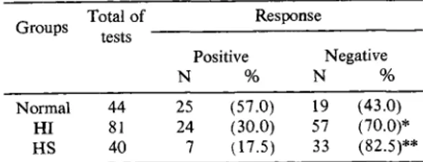
applied in S c h i s t o s o m a - i n f e c t e d patients, only 31 (26% ) were positive, while 25 out of 44 tests (57% ) applied in control individuals were positive (p < 0.001). N o statistical difference was found between H S and H I patients, although positive tests were more fre quently observed among H I (30% ) than is H S indivi duals (17.5% ). E ach group gave statistically different results when compared withnorm al controls (Table 2).
Volumes of 0.1 ml of antigen solution were intradermally injected in the undersurface of the left forearm. Reactions were read 48 hours later and considered as positive when equal or higher than 5 mm in the largest diameter, according to standard techniques27.
The statements of the Helsinki D eclaration were strictly followed throughout the investigation.
The results were statistically analysed by the Chi-square test and the Fisher’s exact te s t
RESU LTS
Fifty percent (5/10) of the individuals with hepatosplenic schistosomiasis (H S) and 26% ( 5 / 1 9 )
of those with hepatointestinal form (H I) presented no response to any of antigens tested. This pattern of response did not occur in any of the 10 normal controls ( P < 0 .05), as showed in Table 1.
Table 2 - Response o f hepatointestinal (HI), hepatosplenic (H S) and normal individuals to recall antigens by intradermal te st a general assessment of positive reactions.
Groups Total o f Response tests
Positive
N %
Negative
N %
Normal 44 25 (57.0) 19 (43.0)
HI 81 24 (30.0) 57 (70.0)*
HS 40 7 (1-7.5) 33 (82.5)**
* Significantly different from normal individuals (p < 0.01) Chi-Square Test. ** Significantly different from nom .al individuais (p < 0.001) Chi-Square 7 e s t
Subjects with schistosomiasis showed a lower frequency of positive tests for each individual antigen as compared to controls, except for vaccinia antigen. The positivity of schistosomiasis patients was 4% (1/26) with trichophytin, 7% (2/29) with streptoki nase-streptodomase, 31% (9/29) with oidiomycin, 45% (13/29) with P P D and 62.5% (5/8) with vacci nia virus. While in normal individuals the following frequency of positive tests was found: 25% (1/4) with trichophytin, 30% (3/10) with streptokinase-strep- todomase, 80% (8/10) to oidiomycin, 80% (8/10) to P P D and 50% (5/10) to vaccinia virus.
From the total of the 121 intradermal tests
M u n i z - J u n q u e i r a M I , T o s t a C E , P r a t a A . 7 c e l l - d e p e n d e n t i m m u n o d e p r e s s i o n in vivo in Schistosoma mansoni in f e c te d p a t i e n t s . R e v i s t a d a S o c i e d a d e B r a s i l e i r a d e M e d i c i n a T r o p i c a l 2 3 : 2 7 - 3 1 , j a n - m a r , 1 9 9 0 .
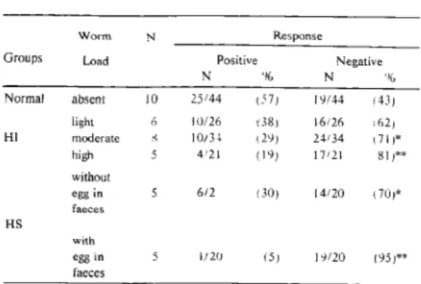
Table 3 - Response o f intradermal test to recall antigens in relation to S . m a n s o n i load in patients with hepa-tointestinal (H I) or hepatosplenic (H S) schisto somiasis.
Worm N Response
Groups Load Positive
N %
Negative
N %
Normal absent 10 25/44 t^ 7 j 19/44 (43,
light 6 i 0/26 (38) 16/26 .62,
Hl moderate .<< 10/34 (29, 24/34 ! 7 1 )* high
without
5 4'21 (19; 17/21 81 ,**
HS
egg in faeces
with
5 6/2 (30, [4/20 ( 70,*
egg in faeces
5 1/20 (5, 19/20 (95)**
* S ignificantly different fron n o rm al (p < 0 .0 5 ) C h i-S q u a re T est. ** S ignificantly different from no rm al ( p < 0 .0 1 ) C h i-S q u are T est.
A complete unresponsiveness to all antigens was detected in 4 out of 5 H S patients passing. S. m a n s o n i
-eggs, and in only one with no eggs in faeces. This latter individual had a palpable spleen 9 cm below the left costal edge.
On the other hand, a complete unresponsiveness to all intradermal tests applied occurred in 3 out of 4 patients with the spleen larger than 6 cm, but in only 2 out of 6 whose spleen was smaller than 6 cm. These two patients also showed the highest worm load of all the H S patients (984 and 672 eggs/g). Moreover, the only non-anergic patients with a spleen larger than 6 cm did not show S . m a n s o n i - eggs in faeces at the moment of the examination.
D IS C U S S IO N
Cellular immunodepression to S c h i s t o s o m a m a n s o n i - e g g antigens during infection may benefit the host, since the granulomatous reaction to the eggs plays a crucial role in the pathogenesis of schistoso m ia sis.^ However, the development of non-specific cellular immunodepression may represent a serious constraint to the elimination of intracellular pathogens during schistosomiasis. 9 12
In our study, 26% of hepatointestinal and 50% of hepatosplenic patients presented T-dependent immu-. nodepression. Similar findings were obtained by Cou- tinho et al8 and Higashil 3 jn hepatosplenic patients. However, our data show that individuals even with mild infection may present cellular immunodepression. It can be postulated that this non-specific t-cell immu
nodepression may make difficult the elimination of pathogens such as salmonellas, since this response is important to immunity to this bacteria4 15.
W e show that patients with schistosomiasis pre sented less positive response to the antigens tested, except for vaccinia virus. However, there was a gradation of positive responses, varying from 4% with trichophytin to 62.5% to vaccinia virus. This indicates that T-cell immunodepression is not an all or none phenomenon. This diversity o f response may occur due to the frequent individual variability of host- parasite relationships in schistosomiasis, which de pend on the duration of infection, worm burden, clinical form, and the individual response of each patient to the pathogen. This results in different degrees of involvement of the immune system of the host.
O ur results show that T-ceil immunodepression was related to S . m a n s o n i load. This indicates that unspecific cellular immunodepression depends on factors related to worm or egg antigens. Accordingly, Ellner et al*0 have demonstrated a decreased blas togénie response in vitro in schistosomiasis patients with high-level infectioa In fact Todd et al28 and Ottesen et al2l have presented evidence of immunodepression in vitro related to humoral factors, possibly antigens or immunocomplexes, present in the sera of schis tosomiasis patients.
The verification that 50% of the hepatosplenic patients did not present S . m a n s o n i - e g g s in their faeces, and that none of them presented high worm burden suggest that besides worm and eggs antigens, other factors may play a role in the immunodepression associated to schistosomiasis.The inverse relationship between T-cell responsiveness and size spleen indi cates that immune response in more altered in those patients presenting more severe anatomical and func tional involvement by the infection.
Although the factors responsible for immuno depression in schistosomiasis are not completely de terminated it has been suggested that both suppressor cells26, or adherent phagocytic cells24 may play a role. O ur data suggest that immunodepression in schistosomiasis depend on the degree of clinical involvement and, probably, on the amount of antigen released by the parasite.
R ESU M O
A imunidade celular foi avaliada em 29 pacientes com as formas hepatoesplênica ou hepatointestinal, da esquistossomose mansoni, através de testes intradér- micos com antígenos não relacionados ao S c h i s t o
M u n i z - J u n q u e i r a M I , T o s ta C E , P r a t a A . I c e l l - d e p e n d e n t im m u n o d e p r e s s i o n in vivo in Schistosoma mansoni in f e c te d p a t i e n t s . R e v i s t a d a S o c i e d a d e B r a s i l e i r a d e M e d i c i n a T r o p i c a l 2 3 : 2 7 - 3 1 , j a n - m a r , 1 9 9 0 .
pacientes com a forma hepatointestinal e em 50% daqueles com a forma hepatoesplênica. A imuno- depressão celular foi relacionada com a carga para sitária e o tamanho do baço. E sta imunodepressão celular pode dificultar a eliminação de patógenos intracelulares tanto na forma hepatoesplênica quanto na forma hepatointestinal da esquistossomose.
Palavras-chaves: Esquistossomose. Imunode pressão celular.
REFER EN C ES
1. Andrade ZA , Warren KS. Mild prolonged schistoso miasis in mice: alterations in host response with time and the development of portal fibrosis. Transactions of the Royal Society of Tropical Medicine and Hygiene 58:53-57, 1964.
2. Attallah AM , Smith A H , Murrell KD, Fleischer T, W oody J, Vannier WE, Scher I, Ahmed A, Sell KW. Characterization o f the immunosuppressive state during S c h i s t o s o m a m a n s o n i infection. Journal of Immunology
122:1413-1420, 1979.
3. Barsoum IS, Gamil FM, Al-Khafif MA, Ramzy RM, El Alamy M A , Colley D G . Immune responses and immu-noregulation in relation to human schistosomiasis in Egypt. I. Effect o f treatment on in vitro cellular respon siveness. The American Journal of Tropical Medicine and Hygiene 31:1181-1187, 1982.
4. Blanden RV, Machaness GB, Collins FM. Mechanisms of acquired resistance in mouse typhoid. Journal of Experimental Medicine 124:585-600, 1966.
5. Boros D L, Pelley RP, Warren KS. Spontaneous mo dulation o f granulomatous hypersensitivity in schisto somiasis mansoni. Journal o f Immunology 114:1437-1441, 1975.
6. Colley D G , Todd CW, Lewis F A , Goodgame RW. Immune responses during human schistosomiasis man soni. VI. I n v itr o nonspecific suppresion o f phytohe magglutinin responsiveness induced by exposure to certain schistosomal preparations. Journal o f Immuno logy 122:1447-1453, 1979.
7. Cottrell BJ, Humber D , Sturrock RF, Seitz H, Rees P. Non-specific cell mediated immunity in patients infected with S c h i s t o s o m a M a n s o n i in Kenya. Transactions of the Royal Society o f Tropical Medicine and Hygiene 76:234-237, 1982.
8. Coutinho A D , Antunes M TA, Domingues ALC. Estudo da imunidade humoral e celular na doença hepática esquistossomótica. Revista do Instituto de Medicina Tropical de São Paulo 24:282-291, 1982.
9. Edelson PJ, Intracellular parasites and phagocytic cell: cell biology and pathophysiology. Reviews o f Infectious D iseases 4:124-135, 1982.
10. Ellner JJ, Olds GR, Osman G S, El Kholy A, Mahmoud A A F. Dichotomies in the reactivity to worm antigen in human schistosomiasis mansoni. Journal oflmmunology 126:309-312, 1981.
11. Gazzinelli G , Katz N, Rocha RS, Colley D G . Immune responses during human schistosomiasis mansoni. VIII. Differential in vitro cellular responsiveness to adult worm and schistosomular tegumental preparations. The American Journal o f Tropical Medicine and Hygiene 32:326-333, 1983.
12. Hahn H, Kaufmann SH. The role of cell-mediated immunity in bacterial infections. Reviews of Infectious Disease 3:1221-1250, 1981.
13. Higashi GI. The interaction o f S c h i s t o s o m a and bac terial infections. WHO. Expert Committee on the Con trol o f Schistosomiasis, Geneva 8-13 november, S C H /E C /W P /84.30, 1984.
14. Katz N , Chaves A, Pellegrino J. A simples device for quantitative stool thick-smear technique in schistoso miasis mansoni. Revista do Instituto de Medicina Tro pical de São Paulo 14:397-400, 1972.
15. Kita E, Emoto M, Yasui K, Yasui K, Katsui N, Nishi K, Kashiba S. Cellular aspects o f the longer-lasting immu nity against mouse typhoid infection afforded by the live cell and ribosomal vaccines. Immunology 57:431-435, 1986.
16. Mota-Santos TA, Gazzinelli G , Ramalho-Pinto FJ, Pellegrino J, D ias de Silva W. Immunodepression in mice following S c h i s t o s o m a m a n s o n i infection. Revista do Instituto de Medicina Tropical de São Paulo 18:246-250, 1976.
17. Mota-Santos TA , Tavares CAP,Gazzinelli G, Pelle grino J. Immunosuppression mediated by adult worms in chronic schistosomiasis mansoni. The American Journal o f Tropical Medicine and Hygiene 26:727-731, 1977.
18. N ash TE, Cheever A W Ottesen EA, Cook JA. Schis tosome infections in humans: perspectives and recent findings. N IH Conference. Annals of Internal Medicine 97:740-754, 1982.
19. N ashT E , Ottesen E A , Cheever AW . Antibody response to a polysaccharide antigen present in the schistosome gut. II. Modulation of antibody response. The American Journal o f Tropical Medicine and Hygiene 27:944-950, 1978.
20. Olds GR, Kholy A E , Ellner JJ. Two distinctive patterns of monocyte immunoregulatory and effector functions in heavy human infections with S c h i s t o s o m a m a n s o n i. Journal oflm m unology 131:954-958, 1983.
21. Ottesen E A , Poindexter RW. Modulation of the host response in human schistosomiasis. II. Humoral factors which inhibit lymphocyte proliferative response to pa rasite antigens. The American Journal o f Tropical M e dicine and Hygiene 29:592-597, 1980.
22. Pelley RP, RuffierJJ, Warren KS. Suppressive effect o f a chronic helminth infection, schistosomiasis mansoni, on the in vitro responses of spleen and lymph node cells to the T cell mitogens phytohemagglutinin and concana-valin A. Infection and Immunity 13:1176-1183, 1976.
23. Prata A. Como caracterizar a forma hepato-esplênica da esquistossomose? In: II Simpósio sobre Esquistosso-mose p. 179, Salvador, 1970.
M u n i z - J u n q u e i r a M l , T o s t a C E , P r a t a A . T c e l l - d e p e n d e n t im n .u n o d e p r e s s i o n in vivo in Schistosoma mansoni in f e c te d p a t i e n t s . R e v i s t a d a S o c i e d a d e B r a s i l e i r a d e M e d i c i n a T r o p i c a l 2 3 : 2 7 - 3 1 , j a n - m a r , 1 9 9 0 .
24. Reiner N E, Kamel R, Higashi GI, El Naggar A, Aguib M, Ellner JJ, Mahmoud A A F. Concurrent responses of peripheral blood and splenic mononuclear cells to an tigenic and motigenic stimulation in human hepatos-plenic schistosomiasis. The Journal o f Infectious D i seases 140:162-168, 1979.
25. Rocklin RE, Brown A P, Warren KS, Pelley RP, Houba V, Siongok TK, Ouma J, Sturrock RF, Butterworth AE. Factors that modify the cellular-immune response in patients infected by S c h i s t o s o m a m a n s o n i. Journal of Immunology 125:1916-1923, 1980.
26. Rocklin RE, Tracy JW, El Kholy A. Activiation of antigen-specific suppressor cells in human schistoso miasis mansoni by fractions of soluble egg antigens
nonadherent to Con A sepharose. Journal o f Immuno logy 127:2314-2318, 1981.
27. Rosen FS, Wedgwood RJ, Eibl M. Primary immuno deficiency diseases. Clinical Immunology and Immu-nopathology 40:166-196, 1986.
28. Todd CW, Goodgame RW, Colley D G . Immune res ponse during human schistosomiasis mansoni. VII. Further analysis o f the interactions between patient sera and lymphocytes during in vitro blastogenesis to schis tosome antigen preparations. The American Journal of Tropical Medicine and Hygiene. 29:875-881, 1980.
29. Warren KS. The secret o f the immunopathogenesis of schistosomiasis: in vivo models. Immunological Reviews 61:189-213, 1982.