Perikardit, Erken Repolarizasyon veya Pnömoperikardiyum / Pericarditis, Early Repolarization or Pneumopericardium
Barış Buğan1, Yalçın Gökoğlan2, Turgay Çelik2 1Malatya Military Hospital, Cardiology Service, Malatya, 2Gulhane Military Medical Academy, Department of Cardiology, Ankara, Turkey
Acute Pericarditis, Early Repolarization or Pneumopericardium:
How Can We Diagnose?
Akut Perikardit, Erken Repolarizasyon veya Pnömoperikardiyum:
Nasıl Tanı Koyabiliriz?
A 21-year-old male patient with no significant past medical history presented to the emergency department three days ater the acute onset of precordial chest pain worsening in the
recumbent position and exacerbated by deep inspiration. He
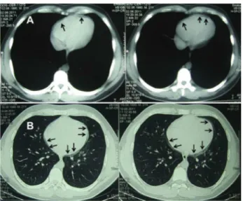
also had cough lasting 1 week. The patient denied having any fevers, chills, hemoptysis, nausea, or vomiting. He did not have any recent trauma or surgeries. He had a pack-day history of smoking cigarettes. On physical examination, the blood pressure was 110/70 mmHg, the pulse was 39 beats/min, respirations were 14/min, temperature was 36.7°C and oxygen saturation was 95% while breathing room air. Electrocardiogram showed sinus bradicardia with early repolarization and ST/T ratio was 0.26 in V5 (Figure 1). The chest radiograph showed linear radiolucency in the let paracardiac region without evidence of pneumothorax (Figure 1). Although computed tomography (CT) scan of the chest revealed pericardial thickening with minimal radiolucency mimicking pneumopericardium, there was no evidence of pneumothorax, bullous changes, or bronchopleural fistula (Figure 2). High resolution computed tomography (HRCT) subsequently depicted that the pericardial thickening caused radiolucency of the chest radiograph which was appeared like pneumopericardium (Figure 2). The patient’s symptoms improved with indomethacin and colchicine therapy. Pericarditis and pneumopericardium are important causes of chest pain. Pneumopericardium is defined as the presence of free air or gas in the pericardial sac. It is a rare, usually self-limited disease primarily afecting young men. Diferentiating the ECG of acute pericarditis from the normal variant with early repolarization is dificult, and serial ECGs in conjuction with the clinical setting are required. Additionally, an ST/T ratio > 0.25 in V5 or V6 may discriminate the ECGs of patients with acute pericarditis from normal variants with early repolarization.
Consequently, pneumopericardium is a rare condition, less common than either isolated pneumothorax or pneumomediastinum. Nevertheless, clinicians should be familiar with this diferential diagnosis of chest pain and its clinical and radiographic manifestations, since prompt recognition and treatment of potential complications may be life saving.
DOI: 10.4328/JCAM.1277 Received: 02.09.2012 Accepted: 17.09.2012 Printed: 01.09.2014 Corresponding Author: Barış Buğan, Malatya Military Hospital, Cardiology Service, Malatya, Turkey.
T.: +905323476096 F.: +90 4223362043 E-Mail: bbugan@hotmail.com
Figure 1. ECG showing ST elevation on admission (A), and basal early repolarization ater treatment (B). Chest radiograph showing linear radiolucency in the let paracardiac region (C), (arrows).