J Bras Pneumol. 2009;35(1):95-98
Case report
We report the case of a 49-year-old female patient, who worked as a maid in a rural area near the city of Dianópolis, Brazil. She presented a history of cough with purulent expectoration, which had intensified in the last four months. She also complained of unconfirmed fever and asthenia. She had received treatment for pneumonia with nonspecific antibiotic therapy in her town, however, with no clinical impro-vement. The patient reported having used a corticosteroid (prednisone), although not preci-sely reporting the reason, dose or duration of the treatment. She denied smoking, alcoholism, and was unaware of previous systemic diseases. Physical examination revealed good general health status, and pulmonary auscultation reve-aled the presence of crackling rales, mainly in
Introduction
Aspergillosis is a disease caused by filamentous fungi of the genus Aspergillus. These organisms can be easily found in the soil, in water and in various types of decomposing organic matter. There are approximately 200 species of Aspergillus, although only a minority are pathogenic for humans, the most common being A. fumigatus, A. flavus, and A. niger. Pulmonary aspergillosis can occur in several forms according to the archi-tecture of the adjacent lung, the host immune response and the level of inhaled inoculum. It is classified as follows: saprophytic aspergillosis or aspergilloma; allergic bronchopulmonary asper-gillosis; semi-invasive or chronic necrotizing pulmonary aspergillosis (CNPA); or invasive asper-gillosis. In this report, we describe a case of CNPA with associated endobronchial lesion.
Chronic necrotizing pulmonary aspergillosis*
Aspergilose pulmonar necrotizante crônica
Eduardo Felipe Barbosa Silva, Melânio de Paula Barbosa, Marco Antônio Alves de Oliveira, Rosane Rodrigues Martins,
Jefferson Fontinele e Silva
Abstract
Chronic necrotizing pulmonary aspergillosis is one of the forms of pulmonary aspergillosis typically found in mildly immunocompromised patients. We report the case of a female patient with complaints of chronic productive cough, fever and asthenia. She reported previous corticosteroid use. Computed tomography of the chest revealed consolidation with interposed cavitation in the right upper lobe. Fiberoptic bronchoscopy revealed purulent fluid within the tracheobronchial tree and an endobronchial exophytic lesion. The results of the biopsy of that lesion and the transbronchial biopsy were consistent with aspergillosis. Based on the clinical, radiological and histopathological findings, the patient was diagnosed with chronic necrotizing pulmonary aspergillosis. Treated with itraconazole, the patient presented a favorable clinical-radiological evolution.
Keywords: Aspergillosis; Lung diseases, fungal; Itraconazole.
Resumo
A aspergilose pulmonar necrotizante crônica é uma das formas de aspergilose pulmonar usualmente encontrada em pacientes com imunossupressão leve. Apresentamos o caso de uma paciente com queixas de tosse produtiva crônica, febre e astenia. Havia utilizado corticóides. A TC do tórax evidenciava consolidação com cavitação de permeio no lobo superior direito. A fibrobroncoscopia demonstrou secreção purulenta em árvore traqueobrônquica e lesão vegetante endobrônquica. Biópsias desta lesão e biópsia transbrônquica foram compatíveis com aspergi-lose. Diante do quadro clínico, radiológico e histopatológico, o diagnóstico de aspergilose pulmonar necrotizante crônica foi realizado. Tratada com itraconazol, a paciente apresentou boa evolução clínico-radiológica.
Descritores: Aspergilose; Pneumopatias fúngicas; Itraconazol.
* Study carried out at the Hospital de Base of the Federal District of Brasília, Brasília, Brazil.
Correspondence to: Eduardo Felipe Barbosa Silva. QRSW 07, Bloco B2, apto. 301, Sudoeste, CEP 70675-722, Brasília, DF, Brasil. [email protected] Tel: 55 61 3325-4525. Email: [email protected]
Submitted: 20 March 2008. Accepted, after review: 30 April 2008.
96 Silva EFB, Barbosa MP, Oliveira MAA, Martins RR, Silva JF
J Bras Pneumol. 2009;35(1):95-98
December of 2007, revealed total regression of the consolidation and cavitation in the right upper lobe, only residual parenchymal bands, translating to parenchymal scars, remaining (Figure 3).
Discussion
Initially described in 1981 by Gefter et al., CNPA or semi-invasive pulmonary aspergillosis is characterized as an indolent, subacute infec-tion that leads to a destructive process of the lung.(1,2) It is transmitted by inhaling the spores
of Aspergillus sp. (generally A. fumigatus) which, after being transported by air from an inanimate reservoir, are deposited in the host lungs, resulting in a local or systemic decrease in immunocom-petence.(1) Unlike other forms of aspergillosis,
CNPA does not require a preexisting cavity within the lungs. Local fungal invasion occurs in the lung parenchyma, with no vascular invasion or dissemination to other organs.(1) Among the
causes of decrease in local immunocompetence are pulmonary interstitial diseases, pulmonary resection, pulmonary TB sequelae, cystic fibrosis, pneumoconioses and COPD. Among the causes of decrease in systemic immunocompetence are the use of corticosteroids—even at lower doses—chemotherapy, radiotherapy, alcoholism, malnutrition and diabetes mellitus.(2-5) Cases of
CNPA have also been described in immunocom-petent patients.(6)
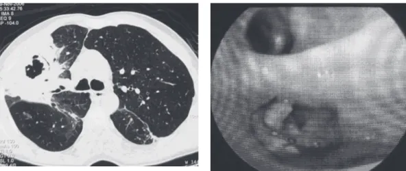
the upper and posterior third of the right hemi-thorax. A high-resolution computed tomography scan of the chest, performed in November of 2006, showed consolidation in the anterior and posterior segments of the right upper lobe, with air bronchograms and interposed cavitation (Figure 1). In view of the clinical-radiological profile, she was submitted to a diagnostic fibe-roptic bronchoscopy, which revealed purulent fluid within the tracheobronchial tree and an exophytic lesion with an irregular surface and yellowish color obstructing the bronchial lumen of the left lower lobe within the upper segment (Figure 2). The results of the biopsy of that lesion and the transbronchial biopsy of the posterior segment of the right upper lobe bronchus reve-aled the presence of septate hyphae with 45º dichotomous ramification, which are charac-teristic of Aspergillus sp. Direct testing and culture for acid-fast bacilli (Löwenstein-Jensen) and fungi (Sabouraud) of the bronchial lavage and bronchial brush samples were negative. No neoplastic cells were observed in this material. Blood workup, biochemical analysis and immu-noglobulin levels were normal. Serology for HIV was negative. Based on the clinical, radiological and histopathological findings, a diagnosis of CNPA was made. Treatment was initiated with 200 mg/day of itraconazole, and significant clinical improvement was observed after twelve months of treatment. A control high- resolution computed tomography scan, performed in Figure 1 - High-resolution computed tomography slice at the level of the carina, performed in November of 2006, showing consolidation in the right upper lobe with interposed cavitation.
Chronic necrotizing pulmonary aspergillosis
J Bras Pneumol. 2009;35(1):95-98
97
Half of all CNPA diagnoses are made without histopathological confirmation and are based on compatible clinical-radiological findings, together with the growth of Aspergillus sp. in cultures of sputum, bronchoscopic samples or transthoracic puncture samples, as well as by the clinical response to specific antifungal therapy and by excluding other diseases, such as TB, chronic histoplasmosis and neoplasias . (1-4,9)
Although determination of the serum anti-body (IgG) levels and skin tests with antigens to Aspergillus sp. are not diagnostic, they can inform the diagnosis.(2)
In the therapeutic field, the triazole anti-fungal itraconazole is currently a good option due to its excellent efficacy, low toxicity and easy administration. It works on the enzymes of the cytochrome P450 of the fungi, blocking the demethylation of lanosterol and synthesis of ergosterol, thereby altering the permeabi-lity of the membrane and, consequently, the fungal viability. The absorption and solu-bilization of this drug are better when it is administered with a gastric pH lower than 3, achieving good concentrations in the lungs, liver and bones. It is metabolized in the liver and excreted by the bile ducts. Among the principal side effects are gastrointestinal intolerance, hepatotoxicity and hypersensiti-vity. Drug interactions occur with rifampicin, isoniazid, carbamazepine, phenytoin, and phenobarbital, which reduce the antifungal serum level, and with cyclosporine, digoxin, warfarin, benzodiazepines, protease inhibi-tors, and terfenadine, which increase its serum level. Due to its teratogenicity, it is contrain-dicated for use in pregnant women. The appropriate duration of itraconazole adminis-tration is indeterminate and depends on the clinical-radiological response. Recurrence can occur after the discontinuation of the medi-cation. Amphotericin B deoxycholate and its lipid formulations are indicated for patients in whom the treatment with itraconazole has failed, as well as for patients with lung dise-ases. Due to their costs, new drugs, such as caspofungin and voriconazole, are reserved to the treatment of severe infections or infec-tions that do not respond to other antifungal agents. Surgical treatment can be considered in patients with the localized form of the disease and presenting no clinical-radiological Middle-aged and elderly individuals are those
typically affected by CNPA. The great majority of patients present early pulmonary and systemic symptoms, and this symptomatology lasts from one to six months. Among the complaints are nonspecific symptoms such as cough, expec-toration, chest pain, fever and weight loss. Although rare, hemoptysis and dyspnea have been reported.(2,3)
The most common radiological profile is characterized by the presence of unilateral or bilateral segmental consolidations, with or without the presence of cavitation or adjacent pleural thickening. These lesions preferably affect the upper lobes or the upper segments of the lower lobes. Mycetomas are observed in half of all cases. These features tend to evolve slowly for months.(5) Endobronchial lesions can also be
present in CNPA.(5-8)
The diagnosis of CNPA is difficult to make and is often delayed (by an average of three months),(3) contributing to increasing its
98 Silva EFB, Barbosa MP, Oliveira MAA, Martins RR, Silva JF
J Bras Pneumol. 2009;35(1):95-98
References
1. Gefter WB, Weingrad TR, Epstein DM, Ochs RH, Miller WT. “Semi-invasive” pulmonary aspergillosis: a new look at the spectrum of aspergillus infections of the lung. Radiology. 1981;140(2):313-21.
2. Soubani AO, Chandrasekar PH. The clinical spectrum of pulmonary aspergillosis. Chest. 2002;121(6):1988-99. 3. Saraceno JL, Phelps DT, Ferro TJ, Futerfas R, Schwartz
DB. Chronic necrotizing pulmonary aspergillosis: approach to management. Chest. 1997;112(2):541-8. 4. Kato T, Usami I, Morita H, Goto M, Hosoda M, Nakamura
A, et al. Chronic necrotizing pulmonary aspergillosis in pneumoconiosis: clinical and radiologic findings in 10 patients. Chest. 2002;121(1):118-27.
5. Franquet T, Müller NL, Giménez A, Guembe P, de La Torre J, Bagué S. Spectrum of pulmonary aspergillosis: histologic, clinical, and radiologic findings. Radiographics. 2001;21(4):825-37.
6. Kadakal F, Uysal MA, Ozgül MA, Elibol S, Urer N, Gürses A, et al. A case report of endobronchial semi-invasive aspergillosis. Tuberk Toraks. 2004;52(2):179-82. 7. Kim SY, Lee KS, Han J, Kim J, Kim TS, Choo SW, et al.
Semiinvasive pulmonary aspergillosis: CT and pathologic findings in six patients. AJR Am J Roentgenol. 2000;174(3):795-8.
8. Dar KA, Shah NN, Bhargava R, Ahmed Z, Pandey DK, Dar NH, et al. Endobronchial Aspergilloma in a 30 year old man. J Bronchol. 2007;14(3):207-9.
9. Wallace JM. The Role of Bronchoscopy in Pulmonary Mycoses: Part 2: Aspergillus Species and Other Phagocyte Opportunists. J Bronchol. 2001;8(3):213-21.
10. Martinez R. An update on the use of antifungal agents. J Bras Pneumol. 2006;32(5):449-60.
response to antifungal agents. However, it should be emphasized that the rate of posto-perative complications is high.(3,10)
Our patient was middle-aged and presented complaints of chronic productive cough, fever and asthenia. The radiological findings included consolidation with cavitation predominantly affecting the upper lobes. This is the pattern most often reported in the literature. Although there have been cases of CNPA in immunocom-petent individuals, the corticosteroid use by the patient probably caused a degree of immu-nosuppression that predisposed her to this disease. The diagnosis was made based on the analysis of pulmonary samples from fiberoptic bronchoscopy (endobronchial and transbron-chial biopsies), despite the reportedly low yield of these procedures. She evolved with a good response to the use of itraconazole, which is in accordance with the literature. Finally, we emphasize that in patients with predisposing factors, suggestive clinical-radiological profile and positive culture for Aspergillus sp. in pulmonary samples, the hypothesis of CNPA should be considered. This is a determining factor to reduce the morbidity and mortality that frequently occurs as a consequence of the delayed diagnosis of this disease.
About the authors
Eduardo Felipe Barbosa Silva
Attending Physician. Department of Bronchoesophagology, Hospital de Base of the Federal District of Brasília, Brasília, Brazil.
Melânio de Paula Barbosa
Attending Physician. Department of Pulmonology, Hospital de Base of the Federal District of Brasília, Brasília, Brazil.
Marco Antônio Alves de Oliveira
Attending Physician. Department of Bronchoesophagology, Hospital de Base of the Federal District of Brasília, Brasília, Brazil.
Rosane Rodrigues Martins
Radiologist. Hospital das Forças Armadas – HFA, Armed Forces Hospital – Brasília, Brazil.
Jefferson Fontinele e Silva