Hidrokarbon Pnömonisi / Hydrocarbon Pneumonitis
Hydrocarbon Pneumonitis; Clinical and Radiological Variability
Hidrokarbon Pnömonisi; Klinik ve Radyolojik Değişkenlik
DOI: 10.4328/JCAM.869 Received: 17.12.2011 Accepted: 19.01.2012 Printed: 01.01.2015 J Clin Anal Med 2015;6(1): 117-9 Corresponding Author: Bunyamin Sertogullarindan, YYU Arastirma Hastanesi, K.Karabekir Caddesi 65200 Van, Turkey.
T.: +90 4322150470-6135 GSM: +905336584452 E-Mail: [email protected]
Özet
Hidrokarbonların aspirasyonu basitten kritik düzeye kadar solunumsal patolojilere neden olur. Amacımız bu olgulardaki radyolojik ve klinik değişkenliğe dikkati çek
-mektir. Yirmi iki yaşında kaza ile dizel yakıt aspire eden genç bir erkek olguyu sun
-duk. Olgu Aspirasyondan üç gün sonra klinik kötüleşme nedeni ile acil servisimize başvurdu. Başvuru sırasına ait güğüs grafisinde sağ orta lobda infiltrasyon sap
-tandı. Fakat iki gün sonra sağ alt lob atalektazisi gelişti. Bronkoskopik inceleme
-de mukozal hiperemi ve ö-dem izlendi, bronş obstrüksiyonu yoktu. Sekon-der bakte
-riyel pnomoni saptanmadı. Hasta sistemik kortikosteroid ve antibiyotik ile teda
-vi edildi. Teda-viden iki hata sonra tam klinik iyileşme ve önemli radyolojik gerile
-me vardı. Hidrokarbon aspirasyon vakalarında klinik tablo değişkendir. Radyolojik görünüm gecikme ile gelişebilir. Önemli bir komplikasyon için yeni bir delil saptan
-madıkça steroid tedaviye devam edilmelidir.
Anahtar Kelimeler
Hidrokarbonlar; Aspirasyon
Abstract
Aspiration of hydrocarbons causes respiratory pathologies from simply to critical.
Our aim is to attract attention to clinical and radiological variability in these
cas-es. We presented a 22-year old young case exposed to diesel fuel by accidental as
-piration. Three days ater the aspiration, because of clinical deterioration, he was admitted to our emergency clinic. In the chest radiograph on admission showed iniltration in the right middle lobe. But a right lower lobe atelectasis emerged two days later. Bronchoscopy revealed inlamed and hyperemic mucosa and no bronchial obstruction. No secondary bacterial pneumonia was seen. The patient was treated with systemic steroids and antibiotics. Ater two weeks of treatment there was complete clinical improvement and signiicant radiologic regression. In
the hydrocarbon aspiration cases clinical picture is variable. Radiological picture
may develop with a delay. Unless there is a new evidence for an important com
-plication, the steroid treatment should be followed.
Keywords
Hydrocarbons; Aspiration
Bunyamin Sertogullarindan1, Aydin Bora2, Fuat Sayır3, Bulent Ozbay1 1Department of Pulmonary Medicine, 2Department of Radiology, 3Department of Chest Surgery, Yuzuncu Yil University Medical Faculty, Turkey
| Journal of Clinical and Analytical Medicine
Introduction
Products of petroleum have been used commonly in daily life. They are members of hydrocarbons. Aspiration of hydrocarbons causes respiratory pathologies from simple symptoms to criti -cal result. Hydrocarbon pneumonitis, known also as ire-eater pneumonia, always develops ater aspiration of low viscosity, volatile hydro carbides [1]. In the current article, we report a case of hydrocarbon pneumonitis of a 22-year-old young truck driver ater petroleum aspiration by siphonage.
Case Report
A 25-year-old a truck driver young man was admitted to our emergency department for evaluation of moderate dyspnea, severe right-side chest pain, hemoptysis and cough. His general condition was poor. There was no previous medical history. The symptoms began ater an episode of accidental diesel aspira -tion while siphoning from the fuel tank of a truck. His personal history was unremarkable. Clinical examination revealed chest wall tenderness and ine rales over the right midlung ield. How -ever prominent decreased breath sound was developed in the right base ater two day admission. He was normotensive with a heart rate of 104 per minute and a respiratory rate of 25 breaths per minute. The remaining physical examination ind -ings were normal.
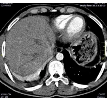
On laboratory examination, complete blood count revealed ele-vated white blood cells (14.110/μl) and an erythrocyte sedimen -tation rate of 56 mm/h. Serum electrolytes, hepatic and renal function tests were normal. Arterial blood gas measurements; pH = 7.38, pCO2 = 36.3 mmHg, pO2 = 55 mmHg and SaO2 = 85%. Spirometer revealed moderate restriction of lung function. On admission chest radiograph demonstrated a density in the right middle lobe. A Repeated chest radiography on the 3rd day of admission showed consolidation with volume loss and blur -ring of diaphragmatic contour in the right lower lobe (Fig 1A, 1B). A chest computed tomography scan showed a prominent consolidation with an air bronchogram in the right middle lobe, a minimal consolidation in the let lower lobe and atelectasis in
the lower right lobe, and pleural efusion in the right hemitorax (Figs. 2 and 3). Bilateral minimal pleural efusion was seen on thoracic ultrasonography. On bronchoscopic examination, in-lamed and hyperemic mucosa especially on the right side was noted. Signs of bronchial purulence or obstruction were not ob -served. On examination of bronchoalveolar lavage (BAL) luid no pathogens, ie, bacteria, mycobacteria or fungi, were dem -onstrated, either microscopically or by culturing. Conservative therapy was initiated, which included 1 g of ampicilline/sulbac -tam i.v. tid, and 40 mg of metilprednisolone/day. Conservative therapy led to a marked decline of the symptoms and radiologic indings (Fig 1C), accompanied by an obvious improvement of the patient’s general condition.
Discussion
Exposure to hydrocarbons can happen in many forms, but it causes the most destruction with aspiration [1]. Aspiration of hydrocarbons is an acute hydrocarbon pneumonitis which may happen accidentally while siphoning from fuel tank or motor ve -hicles. It is occasionally seen in adults, especially drivers, farmers and auto mechanics [2-4]. The toxic potential of hydrocarbons is directly related to their physical properties. Highly volatile with low viscosity and lower surface tension hydrocarbons are more likely to be inhaled or aspirated into the respiratory system [1]. The hydrophobic nature of them let them to penetrate deep into the tracheobronchial tree. Direct contact with alveolar mem -branes can lead to hemorrhage, hyperemia and edema. They provoke the activation of macrophages, leading to an increased release of cytokines and a prolonged inlammatory reaction [5]. The type II pneumocytes are most afected, resulting in a de -creased surfactant production. The surfactant layer, which is composed of lipids, is made soluble by hydrocarbons, causing further damage [6]. The end results of hydrocarbon aspiration are necrosing chemical pneumonitis, atelectasis, pleural efu -sion and hypoxemia [7].
Typical clinical symptoms and indings of patients are similar with any infectious pneumonia; chest pain, dyspnea, cough, fever, and hemoptysis can be seen [8]. Especially severe chest pain and tenderness of chest wall, which could be related to chemical pleuritis, were remarkable symptoms of the patient. Shortness of breath worsened in late-period due to atelectasis in our patient.
The radiological lesions are generally out of proportion to the clinical indings. Radiographic indings include unilateral or bi -lateral lung consolidation, pneumatoceles, pleural efusion, at -electasis and spontaneous pneumothorax [9]. It was reported that pneumonitis of right middle lobe might occur if the patient bent forward. while siphoning [2]. Our patient also reported that he was in similar position. Ultrasound examination revealed a small amount of bilateral pleural efusion, while there was no pneumonia on the let side on chest radiography. It is sug -gested that a small amount hydrocarbon aspiration can cause
Figure 2. Computerized tomography of the thorax shows atelectasis in the right lower lobe and pleural efusion
Figure 1. Chest radiographs of the patient taken on admission (A), on the 3rd (B) and 15th (C) day of the treatment.
| Journal of Clinical and Analytical Medicine
118
| Journal of Clinical and Analytical Medicine
pleural irritation that leads to pleural efusion. In the hydrocar -bon pneumonia, the afected region can be seen directly, BAL can be performed and microbiological samples can be taken via bronchoscopy [5] .On bronchoscopy we observed bilateral hy -peremia, edema and necrosis, which was more prominent on the right side.
Hydrocarbon pneumonia is a pseudo-infectious lung disease with a severe release of inlammatory cytokines with macro -phage activation. The inlammatory nature of the disease sug -gests that steroid treatment can be useful in these patients. However there have been conlicting reports about the efec -tiveness of steroids [10]. Our patient had signiicantly improved by steroid treatment. The conlicting reports on the use of ste -roids could be due to diferent intensity of exposures and com -plications of pneumonitis such as microbial superinfection and acute respiratory failure. Prophylactic antibiotic therapy is usu -ally applied in these cases. A prophylactic antibiotic therapy was also begun in our patient since he was admitted 3 days ater the event. Because pathogens were not detected in bronchial samples we discontinued antibiotic treatment. The prognosis is generally good in hydrocarbon aspiration and our patient completely improved ater 15 days of treatment. However some studies reported that some complications may develop, which included abscess, bronchopulmonary istula, bacterial superin -fection and acute respiratory failure and death.
Conclusion
Symptoms and radiologic indings may be not clear in early time. Also the relationship between exposure and symptoms may be overlooked in the late period. physician should be aware of that hydrocarbons can provoke a pneumonitis.
Competing interests
The authors declare that they have no competing interests.
References
1. Dyer S. Hydrocarbons. In: Wolfson AB, Editor. Harwood-Nuss’ Clinical Prac
-tice of Emergency Medicine. Philadelphila USA: Lippincott Williams & Wilkins;
2005.p.1590-3.
2. Carlson DH. Right middle lobe aspiration pneumonia following gasoline siphon -age Chest 1981;80(2):246-7.
3. Khanna P, Devgan SC, Arora VK, Shah A. Hydrocarbon pneumonitis following
diesel siphonage. Indian J Chest Dis Allied Sci 2004;46(2):129-32.
4. Sertogullarından B, Ozbay B, Asker S, Ekin S. Hydrocarbon pneumoniae cases. The Archives of Lung 2011;12(1):33-8.
5. Burkhardt O, Merker HJ, Shakibaei M, Lode H. Electron microscopic indings in BAL of ire-eater ater petroleum aspiration. Chest 2003;124(1):398-400. 6. Giammona ST. Efects of furniture polish on pulmonary surfactant. Am J Dis
Child 1967;113(6):658-63.
7. Olsen KR. Poisoning. In: Tierney LM, McPhee SJ, Papadakis MA, editors. Current Medical Diagnosis and Treatment. New York McGraw-Hill Company;
2000.p.1567-8.
8. Kitchen JM, O’Brien DE, McLaughlin AM. Perils of ire eating. An acute form of lipoid pneumonia or ire eater’s lung. Thorax 2008;63(5):401-39.
9. Franquet T, Gómez-Santos D, Giménez A, Torrubia S, Monill JM. Fire eater’s pneu
-monia: radiographic and CT indings. Comput Assist Tomogr 2000;24(3):448–50. 10. Steele RW, Conklin RH, Mark HM. Corticosteroids and antibiotics for the treat
-ment of fulminant hydrocarbon aspiration. JAMA 1972; 219(11):1434-7.
Journal of Clinical and Analytical Medicine | 119