Annica de Barros Rosa
Graduated in Cellular and Molecular Biology
Multifunctional Organometallic
Compounds for Auger Therapy
Dissertation to obtain Master
’
s Degree in
Biochemistry
Supervisor: Dr. António Manuel Rocha Paulo, C
2TN-IST
Jury:
President: Prof. Dr. Pedro António de Brito Tavares Examiner: Prof. Dr. Paula Dolores Galhofas Raposinho Supervisor: Prof. Dr. António Manuel Rocha Paulo
Multifunctional Organometallic Compounds for Auger Therapy
Copyright
Annica de Barros Rosa, FCT/UNL-UNL
To my mentor, Dr. António Paulo, I thank you for the mentoring, transmitted knowledge and incentives. For the availability and accessibility demonstrated.
To Dr. Isabel Rego dos Santos for the opportunity and acceptance in the group Ciências Radiofarmacêuticas of C2TN.
To Letícia Alves do Quental for all the support with the laboratory practices, teaching, availability and all the extra help.
To Dr. Paula Raposinho, Dr. Sofia Gama and Dr. Célia Fernandes for the studies provided, availability and for all the help. To Dr. Goreti Morais and Dr. Elisa Palma for the teaching and friendship.
To Susana, Sofia M., Elisabete R., Inês, Filipe, Maria, Vera, Mariana and Maurício for the NMR spectra, friendship and company. To the rest of the group, I am equally grateful for the welcoming and friendship.
To you my parents, I thank you. For the financial support, but essentially, for the emotional support, and for calling me religiously every Friday night during these 5 years I have been away from home. And thank you dad for coming visit me during my writing.
To my favorite person in this world, my big brother Yannick. To my grandmothers and to all my relatives, for the calls, and for supporting me from the distance.
To my BFF Maira, for the friendship and endless random talking. Thank you to all my other fellow compatriots for bringing a piece of home to this foreign country. In special my relatives, high school buddies and housemates.
Abstract
Auger emitters gained significant interest in the past years due to the favorable suitability for target therapy at cellular level. Among Auger emitters, 99mTc seems like the most appealing candidate for this task due to its great advantages regarding low cost, high availability and almost ideal radiochemical properties. The short range of Auger electrons demands that the radionuclide must be as close as possible to the target, usually the nuclear DNA, in order to cause significant therapeutic effects. Thus, the search for efficient delivery systems is the major challenge for Auger therapeutic applications. Radioactive complexes capable of entering the cell and target the nucleus showing affinity for the DNA molecule have been reported in the literature. Such compounds comprise efficient multifunctional chelators with the ability to stabilize the fac-[99mTc(CO)3]+ core, as well as DNA-intercalating agents that ensure close
proximity to the target. Herein we aimed to synthesize 99mTc complexes (Tc1-4) stabilized by
multifunctional chelators of the pyrazole-diamine type, containing anthracene (L1 and L2) or
acridine orange (L3 and L4) as DNA-binding groups with longer (L2 and L3) or shorter (L1 and L4) spacer between the latter and the chelating agent. The corresponding Re complexes ( Re1-4) were also foreseen for full chemical characterization. Due to the demanding synthesis
processes and available time, only Tc1 and Tc2 could be synthesized, characterized and
biologically evaluated. The Re congeners showed ability to interact with the CT-DNA molecule trough spectroscopic studies. Tc1 and Tc2 also showed great ability to target the nucleus, once
diffused into the cells of B16-F1 murine melanoma and PC3 human prostate cells, but no radio-cytotoxic effect in these cell lines. Although efficient delivery system at subcellular level could be provided in this work, the relevance of the complexes for the design of Auger emitting 99mTc radiopharmaceuticals could not be demonstrated.
Keywords
Nuclear Medicine
Radiotherapy
Radiometal
Auger electrons
Technetium-99m
Resumo
A terapia com emissores Auger tem despertado interesse nos últimos anos devido à possibilidade de explorar terapia direccionadas, a nível celular. Entre os emissores Auger conhecidos, o 99mTc oferece diferentes vantagens: baixo custo, elevada disponibilidade e propriedades radioquímicas favoráveis. O curto alcance dos electrões Auger constitui um factor limitante para a sua eficácia biológica exigindo que o radionucídeo se encontre o mais próximo possível do seu alvo, usualmente o DNA nuclear, de modo a exercer os seus efeitos terapêuticos. Assim, o maior desafio para o uso deste tipo de radionuclídeos em procedimentos terapêuticos é o desenho de vectores de entrega eficientes a nível subcelular. Já se encontra descrito um reduzido número de complexos radioactivos com capacidade de se localizar no núcleo das células e com afinidade para o DNA. Para esse efeito, tais compostos possuem em geral um agente quelante multifuncional com capacidade de estabilizar a unidade fac
-[99mTc(CO)3]+ e, simultaneamente, um agente intercalador do DNA que assegura a proximidade e afinidade para o alvo. Neste trabalho propôs-se a síntese de complexos de tecnécio (Tc1-4)
estabilizados por agentes quelantes do tipo pirazolo-diamina contendo os agentes intercaladores antraceno (L1 e L2) ou alaranjado de acridina (L3 e L4), com um espaçador
metilénico maior (L2 and L3) ou menor (L1 and L4) entre o agente quelante e intercalador.
Perspectivou-se ainda a síntese dos respectivos complexos de rénio (Re1-4) para uma
completa caracterização dos compostos. Devido à elevada exigência da síntese dos ligandos e ao tempo disponível, apenas os complexos Tc1 e Tc2 foram sintetizados e caracterizados.
Estudos espectroscópicos com os congéneres de Re mostraram que os complexos interagem com o DNA. Tc1 e Tc2 demonstraram elevada capacidade de se localizar no núcleo celular em
estudos com células B16-F1 de melanoma de ratinho e células PC3 de cancro de próstata humana. No entanto, não exerceram qualquer efeito radio-citotóxico. Apesar de os complexos apresentarem uma elevada capacidade para atingir o núcleo de células tumorais, não foi possível demonstrar o seu interesse para o desenho de radiofármacos de 99mTc para terapia Auger.
Palavras-chave
Medicina Nuclear Radioterapia Radiometal Electrões Auger Tecnécio-99mIndex
Acknowledgments ... iii
Abstract ... v
Keywords ... v
Resumo ... vii
Palavras-chave ... vii
List of Figures ... xiii
List of Schemes ... xv
List of tables ... xvii
List of symbols and abbreviations ... xix
1 Introduction ... 1
1.1 General aspects about Nuclear Medicine ... 1
1.1.1 Diagnosis in Nuclear Medicine ... 2
1.1.2 Therapy in Nuclear Medicine ... 4
1.1.2.1 Biological effects of ionizing radiation ... 4
1.1.2.2 Systemic cancer therapy in Nuclear Medicine ... 5
1.2 Auger electrons and cancer therapy ... 7
1.2.1 Auger electrons ... 7
1.2.2 Prospective Auger emitters for cancer therapy ... 8
1.3 Design of metal-based radiopharmaceuticals ... 10
1.4 Technetium ... 11
1.4.1 Basic chemical and radiochemical aspects ... 11
1.4.2 99mTc and cancer theranostics ... 15
1.5 Background and aim of the thesis ... 20
1.6 Rationale for the design and evaluation of the proposed complexes ... 22
1.6.1 Pyrazole-diamine BFCs ... 22
1.6.2 DNA-binding groups and DNA interaction ... 23
1.6.2.1 DNA binders and DNA-binding modes ... 24
1.6.2.2 Acridine orange and anthracene ... 25
1.6.2.3 Evaluation of DNA-binding ... 26
1.6.3 Functionalization with a bioactive peptide and a nuclear localization sequence: a perspective ... 27
1.6.3.1 Bioactive peptide ... 27
1.6.3.2 Nuclear localization sequence ... 28
2 Results and discussion: synthesis and characterization of the ligands and complexes ... 31
2.1 Synthesis and characterization of the ligands ... 31
[x]
3 Results and discussion: in vitro and biological studies ... 47
3.1 Spectroscopic studies of the interaction of the ligands and corresponding Re(I) complexes with the DNA molecule ... 47
3.2 Lipophilicity of the 99mTc complexes ... 49
3.3 In vitro stability and protein binding of the 99mTc complexes ... 50
3.4 Cellular uptake and subcellular distribution of the 99mTc complexes ... 51
3.4.1 Cell uptake and cell internalization ... 52
3.4.2 Nuclear and mitochondrial uptake ... 54
3.5 Cytotoxicity of the tricarbonyl complexes and respective ligands ... 56
3.5.1 Cytotoxicity of the ligands and Re complexes ... 57
3.5.2 Radio-cytotoxicity of the 99mTc complexes ... 59
4 Conclusions and future prospects ... 61
5 Experimental section ... 63
5.1 General aspects ... 63
5.2 Materials and methods ... 63
5.2.1 Solvents and reagents ... 63
5.2.2 Techniques for purification and/or characterization ... 64
5.3 Synthesis and characterization of the ligands ... 67
5.3.1 Synthesis and characterization of the pyrazol units ... 67
5.3.2 Synthesis and characterization of the DNA-binding units ... 76
5.3.3 Synthesis and characterization of Ant-CH2-pzNN (L1) ... 80
5.3.4 Synthesis and characterization of Ant-(CH2)3-pzNN (L2) ... 82
5.3.5 Synthesis and characterization of AO-(CH2)3-pzNN (L3) ... 83
5.4 Synthesis and characterization of the Re complexes ... 85
5.4.1 fac-[Re(CO)3(Ant-CH2-pzNN)]Br (Re1) ... 85
5.4.2 fac-[Re(CO)3(Ant-(CH2)3-pzNN)]Br (Re2) ... 85
5.5 Synthesis and characterization of the 99mTc complexes ... 86
5.5.1 Precursor fac-[99mTc(H2O)3(CO)3]+ ... 86
5.5.2 fac-[99mTc(CO)3(Ant-CH2-pzNN)]+ (Tc1) ... 87
5.5.3 fac-[99mTc(CO)3(Ant-(CH2)3-pzNN)]+ (Tc2)... 87
5.6 Spectroscopic studies of the interaction of the ligands and Re complexes with the DNA molecule... 88
5.7 In vitro studies of the 99mTc-Ant-(CH2)3-pzNN (Tc2) ... 89
5.7.1 Lipophilicity ... 89
5.7.2 In vitro stability ... 89
5.7.2.1 Stability in the presence of histidine ... 89
5.8 Cell studies ... 90
5.8.1 Cell lines and cultures ... 90
5.8.2 Cell internalization of the 99mTc complexes ... 90
5.8.3 Cellular and nuclear uptake of the 99mTc complexes ... 91
5.8.4 Mitochondrial and nuclear uptake of the 99mTc complexes ... 92
5.8.5 Radio-Cytotoxicity of the 99mTc complexes ... 92
5.8.6 Cytotoxicity of the ligands and Re complexes ... 93
List of Figures
Figure 1.1 Representation of a target-specific or 2nd generation radiopharmaceutical and its target4 ... 2 Figure 1.2 Direct vs indirect effects of ionizing radiation on DNA molecule7 ... 5
Figure 1.3 Representation of the path lengh of the α-, β- and Auger radiation at cellular and subcellular levels. The Auger emitter requires close proximity to the target in order to have high biological effectiveness ... 7 Figure 1.4 Left: schematic representation of internal conversion (IC) process. The de-excitation
of the radionuclide may occur by γ-ray or IC electron emission. The gap created can be filled by an electron of a higher electronic shell, with energy release.1 Right: schematic representation of Auger electron emission. A gap in eletronic shell originated by IC or by electron capture is filled by an electron of a higher shell. The energy is transfrerred to another electron of the higher shell which is emitted. ... 8 Figure 1.5 Schematic representation of the bifunctional chelator (BFC) approach ... 10 Figure 1.6 Schematic representation of SPECT imaging with the brain perfusion agent 99m Tc-ethyl cysteinate dimer (ECD, or Bicisate) in patient. Right: molecular structure of ECD; Center: schematic representation of a gamma camera image acquisition; Left: SPECT image obtained 12 hours after ECD administration in a patient with a cerebral embolism (adapted)27–29 ... 12 Figure 1.7 Obtention of 99mTc from a 99Mo/99mTc generator followed by 99mTc
radiopharmaceutical preparation and administration in patient (adapted) 30 ... 12 Figure 1.8 Molecular scrutures of some of the currently approved perfusion 99m Tc-radiopharmaceuticals29 ... 13 Figure 1.9 Section of the Periodic Table with the group 7 elements Tc and Re ... 15 Figure 1.10 99mTc-tricarbonyl complexes containing pyrene (left) or anthraquinone (right) DNA-binding groups12 ... 17 Figure 1.11 Re-tricarbonyl complexes containing acridine orange as DNA-binding group (left)
functionalized with bombesin (BBN) bioactive peptide (right) introduced by Alberto and coworkers43 ... 18 Figure 1.12 99mTc-tricarbonyl complexes containing anthracene DNA-binding group at terminal
amine of the pyrazole-diamine chelator (left) or at 4-position of the pyrazolyl ring (right) introduced by Vitor et al48 ... 19
[xiv]
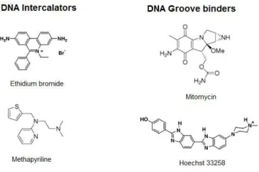
Figure 1.17 The three main binding modes of small molecules with the DNA with biomedical interest for drugs containing DNA-binding units (A-intercalation; B-minor groove binding;
C-major groove binding) ... 24
Figure 1.18 Molecular structure of some DNA-binding molecules56,57 ... 25
Figure 1.19 Molecular structures of acridine orange and antracene ... 26
Figure 2.1 1H-NMR spectrum of ethyl 1-(2-bromoethyl)-1H-pyrazole-4-carboxylate (22) ... 38
Figure 2.2 13C-NMR spectrum of ethyl 1-(2-bromoethyl)-1H-pyrazole-4-carboxylate (22) ... 38
Figure 2.3 1H-NMR spectrum of ethyl 1-(2-(tert-butoxycarbonyl(2-(tert-butoxycarbonylamino)ethyl)amino)ethyl)-1H-pyrazole-4-carboxylate (25) ... 39
Figure 2.4 1H-NMR spectrum of 1-(2-(tert-butoxycarbonyl(2-(tert-butoxycarbonylamino)ethyl)amino)ethyl)-1H-pyrazol-4- hydroxymethyl (26) ... 39
Figure 2.5 ESI-MS spectrum of Re-Ant-(CH2)3-pzNN (Re2) ... 42
Figure 2.6 [1H, 1H] COSY spectra of Re2 in CD3OD (ampliation of the aliphatic region) ... 42
Figure 2.7 1H-NMR spectra of Re2 in CD3OD ... 43
Figure 2.8 [1H, 13C] HSQC spectra of Re2 in CD3OD (ampliation of the aliphatic region) ... 43
Figure 2.9 13C-NMR spectra of Re2 in CD3OD ... 43
Figure 2.10 HPLC trace of the precursor [99mTc(H2O)3(CO)3]+ ... 44
Figure 2.11 HPLC chromatograms of Tc1 and Re1 obtained by γ- and UV-detection, respectively ... 45
Figure 2.12 HPLC chromatograms of Tc2 and Re2 obtained by γ- and UV-detection, respectively ... 45
Figure 3.1 Absorption spectra of solutions containing 50 µM of L2 (left) or 100 µM of Re2 (right) in PBS (10 mM, pH = 7.2) with different equivalent amounts of CT-DNA. ... 47
Figure 3.2 Fluorescence spectra of solutions containing 8 µM of L2 (left) or Re2 (right) in PBS (10 mM, pH = 7.2) with different equivalent amounts of CT-DNA ... 48
Figure 3.3 Stability of Tc2 in presence of histidine in PBS (pH = 7) after 2 hours of incubation at 37°C ... 50
Figure 3.4 Stability of Tc2 in presence human serum after 6 hours of incubation at 37°C ... 51
Figure 3.5 Cell internalization of Tc1 in function of time, after 4 hours incubation at 37°C, in B16-F1 cells (left) and PC-3 cells (right) ... 53
Figure 3.6 Cell internalization of Tc2 in function of time, after 4 hours incubation at 37°C, in B16-F1 cells (left) and PC-3 cells (right) ... 53
Figure 3.7 Cell uptake of Tc1 and Tc2 as a function of time, after 4 hours incubation at 37°C, in B16-F1 and PC-3 cells ... 54
Figure 3.8 Cellular, nuclear and mitochondrial uptakes of Tc1 and Tc2 by B16-F1 and PC-3 cells per million cells after 2 hours incubation at 37°C ... 56
Figure 3.9 Cytotoxicity of L1 and Re1 at different concentrations, after 40 hours incubation at 37°C, in B16-F1 cells (left) and PC-3 cells (right) ... 58
Figure 3.11 Radio-cytotoxicity of Tc1 at different activities, after 40 hours incubation at 37°C, in
B16-F1 cells (left) and PC-3 cells (right) ... 60
Figure 3.12 Radio-cytotoxicity of Tc2 at different activities, after 40 hours incubation at 37°C, in B16-F1 cells (left) and PC-3 cells (right) ... 60
List of Schemes
Scheme 1.1 Simplified scheme of 99Mo/99mTc generator nuclear decays (adapted)31 ... 13Scheme 1.2 Aqueous-based kit preperation of fac-[99mTc(H2O)3(CO)3]+ precursor by Alberto et al, 200136 ... 14
Scheme 2.1 Synthesis of Ant-CH2-pzNN (L1) ... 31
Scheme 2.2 Synthesis of the N-hydroxysuccinimine (NHS) activated ester pyrazole-diamine derivative (13) ... 33
Scheme 2.3 Synthesis of 3-(anthracen-9-yl)propan-1-amine (14) ... 34
Scheme 2.4 Syntheis 10-(3-aminopropyl)-3,6-bis(dimethylamino)acridinium (18) ... 34
Scheme 2.5 Synthesis of Ant-(CH2)3-pzNN (L2) and AO-(CH2)3-pzNN (L3) ... 35
Scheme 2.6 First strategy for the synthesis of AO-CH2-pzNN (L4) ... 36
Scheme 2.7 Second strategy for the synthesis of AO-CH2-pzNN (L4) ... 36
Scheme 2.8 Synthesis of tert-butyl 2-(2-(4-(hydroxymethyl)-1H-pyrazol-1-yl)ethylamino)ethylcarbamate (24) and 1-(2-(tert-butoxycarbonyl(2-(tert-butoxycarbonylamino)ethyl)amino)ethyl)-1H-pyrazol-4- hydroxymethyl (26) ... 37
Scheme 2.9 Synthesis of 4-(bromomethyl)-1-trityl-1H-pyrazole (28) ... 40
Scheme 2.10 Synthesis of Re-Ant-CH2-pzNN (Re1) by reaction of L1 with the precursor [Re(CO)5]Br ... 41
Scheme 2.11 Synthesis of Re-Ant-(CH2)3-pzNN (Re2) by reaction of L2 with the precursor [Re(H2O)3(CO)3]Br... 41
List of tables
Table 1.1 Commonly used β+
emitters for PET imaging6 ... 3
Table 1.2 Commonly used γ-ray emitters for SPECT imaging6 ... 4
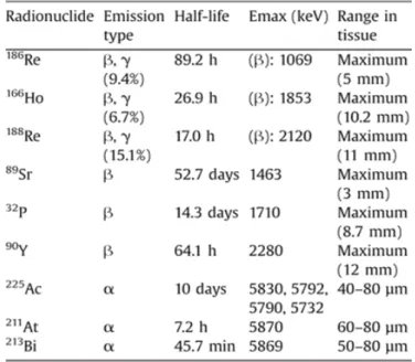
Table 1.3 Examples of α- and β-emitters radionuclides for therapeutic applications3 ... 6
Table 1.4 Examples of Auger electron emitters with potential interest for radiotherapy10 ... 9
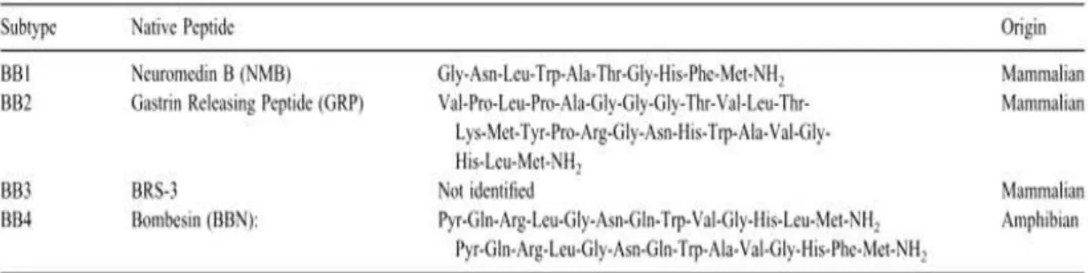
Table 1.5 Bombesin receptor subtypes and the respective native peptide sequences77 ... 28
Table 1.6 Exemples of nuclear localization sequences of the different classes80,81 ... 29
Table 3.1 Intrinsic binding constants (k) of L1, Re1, L2 and Re2 ... 48
Table 3.2 Stern-Volmer constant (Ksv) of L2 and Re2 ... 49
Table 3.3 Partition coefficient (log Po/w) values of Tc1 and Tc2 ... 49
Table 3.4 Cellular, nuclear and mitochondrial uptakes of Tc1 in B16-F1 and PC-3 cells after 2 hours incubation at 37°C ... 55
Table 3.5 Cellular, nuclear and mitochondrial uptakes of Tc2 by B16-F1 and PC-3 cells after 2 hours incubation at 37°C ... 55
List of symbols and abbreviations
ACN - Acetonitrile
Ant - Anthracene
AO - Acridine orange
aq - Aqueous
BBN - Bombesin
BFC - Bifunctional chelator
BM - Biomolecule (s)
BOC - tert-Butyloxycarbonyl
Boc2O - Di-tert-butyl dicarbonate
br- Broad
C2TN - Centro de Ciências e Tecnologias
Nucleares
CD3OD - Deuterated methanol
CDCl3 - Deuterated chloroform
Ci - Curie
cpm - Counts per minute
CR group - Ciências Radiofarmacêuticas
group
CT-DNA - Calf Thymus DNA
d - Doublet
DCM - Dichloromethane
DIPEA - N,N-Diisopropylethylamine
DMF - Dimethylformamide
DMSO - Dimethyl Sulfoxide
DNA - Deoxyribonucleic acid
DSB - Double strand break
EDC - 1-Ethyl-3-(3-dimethylaminopropyl)-
carbodiimide
ESI/MS - Electrospray ionization Mass
spectrometry
Et - Ethyl
Et2O - Diethyl ether
EtOH - Ethanol
fac -facial
h - Hour (s)
HPLC - High-performance liquid
chromatography
IC - Internal conversion
IST - Instituto Superior Técnico
IT - Isomeric transition
K - Intrinsic binding constant
LET - Linear energy transfer
Log P - Partition coefficient
m - Multiplet
MeOH - Methanol
Met - Methyl
min - Minute (s)
MTT - 3-(4,5-dimethylthiazol-2-yl)-
2,5-diphenyltetrazolium bromide
NHS - N-Hydroxysuccinimide
NLS - Nuclear Localization Sequence
[xx]
O-PPh3 - Triphenylphosphine oxide
PBS - Phosphate Buffered Saline
PET - Positron emission tomography
PPh3 - Triphenylphosphine
ppm - Parts per million
pz - Pyrazole
pzNN - Pyrazole-diamine
q - Quartet
r.t. - Room temperature
Rf - Retardation factor
ROS - Reactive oxygen species
rpm - Rotation per minute
s - Singlet
SPECT - Single photon emission computed
tomography
SSB - Single strand break
t - Triplet
t1/2 - Half life time
TFA - Trifluoroacetic acid
THF - Tetrahydrofuran
TLC - Thin layer chromatography
tR - Retention time
Tr - Trityl
UV - Ultraviolet
UV-Vis - Ultraviolet Visible
α - Alfa
β - Beta
γ - Gamma
δ - Chemical shift
1.1
General aspects about Nuclear Medicine
Nuclear Medicine is a specialized medical area for development and application of pharmacological agents (carrier) carrying radioactive atoms (radioisotopes) that emit penetrating and/or non-penetrating radiation. Both components, the carrier and the radioisotope, constitute the radiopharmaceutical agent and are determinant for its effectiveness. The characteristics of the carrier should provide tissue/organ specificity and good clearance from the organism, while the radionuclide should provide adequate amount and type of energy for the intended procedure. The different characteristics of the radiation emitted by the radioisotopes define if they can be applied for diagnostic or therapeutic purposes. However, a given radionuclides can be used to obtain therapeutic and diagnostic radiopharmaceuticals, and can also provide in vivo monitoring of therapeutic procedures. Its half-life (t1/2) is also very
important for the data acquisition in diagnosis and for the dose-limiting in therapy.1–3 Theranostics is another, very attractive, modality for the use of the radiopharmaceutical agents, as many radionuclides or matched/pairs of radionuclides have physical properties suitable both for imaging and therapeutic applications.
Depending on the mode of their biodistribution, the radiopharmaceutical agents can be classified as perfusion or 1st generation radiopharmaceuticals and target-specific or 2nd generation radiopharmaceuticals. The biodistribution of the former depends primarily on its
[2]
Figure 1.1 Representation of a target-specific or 2nd generation radiopharmaceutical and its target4
The targets for the target-specific radiopharmaceuticals are usually receptors but they can also be other biological entities. The BMs are referred as vehicle molecules and they can be enzyme substrates, agonists or antagonists for receptors and transporters or metabolites. A linker is usually used to minimize eventual structural-related changes in the properties and specificity of the BM with the introduction of the radionuclide. It can also be used as pharmacokinetics modulator of the final radiopharmaceutical.
The final radiopharmaceutical should have good physical-chemical properties and pharmacokinetics, preferably with high metabolic stability; short but effective biological half-life, which represents the time needed for half of the radiopharmaceutical disappear from the biologic system; high specificity for the target and minimal accumulation in non-target tissue (ensured by the carrier); and should be easily produced with ready and cheap availability in clinical facilities (highly dependent on the t1/2 and availability of the radionuclide). If it is a radiotherapeutic agent it should also present emission of ionizing particles that can cause damage to the target biological system.1,4,5
The use of radiopharmaceuticals for cancer diagnosis and/or therapy is of great interest. Early detection and targeted therapy can be provided (and possibly monitored simultaneously); however, as we will see throughout this thesis, it is a very demanding and complex process involving multiple areas such as molecular biology, organic and inorganic chemistry, biomedicine, etc.
1.1.1
Diagnosis in Nuclear Medicine
The big majority (> 90%) of the existing radiopharmaceuticals are for diagnostic applications, through molecular imaging procedures. This procedure is non-invasive and allows in vivo
are obtained through externally applied radiation, in Nuclear Medicine, the images are obtained by equipments that detect the tissue-penetrating radiation of the administrated (usually intravenously) radiopharmaceutical from the inside the body. Two imaging modalities, single photon emission computed tomography (SPECT) and positron emission tomography (PET) are the basis of Nuclear Medicine. These techniques are often coupled to computed tomography (CT) or to magnetic resonance imaging (MRI) to give hybrid imaging systems such as PET/CT, SPECT/CT or PET/MRI. This is because the SPECT and PET techniques have poor anatomic special resolution, contrary to the CT and MRI. The hybrid techniques provide acquisition of both anatomical and molecular/functional information with high resolution.1–3,6
The radionuclides for PET imaging decay by emission of positron. This positively charged particle is annihilated by an electron in the matter originating two 511 keV γ-ray at opposite directions which are then detected simultaneously.
Table 1.1 Commonly used β+ emitters for PET imaging6
[4]
Table 1.2 Commonly used γ-ray emitters for SPECT imaging6
The radionuclides for SPECT imaging are γ-photon emitters with an energy range about 30-300 keV (minimal and maximum energies detectable using the commonly available collimators) with an optimal range around 150 keV. The collimator is positioned in front of the gamma camera and it defines the angle of incidence of the emitted γ-rays. In SPECT, the detection system is less sensitive and less efficient than in PET, thus presenting images with lower resolution. The major advantage for the use of SPECT over PET is the higher availability of suitable radionuclides, with longer half-lives at low cost. However, the SPECT radionuclides are mostly metallic elements (Table 1.2) requiring different and more difficult approaches for the synthesis of efficient radiopharmaceuticals compared to PET (see Section 1.3).1,6
1.1.2
Therapy in Nuclear Medicine
1.1.2.1 Biological effects of ionizing radiation
Figure 1.2 Direct vs indirect effects of ionizing radiation on DNA molecule7
In direct effect the energy is directly transferred to the target molecule while in indirect effect
an intermediate molecule is ionized leading to generation of highly reactive species which react with the target. The most important intermediate molecule for indirect effect is the extremely abundant H2O (about 80% of the cell mass) which produces reactive oxygen species (ROS), especially the hydroxyl radical (OH.), by radiolysis. It is estimated that the indirect effect occurs twice as much as the direct effect of radiation.7,8
The main DNA lesions introduced by ionizing radiation are bases and sugar damage, single strand breaks (SSB) and double strand breaks (DSB). The damages can lead to cell death by necrotic or apoptotic activation pathways. The presence of scavengers or DNA-repair mechanisms may reduce significantly the potential of radiation damage. But they are not very effective for high radiation deposition which is associated with the direct effect and DSB lesions and present reduced rate of cell survival.7
1.1.2.2 Systemic cancer therapy in Nuclear Medicine
Cancer is one of the most common and challenging diseases of today’s society. The search for
highly specific and efficient therapeutic agents is incessant with two major modalities: chemotherapy and radiotherapy. The reduced blood supply and biological transformation usually observed in cancer cells are limiting for systemic targeted therapy.9 Systemic radiotherapeutic procedures in Nuclear Medicine are still less developed, and the majority of the available radiopharmaceutical agents are yet at pre-clinical stage. This field is more challenging due to the poor availability of suitable therapeutic radionuclides and the need of developing highly specific delivery systems. The lack of specificity makes the determination of the administration dose a very difficult task.5,10,11
[6]
radionuclides for therapeutic use: the α-emitters, β-emitters and Auger emitters. The overwhelming majority of the current radiotherapeutic agents in clinical use are β-emitters. Until recently, only α- and β-emitters radionuclides have been explored for therapeutic application (Table 1.3) but nowadays the potential of Auger electrons is gaining more attention.3,10,12
Table 1.3 Examples of α- and β-emitters radionuclides for therapeutic applications3
Besides the type of energy, the three classes of therapeutic radionuclides differ in the magnitude of the linear energy transfer (LET) and therefore in radiation range/penetration and
biological effects. LET corresponds to the average energy deposition per unit distance along the path of the radiation and it is expressed as keV/ µm. The density of the energy deposition in biological tissue or cell determines the biological effectiveness of the radiation.7
β-particles have low LET and long path length in order of millimeters, compared to the 20 µm diameter of the average cell. The biological effects are due to indirect effects. The LET of these particles is so low that the targeted therapy towards a specific macromolecule is not viable, because the energy deposition is not high enough to cause significant damage. The long range of β
particles can be advantageous for the treatment of big metastases even if there is a non-uniform drug distribution. The big disadvantageous is that there is an enhanced irradiation of the surrounding healthy cells.
Figure 1.3 Representation of the path lengh of the α-, β- and Auger radiation at cellular and subcellular levels. The Auger emitter requires close proximity to the target in order to have high biological
effectiveness
Similarly to the α-particles, Auger electrons present high LET but lower range, in order of nanometers (<100 nm), and very short penetration in biological systems. It is also suitable for the treatment of small metastases at single-cell level, preferentially by direct energy deposition on a cell target-component such as the DNA (Figure 1.3). Contrary to the case of β- emitters, the high LET of α- and Auger emitters allow higher biological effectiveness by causing significant DSB through direct effect on the DNA molecule. The short range would provide a more efficient target therapy for the radiopharmaceuticals with minimal burden for the healthy tissue.10,12
1.2
Auger electrons and cancer therapy
1.2.1
Auger electrons
Daughter nuclides in excited states may be generated after radioactive decay by the parent radionuclides. The de-excitation by isomeric transition (IT) to the ground state may occur by γ -ray emission or by direct transfer of photons to an orbiting electron leading to its ejection in a process called internal conversion (IC) (Figure 1.4, left). This process creates a vacancy in the internal atomic shell. Another process that also generates this gap is the electron capture process in which an electron is transferred from the internal atomic shell into the nucleus.1,7,10,13
[8]
shell but the filling electron comes from the same shell of the vacancy or if both the filling and ejected electrons came from the same shell of the vacancy, respectively. All these low-energy electrons are generally referred as Auger electrons.10,13
Figure 1.4 Left: schematic representation of internal conversion (IC) process. The de-excitation of the radionuclide may occur by γ-ray or IC electron emission. The gap created can be filled by an electron of a higher electronic shell, with energy release.1Right: schematic representation of Auger electron emission.
A gap in eletronic shell originated by IC or by electron capture is filled by an electron of a higher shell. The energy is transfrerred to another electron of the higher shell which is emitted.
1.2.2
Prospective Auger emitters for cancer therapy
The recent interest for Auger electron emitters in nuclear medicine is because α and β
emitters failed as therapeutic agents for the treatment of small metastases due to the lack of ideal nuclides, low specificity or high potential for causing unwanted side effects. The therapeutic potential of Auger electrons was once neglected because of its short range, low penetration and low energy yield per decay. But since Carlson and White14 in 1963 demonstrated that the Auger-emitter 125I could cause molecular fragmentation, and with the advances in molecular biology, targeted therapy at subcellular level gained much interest and potentiated the exploitation of Auger emitting radiopharmaceuticals. The use of Auger electrons emitters for targeted therapy presents great advantages. The short range and high LET (4-26 keV/μm)
provide high effectiveness in the irradiation of the target with low toxicity for the non-target molecules/cells.10,13,15
The DNA is the main target for targeted therapy at subcellular level and most of the cancer therapeutic agents prevent or reduce tumor grow by interfering with the DNA integrity directly or through inhibition of DNA enzymes. The radiotoxicity of Auger electrons by indirect effect
towards DNA molecule has been reported and it’s the most common effect caused by this type
nuclide to the molecule target and with high specificity and clearance from the body in order to reduce the dose limiting toxicity. This would provide administration of adequate doses without accumulation in non-target tissue and efficient tumor-cell killing as the absorbed doses tend to be non-uniform. In other words, this type of radiopharmaceuticals require uptake by all the cells of the tumor in order to exert its therapeutic functions.13,16
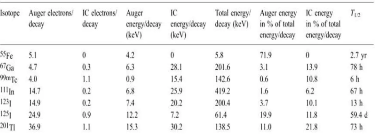
Table 1.4 Examples of Auger electron emitters with potential interest for radiotherapy10
Data from American Association of Physicists in Medicine (AAPM). The total energy per decay also includes γ and X-ray contributions.
The efficiency of Auger emitter-based radiopharmaceuticals is also dependent on the characteristics of the radionuclide. As seen in Table 1.4, there are different Auger emitters of different chemical nature (halogens and metals) with different energy range and Auger electrons yield per decay. The IC electrons energies are presented separately because, despite being higher than those of most Auger electrons, IC electrons present low LET and high penetration
(in μm range), thus presenting different biological effectiveness. Herein we only consider the
Auger electrons for therapy at single-cell level. Other characteristics like half-lives, availability or suitability for functionalization are also critical for the use of such radionuclides. For example, 55
Fe present one of the highest Auger electrons yield per decay but its t1/2 makes it unsuitable for clinical use.10
The Auger emitters 111In, 125I, and 123I are already considered as potential therapeutic radionuclides showing great results in in vitro cytotoxic studies by causing DSB or SSB in DNA.
The toxicity at single-cell level is comparable to that of α and higher than that of β radionuclides
as the low LET of the latter provide predominantly non-specific irradiation effects.13,17–19 Still, the reduced availability (cyclotron produced radionuclides)1 is limiting for the use of these radioisotopes.
[10]
the distance augmentation and the shift on the mechanism, as well as on the magnitude of DSB yield, was quite obvious. The evaluated compounds were derivatives of the DNA groove binder Hoechst 33258, which were labeled with the radionuclide 125I. The compounds only differed on the spacer size between the DNA binder and the radionuclide, which positioned the latter at different distances from the DNA molecule as estimated by molecular modeling. They noticed that in absence of the *OH scavenger dimethyl sulfoxide (DMSO), all the compounds were able to cause DSB although the DSB yield was lower for those compounds that placed the 125I at higher distances from the DNA. However, in the presence of DMSO, the DSB yield remained nearly unchanged for the compounds that ensured closer proximity of 125I to the DNA while the ones with higher 125I distances basically were not able to cause DSB. They concluded that for Auger emitters, the occurrence of direct effect requires a closer proximity at certain distance, even if the potential radiopharmaceutical is bound to the DNA target molecule.
1.3
Design of metal-based radiopharmaceuticals
As seen previously, there is available a large variety of metallic radionuclides with favorable features to be used in the development of diagnostic and/or therapeutic radiopharmaceuticals. For this reason, inorganic chemistry is an important area for the development of radiopharmaceuticals. In fact, most of the available radiopharmaceuticals are 1st or 2nd generation metal-based agents. The metallic radionuclides present great advantages because of its high availability at low cost, rich coordination chemistry and generally adequate characteristics such as half-lives, magnitude/type of the energy emitted or particle emission.5,11,21
The major limitation for the use of these radionuclides is its incorporation into the pharmacologic agents to obtain specific and efficient final radiopharmaceuticals. For design of target-specific compounds, usually a bifunctional chelator (BFC) is needed to provide the attachment of the radiometal to the BM (Figure 1.5).
Figure 1.5 Schematic representation of the bifunctional chelator (BFC) approach
radiometal to the carrier to obtain kinetically inert and termodinamically stable complexes to avoid unbundling or transchelation by the natural chelators, which would lead to accumulation in non-target tissue. It also must retain the specificity of the BM for its target, which is usually ensured by linkers of appropriate length to move away the BM from the radioactive metal. The synthesis of the BFC usually includes the functionalization with the BM before coordination with the radiometal. Both steps must be feasible with reasonable cost and production time, but the latter is limiting, meaning rapid complexation kinetics and high purity of the final radio-conjugate is needed for viable application. Thus, the BFC should provide versatility for chemical conjugation with both radiometal and BM and high selectivity for the radiometal.2,5,11,21–23
1.4
Technetium
1.4.1
Basic chemical and radiochemical aspects
Technetium, from the Greek technetos (artificial), is the first artificially made chemical element
by man before its discovery in nature. It was first predicted by Mendeleev in 1869 as a second row transition metal with the atomic number of 43 but only discovered in 1937 by Perrier and Segrè, from a molybdenum plate bombarded with deuterons accelerated in a cyclotron. This metallic element belongs to the group 7 of the Periodic Table, presenting eight different oxidation states (+7 to -1) and diverse coordination chemistry that allows coordination with several chelators or BFC ligand systems with different denticity.24,25
At least thirty-five isotopes of Tc (86Tc-120Tc) have been identified and none of them is stable.26 The most relevant Tc radioisotope in Nuclear Medicine is the 99mTc, being applied in over 70% of all procedures in this field. This metastable radionuclide has almost ideal nuclear properties for medical imaging and till date it has only been applied for diagnosis. 99mTc emits a highly
abundant 140 keV γ-ray (90%), very close to the optimal energy required for detection by the
gamma cameras, hence providing images with high resolution (see Figure 1.6). This energy is ideal to penetrate biological tissue without overexposure of the patient to radiation. Also, the radiation from the daughter nuclide is not preoccupant as the β
[12]
Figure 1.6 Schematic representation of SPECT imaging with the brain perfusion agent 99mTc-ethyl cysteinate dimer (ECD, or Bicisate) in patient. Right: molecular structure of ECD; Center: schematic representation of a gamma camera image acquisition; Left: SPECT image obtained 12 hours after ECD
administration in a patient with a cerebral embolism (adapted)27–29
The favorable features, together with the ready and inexpensive availability of 99mTc by elution of the 99Mo/99mTc generator and introduction of kits that allow fast and effective synthesis of the radioconjugates (Figure 1.7), justifies the success of 99mTc in diagnostic Nuclear Medicine.1,25,30,31
Figure 1.7 Obtention of 99mTc from a 99Mo/99mTc generator followed by 99mTc radiopharmaceutical preparation and administration in patient (adapted) 30
Scheme 1.1 Simplified scheme of 99Mo/99mTc generator nuclear decays (adapted)31
Scheme 1.1 summarizes the relationship between the nuclear decay of the radionuclides present on the 99Mo/99mTc generator. 99Mo decays with a half-life of 66 hours by β- emission to 99m
Tc (≈87%) and 99
Tc (≈13%). The metaestable 99m
Tc (t1/2 = 6h) decays by isomeric transition of 140 keV to the ground state 99Tc (t1/2 = 2.1 x 10⁵ years) which decays to the stable 99Ru by β -emission.1,30,31
The concentration of [TcO4]- obtained from each elution is usually about 10-7 to 10-10 M, thus the synthesis of the radiopharmaceutical agents has to be done directly from the eluate aqueous solution. As the 99mTcO4- is at +7 oxidation state it first has to be reduced to lower oxidation states by adequate reducing agents under adequate reaction conditions (temperature, pH, etc.), in order to react with the ligands to produce the radiopharmaceutical agent. The final 99mTc oxidation state determines the choice of the donor atoms and denticity of the chelator, as well as the type of metallic core.25 Several different cores with different oxidation states and ligands of different denticity were used to obtain a variety of 99mTc agents applied in diagnostic imaging of brain, skeleton, heart, lung, bone marrow, infection, inflammation, etc., as described in the literature.25,29,31–33 Some examples of the current approved 99mTc-radiopharmaceuticals for kidney, brain and heart imaging can be seen in Figure 1.8.
Figure 1.8 Molecular scrutures of some of the currently approved perfusion 99mTc-radiopharmaceuticals29
-[14]
[99mTc(H2O)3(CO)3]+ precursor, which can be obtained directly from [TcO4]-. Initially, the synthesis of this precursor was performed with the reducing agent NaBH4 under 1 atm of CO.34 This method is not very feasible as the gaseous CO source is considered toxic and also limiting for the fast and ready availability of the precursor in commercial radiopharmaceutical kits. Today
this problem is overcome by the use of a boranocarbonate reducing agent K2[H3BCO2] that works, simultaneously, as a source of CO for the precursor (see Scheme 1.2).35 The available
kits allow fast (20-30 min) preparation of the tricarbonyl precursor in aqueous solution with high
yield (>95%), in a single step.
Scheme 1.2 Aqueous-based kit preperation of fac-[99mTc(H2O)3(CO)3]+ precursor by Alberto et al, 200136
The fac-[99mTc(H2O)3(CO)3]+ precursor is air and water stable in a broad range of pH (1-13) for
hours.34 It is an octahedral complex with three CO ligands facially coordinated to the metal center and each one trans to a water molecules. The water molecules are highly labile and can easily be substituted by a variety of functional groups of different mono, bi or tridentate ligands, while the carbonyl groups are strongly bound to the metal center enabling stable and inert fac
-[99mTc(CO)3(ligand)] complexes.34,36
Although none radiopharmaceutical containing the fac-[99mTc(CO)3] core has yet been applied in
clinical trial, this core is of great interest due to the small and compact size, possibility of being stabilized with a large variety of ligand systems, which allows the formation of complexes with tuned physical-chemical properties, namely in terms of hydrophilicity and lipophilicity.36
The characterization of the 99mTc compounds is a limited process due to the very low concentration of this nuclide in aqueous solution. The only method sensitive enough to analyze these much diluted compounds is the HPLC (or other chromatographic methods) coupled to a
γ-radiation detector. Otherwise only by use of 99Tc nuclide it is possible to perform the characterization with conventional analytical methods such as NMR, mass spectrometry (MS) or X-ray crystallography. The comparison of the HPLC chromatograms of the 99mTc compounds with respective 99Tc counterparts allows their chemical identification.25,30,31
99
Tc is a long-lived isomer of Tc, being the most abundant technetium radioisotope. It is available in macroscopic amounts, and is the most significant byproduct of 235U fission. 99Tc is a weak β
emitter (0.292 MeV) with no accompanying γ-radiation, thus the HPLC studies of 99Tc compounds are performed using UV and β
performed under good laboratory practices, using glassware to block the β-
particles and taking appropriate precautions.25,30,31
The chemical identification of 99mTc complexes can be done by HPLC comparison with Re congeners, in alternative to the use of 99Tc compounds. The use of non-radioactive Re as 99mTc analogous is advantageous as it is easier to manipulate without major precautions. The HPLC studies of Re complexes usually involves UV detection.
Rhenium also belongs to the group 7 of the Periodic Table, with atomic number of 75 (Figure 1.9), thus having rich coordination chemistry comparable to that of technetium. The existence of rhenium element, named after the river “Rhine”, was confirmed by Noddack-Tacke in 1925 by X-ray spectroscopy. Soon, this element was being produced in macroscopic scale.24 Re is one of the rarest elements in nature, consisting of a mixture of two isotopes, 185Re (37.4%) and 187
Re (62.6%).31 Due to the lanthanide contraction, the chemistry of Tc and Re is very similar and the respective complexes present similar physical properties, such as size and lipophilicity. Nevertheless, some differences have to be taken into consideration. The technetium compounds are easier to reduce providing faster complexation reactions with the ligands. The rhenium complexes on the other hand are easier to oxidize and more kinetically inert providing more stable complexes.31,36
Figure 1.9 Section of the Periodic Table with the group 7 elements Tc and Re
Rhenium possesses two important radioactive isotopes, 188Re (t1/2 = 17 h; β
-, 2.1 MeV) and 186
Re (t1/2 = 90 h; β
-, 1.1 MeV)-, which can be obtained by elution of the 188W/188Re generator or by neutron irradiation of 185Re, respectively. These metallic radioisotopes have great interest in Nuclear Medicine and are being explored based on coordination or organometallic complexes similar to those of the congener 99mTc, in terms of available cores and chelators.30,31,37
1.4.2
99mTc and cancer theranostics
[16]
the evaluation of the potential therapeutic properties of this radionuclide, as is the case of the present thesis. In particular, these studies intend to prove the relevance of 99mTc for Theranostics, which requires the combination of diagnostic and therapeutic capabilities in the same entity.
As seen in Section 1.2.2 - Table 1.4, the 99mTc nuclide is not the most promising Auger emitter candidate for therapy in terms of yield per decay. 99mTc decays by isomeric transition predominantly by γ-ray emission and the total energy per decay is about 142.6 keV, where only 0.6% of this energy is due to Auger emission and 10.8% due to IC. The number of IC and Auger electrons per decay are about 1.1 and 4, with energies around 15.4 and 0.9 keV, respectively. Only few studies have shown results that indicate some potentiality of this radionuclide for the development of therapeutic agents. Yet, the search is constant and motivated by the fact that along with all the advantages of using an Auger emitter for targeted therapy, 99mTc is the most easily available at low cost among all the known Auger-emitting radionuclides. The cheap and ready availability, versatility, ideal t1/2 and the possibility to monitor the treatment through high resolution imaging are highly favorable characteristics of 99mTc. The fact that 99mTc is widely used in diagnosis, being available a large number of approved radiopharmaceuticals obtained with a variety of chelators and based on a well established radiochemistry, also facilitates the development of new and more efficient radiopharmaceutical agents.10,13 These favorable features prompted several research groups to evaluate 99mTc complexes as potential radiopharmaceuticals for Auger therapy of cancer. Some of the most important results of such research work are presented bellow.
The first reported in vitro study involving pertechnetate as a radiocytotoxic agent was performed
with a bacterial strain of Escherichia coli, K12S, by Silva and co-workers in 1998. The studies
consisted on two assays where the bacteria were directly (in growth medium) or indirectly (separated by glass-wall) exposed to [99mTcO4]-. The glass attenuates efficiently the Auger and IC electrons but not the γ-rays. They have noticed that the cells directly exposed to [TcO4] -presented significant reduced survival fraction relative to the ones exposed only to gamma radiation (5.6 and 65% respectively, after 180 min incubation with a 37mBq/mL final activity). Thus, they concluded that the Auger and/or IC electrons were responsible for the cell cytotoxicity rather than the gamma radiation of the 99mTc decay. The high radio-cytotoxic effect in this case might be because there is greater access to the DNA in prokaryotes. Also, they showed that the introduction of free radical scavengers or metal ion chelators protected the cells against the radiotoxic effects of the 99mTc decay indicating that these effects occur partially through indirect mechanisms.38
In year 2000, Pedraza-López and co-workers reported in vitro DNA damage induced by 99m
(99mTc-HMPAO) and 99mTc-2,5-dihydroxybenzoic acid (99mTc-gentisic acid). As expected, the 99m
Tc-HMPAO diffused into the cytoplasm (19.6%; with final activity ≈ 5.3 Bq per cell) while the 99m
Tc-gentisic acid remained bonded to the cell membrane (25.6%; with final activity ≈ 4.0 Bq per cell). Despite DNA breaks and alkali labile sites were observed in almost 100% of the cells, no cytotoxic effect was noticed. The results are consistent with the fact that, although the radio-conjugates were close to the nucleus, as it occupies a large part of lymphocyte cells, this proximity was not enough to cause solid and irreparable DNA damages.39
The first in vivo study showing cancer therapy properties of 99mTc was performed with
pertechnetate, which was administred to nude mice carrying NIS (sodium/iodide symporter)-expressing neuroendocrine tumor, as reported by Behr and coworkers in 2007. They showed that the volume of the tumors decreased significantly in the presence of the radioactive compound, indicating potentiality of the this Auger emitter in therapy.40 More recently, successful studies with 99mTc incorporated in multifunctionalized gold nanoparticles provide an extending on the use of this nuclide as a targeted therapeutic agent, through a variety of approaches and delivery systems.41
The earlier studies did show that the Auger electrons emitted by 9mTc can induce DNA damage but mainly emphasized the need for new complexes with the ability to target the cell DNA, in order to function as efficient therapeutic agents. Alberto and co-workers have pioneered the studies with such complexes by introducing multifunctionalized 99mTc compounds with the ability to target the cell nucleus and enhanced radiotoxic effects in eukaryotic cells. First they synthesized two 99mTc(I) complexes containing the
fac-[99mTc(CO)3] core coordinated by a
triamine ligand, and functionalized with two different DNA binders, pyrene and anthraquinone (Figure 1.10). They evaluated the ability of these complexes to cause DSB as an indicator of cytotoxicity.12
Figure 1.10 99mTc-tricarbonyl complexes containing pyrene (left) or anthraquinone (right) DNA-binding groups12
[18]
in its direct vicinity. To evaluate the cytotoxicity at cellular level, they functionalized the pyrene-containing 99mTc complex with the SV40 nuclear localization sequence (NLS) peptide to allow nuclear targeting and hence, the proximity to the DNA molecule.42 They showed that the complex was able to accumulate in the nucleus of B16-F1 murine melanoma cells and induce a dose-dependent radiotoxic effect much more significant compared to that of non-nucleus/DNA-targeting complexes (such as [99mTcO4]-). The cytotoxic effects through DNA damage were evident by the necrotic cell death through mitotic catastrophe pathway. Still, the doses of 99mTc radioactivity applied were too high with small/slow uptake, which can be limiting for the use of this radionuclide in therapy. This problem had to be overcome by the use of efficient delivery systems that can provide both fast and high uptake by the cells in order to minimize the applied doses.
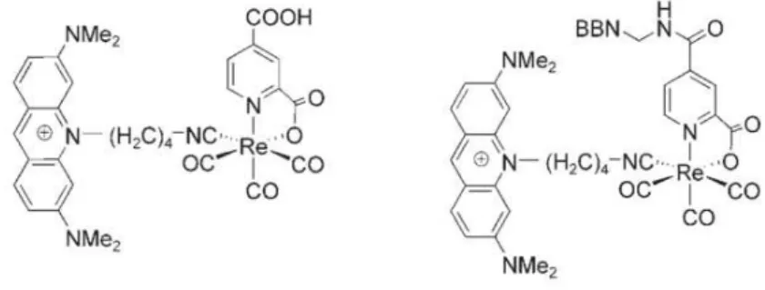
In another study, Alberto and coworkers found out that related tricarbonyl M(I) (M = 99mTc, Re) complexes bearing acridine orange as DNA-binding group were able to target the nucleus of B16-F1 and PC3 human prostate cancer cells without the presence of the NLS peptide.43 The complexes functionalized with a bioactive peptide bombesin (BNN) showed high specific uptake by the PC3 cells through receptor-mediated mechanisms but were not able to reach the nucleus. All the results together indicate that an efficient therapeutic 99mTc radiopharmaceutical has to be at least functionalized with a DNA-binding group, a NLS and a tumor seeking-vector. Another option is the so-called [2 +1] approach, where the bioactive peptide could be released once inside the cell, while the 99mTc complex would then reach the nucleus without the presence of the NLS.44
Figure 1.11 Re-tricarbonyl complexes containing acridine orange as DNA-binding group (left) functionalized with bombesin (BBN) bioactive peptide (right) introduced by Alberto and coworkers43
The group of Ciências Radiofarmacêuticas (CR) from C2TN – IST, where the experimental work on the basis of this MSc thesis has been performed, has also been involved in the study of multifunctional 99mTc tricarbonyl complexes for specific targeting of the nucleus of cancer cells, as summarized below.45–48
Victor et al, 2008, verified by fluorescence microscopic studies that two novel Re(I) complexes
target and enter the nucleus of B16-F1 murine melanoma cells. The characterization of the DNA interaction of these complexes by different spectroscopic techniques has shown a moderate affinity of the compounds towards CT-DNA. Later, in 2009, the authors synthesized the congener complexes with 99mTc and demonstrated that the complexes were also able to enter the nucleus of B16-F1 cells, causing in some cases significant radio-cytotoxic effects and cell death. Most importantly, the radiotoxicity found for these anthracenyl-containing pyrazolyl-diamine 99mTc(I) tricarbonyl complexes containing against B16F1 murine melanoma cells was well correlated with their nuclear uptake. The 99mTc complex having the anthracenyl group at the 4- position of the azolyl ring (complex Tc1, Figure 1.12 (right)) showed the highest
radio-cytotoxicity and induced cell death apparently through caspase-3 apoptotic pathways.
Figure 1.12 99mTc-tricarbonyl complexes containing anthracene DNA-binding group at terminal amine of the pyrazole-diamine chelator (left) or at 4-position of the pyrazolyl ring (right) introduced by Vitor et al48
Esteves et al (2010) have introduced a pyrazole-diamine 99mTc(I) complex bearing acridine
orange (AO) as a DNA binding group, using a butylenic spacer to attach the AO to the 4- position of the pyrazolyl ring. The authors showed that this complex was also able to target the nucleus of B16-F1 cells. Moreover, Esteves et al (2011) further functionalized the AO containing
99mTc(I) complex with the bombesin analogues SGS-BNN and GGG-BNN as tumor-seeking
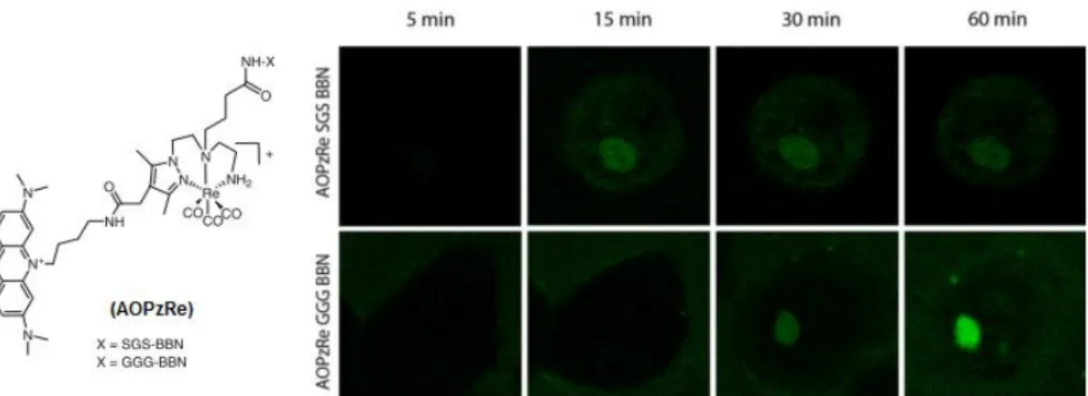
peptides. The Re and 99mTc complexes containing the GGG-BNN showed high cellular internalization and nuclear uptake in PC-3 cells expressing the GRP-receptor (see Figure 1.13). The observed cell uptake was receptor-mediated showing that the presence of the metal-complexes and/or DNA intercalator did not compromise the ability of specific cell targeting by the BBN vector. Another interesting result is the fact that the GGG-BNN complex can still reach the nucleus, most probably following the lysosomal degradation of the BBN peptide. These are the first examples of 99mTc complexes that can simultaneously have specific cell targeting and nuclear internalization, a quite encouraging result to further explore this class of compounds in the design of radiopharmaceutical agents for Auger therapy of cancer. But, unlike the Tc1
[20]
Figure 1.13 Left: Molecular structure of Re-tricarbonyl complexes containing acridine orange DNA-binding group at 4-position of the pyrazolyl ring and functionalized with bombesin analogues. Right: Single-cell fluorescence distribution of the Re-complexes in PC-3 cells, visualized by time-lapse cofocal microscopy
imaging at time points of 5, 15, 30 and 60 min.47
1.5
Background and aim of the thesis
As mentioned before, the potential capability of 99mTc-labeled compounds to simultaneously function as therapeutic and diagnostic chemical entities has gained interest in recent years. To explore this potential quality, also known as theranostic property, several complexes have been synthesized and characterized chemically and biologically. For this purpose, the major challenge is to obtain compounds with selective uptake by tumor cells and with the ability to interact with its nuclear DNA, thus leading to cell death as a result of DNA damage induced by Auger electrons emitted by 99mTc. As this radionuclide is a gamma emitter as well, it could also be possible to monitor the treatment with such complexes using SPECT techniques in patients. Despite some promising in vitro results that have been reported, to date there is no example of
99mTc that has fulfilled the necessary requisites to be used in theranostic applications,
particularly in Auger therapy of cancer.
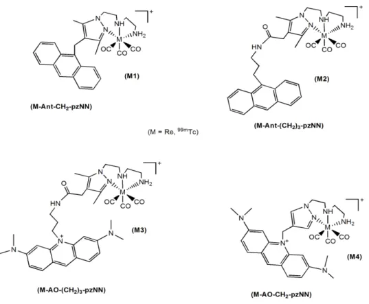
Following some of the previous studies started by the CR group and inspired by several other studies in this field; in this project it was aimed to synthesize, characterize and evaluate biologically 99mTc(I) tricarbonyl complexes stabilized by pyrazole-diamine units functionalized with two different DNA-binding moieties, anthracene or acridine orange. In particular, it was intended to test the effect of the distance between the decaying 99mTc and the intercalator on the DNA damage and radio-cytotoxicity induced by the radiocomplexes. For these studies, it was decided to further evaluate the best performing complex introduced by the CR group, i.e.
Tc1 (see Fig. 1.12) extending its in vitro biological evaluation to PC-3 human prostate cancer
groups as DNA binders and different linkers to attach these groups to the pyrazolyl ring (see Figure 1.14).
Figure 1.14 Molecular structures of the Re and 99mTc complexes proposed in this thesis
After identifying the best performing complex from those proposed in Figure 1.14, a next step would be its functionalization with a bioactive peptide (BM), such as bombesin, and a NLS, as proposed in Figure 1.15. The multi-functionalization with a NLS along with the tumor-seeking vector should improve the ability of the complexes to target the nucleus of tumor cells, in close proximity with DNA, which is a crucial issue to explore 99mTc in Auger therapy.
[22]
Briefly, a 99mTc compound that can combine specific cell targeting, nuclear internalization and significant cell killing due to the Auger electron effect is yet to be introduced. Herein, it was intended to contribute for a better understanding of the structural characteristics (e.g. nature of DNA binder, spacer length, presence of NLS and/or bioactive peptide) that are more determinant of their ability to induce DNA damage and cause radiotoxic effects against tumor cells.
1.6
Rationale for the design and evaluation of the proposed complexes
1.6.1
Pyrazole-diamine BFCs
A large variety of mono, bi or tridentate N, S, P or O-donor ligands can stabilize the fac
-[M(CO)3]+ (M = Re, Tc) core.49–52 Pyrazole derivatives are very versatile, have been widely used for biomedical applications and in some cases can also coordinate this organometallic core.53 Mono and bidentate chelators give M(I) (M = Re, Tc) tricarbonyl complexes that show reduced
in vivo and in vitro stability. By contrast, it has been established that tridentate NNN-donor
ligands give more stable complexes.51 In fact, various studies showed that pyrazole-diamine (pzNN) ligands are ideal tridentate BFCs for the stabilization of fac-[M(CO)3]+, leading to
complexes with high kinetic inertness and thermodynamic stability that are obtained in high specific activity, with high yield and radiochemical purity,.45–52,54 Pyrazolyl-diamine chelators provide three N donor atoms with great capacity to replace the water molecules of the fac
-[M(CO)3]+ (M = Re, Tc) core. More details about its coordination chemistry can be found elsewhere.49,50
Figure 1.16 Schematic representation of different possibilities for functionalization of pyrazole-diamine BFCs with biomolecules and/or DNA-binding groups. The N donor atoms for stabilization of fac-[M(CO)3]+
are also featured.
So far, the reported studies showed that their coordination capability towards the fac-[M(CO)3]+
(M = Re, Tc) is not affected by their functionalization with BMs or DNA. Usually, the resulting radio-conjugates have great in vitro and in vivo stability, and still retain its biological
functions.49,50,52,54 Favorable pharmacokinetics (i.e. fast clearance from the blood, minimal retention in the hepatobiliary tract and kidneys with rapid urinary excretion) have also been reported in mice for several pyrazole-diamine 99mTc tricarbonyl complexes bearing bioactive peptides, such as bombesin or melanocortin analogues.52,55
Herein, we will use pyrazole-diamine ligands containing anthracenyl or AO groups, as DNA binders that are introduced at the 4- position of the pyrazolyl ring using appropriate linkers. Then, the secondary amine can be used for further functionalization with the BBN analogue and with a nuclear localization sequence (NLS).47,52,54 It is worthwhile to mention that in the case of anthracene derivatives of pyrazolyl-diamine ligands it has been shown that the corresponding 99mTc complexes have higher nuclear uptake and enhanced radio-cytotoxic effects on B16-F1
murine melanoma cells when the DNA binder is attached through the azolyl ring, if compared with the complexes having the anthracenyl group at the terminal amine. Studies of the interaction of this type of anthracenyl-containing ligands and their Re complexes with calf thymus (CT) DNA showed that the position used to introduce the DNA binder also influences the way how the compounds interact with DNA.
1.6.2
DNA-binding groups and DNA interaction
As said before, for Auger therapy it is mandatory that the radionuclide stays as close as possible to the target DNA in order to cause a significant damage by direct effect of the Auger electrons. By functionalization of the chelate with an aromatic DNA-binding group it is expected that once inside the cell nucleus, the radio-conjugate will be able to have a strong interaction with DNA and provide an enhancement on the radio-cytotoxicity effect by the short-ranged Auger electrons. The introduction of aromatic DNA-binding group in 99mTc radiopharmaceuticals is also very convenient, as its fluorescence properties allow in vitro studies with the Re
congeners by different microscopy techniques, at cellular and subcellular level where the in vivo
imaging by 99mTc has very poor resolution.43