Diana Isabel Pacheco de Sousa
Bachelor Degree in Biochemistry
Improving the anti-tumor immune responses against
cancer cells
Dissertation to obtain the Master Degree in
Biochemistry for Health
Supervisor: Professor Paula Videira, PhD, FCT/UNL
Co-supervisor: Zélia Silva, PhD, FCT/UNL
Diana Isabel Pacheco de Sousa
Bachelor Degree in Biochemistry
Improving the anti-tumor immune responses against
cancer cells
Dissertation to obtain the Master Degree in
Biochemistry for Health
Supervisor: Professor Paula Videira, PhD, FCT/UNL
Co-supervisor: Zélia Silva, PhD, FCT/UNL
Jury:
President: Professor Maria Teresa Nunes Mangas Catarino, PhD, FCT-UNL Arguer: Ana Lúcia Freitas de Mesquita Barbas, PhD, iBET
Supervisor: Professor Paula Alexandra Quintela Videira, PhD, FCT-UNL
Faculty of Sciences and Technology, NOVA University of Lisbon
iii
Copyright
Improving the anti-tumor immune responses against cancer cells
Diana Isabel Pacheco de Sousa
FCT-UNL, UNL
The Faculty of Sciences and Technology and the NOVA University of Lisbon have the right, forever and without geographical limits, to file and publish this dissertation through printed copies reproduced in paper or by digital means, or by any other mean known or that is invented, and to disclose it through scientific repositories and to allow its copying and distribution for non-commercial educational or research purposes, provided that the author and editor are credited.
v The work developed until the present has originated:
- Two oral presentations:
- Diana Sousa, Paula Videira, Zélia Silva (2016). Improving the anti-tumor immune responses against cancer cells. Jornadas Intercalares das Dissertações anuais dos Mestrados do DQ e do DCV,
January 28th-29th, Caparica, Portugal.
- Diana Sousa, Paula Videira, Zélia Silva (2016). Improving the anti-tumor immune responses against cancer cells. Seminários do Departamento de Ciências da Vida, FCT-UNL, November 16th, Caparica,
Portugal.
- One poster:
- Liliana Loureiro, Carlos Novo, Diana Sousa, Angelina Palma, José Alexandre Ferreira, Ana Barbas, Paula Videira (2016). Development and characterization of a novel antibody against the tumour-associated carbohydrate antigen Sialyl-Tn. PEGS Europe, October 31st to November 4th, Lisbon,
Portugal.
Part of the introduction of this thesis was adapted from an Ebook chapter co-authored by
the author of this thesis and her supervisor, amongst others, already submitted and accepted and with expected publication by the end of 2016:
- Paula A. Videira, Sylvain Julien, Liliana R. Loureiro, Diana P. Sousa, Mylène A. Carrascal, Philippe Delannoy (2016). Vaccine And Antibody Therapy Against Thomsen–Friedenreich Tumor-Associated Carbohydrate Antigens. In Recent Advances in Biotechnology (volume 3): Recent Progress in
vii
ix
“The thing I really want to emphasize is, I didn't have a choice. I didn't have a choice (…) the dream is something you never knew was going to come into your life. Dreams always come from behind you, not right between your eyes. It sneaks up on you. But when you have a dream, it doesn't often come at you screaming in your face, "This is who you are, this is what you must be for the rest of your life". Sometimes a dream almost whispers. And I've always said to my kids, the hardest thing to listen to - your instincts, your human personal intuition - always whispers; it never shouts. Very hard to hear. So you have to every day of your lives be ready to hear what whispers in your ear; it very rarely shouts. And if you can listen to the whisper, and if it tickles your heart, and it's something you think you want to do for the rest of your life, then that is going to be what you do for the rest of your life, and we will benefit from everything you do.”
xi
AKNOWLEDGEMENTS
Everyday my goal is to make someone smile. For that reason, I can’t help but express my most sincere acknowledgements to all those that, in some way, contributed to put a smile on my face every day for the past two years:
To my supervisor, professor Paula Videira, for receiving me in the Glycoimmunology group once again, for allowing me to perform the experimental part of the work on her lab (and for the patience in all the times I asked her if I could go back in her lab, during the first year of the master course), for all the guidance, advice and opportunities.
To Zélia Silva, for accepting the co-supervision of this thesis, for all the teachings about dendritic cells and flow cytometry, and for all the help and discussions during the co-culture procedures and analysis of the results.
To professor Carlos Novo, for all the work and help with the culture of the hybridoma cells. To Liliana Loureiro, for being an excellent (the best!) and patient teacher, for teaching me so many useful tricks, for pushing me over my limits (all those days saying “Come on, come on, come on!”, behind me when I was learning ELISA and was still too slow ); I consider you as my co-supervisor as well.
To all the lab colleagues, the ones that are still present and the ones that already left, for all the help and for all the good and fun moments at the lab: Inês Silva, Graça Marques, Inês Ferreira, Leonor Rodrigues, Márcia Gonçalves, Mauro Monteiro, Mariana Silva, Maria Monticelli, Luiz Neto, Edson Teixeira, Constança Amaral, Carlota Pascoal, Fanny Deschepper, Roberta Zoppi, and especially to Tiago Ferro for all the teachings regarding the isolation of PBMCs and monocytes and Mylène Carrascal for all the teachings regarding the RT-PCR procedures.
To our collaborators, for providing the mouse anti-STn antibodies and also the humanized anti-STn antibodies, without which the work of this thesis could not have been fully performed.
To all my master colleagues, for sharing so many laughs, craziness and despair during our first year, especially during lunch time at ITQB, and for creating memories that I will surely not forget.
To Carla Ferreira, for being the best academic mom ever, for always checking on me and asking if everything was alright and for and showing her support, even with the distance.
To Ricardo (das Neves, HAHA!) Ferreira for reminding me too many times in the last year that I couldn't be a proper scientist if I always put work before sleep, for saying silly (and sometimes serious) things and getting a smile on my face, and for sharing awesome music and getting me into the world of the mad man with a blue box, both of which turned out to be healthy addictions.
To Bruno Guerreiro, the ultimate biochemist (whom I believe will win a Nobel Prize one day - 2087, to be exact), for keeping me going, for always inspiring me to learn more and do more and for showing me that if we work hard enough, we can do whatever we want in life and that everything is possible; for being my special “kid” and always making fun of everything. I’m really proud of you.
xii To Joana Guerreiro, for being one of my best friends, for being there each and every day throughout these two years, for hearing me out, for keeping my thoughts positive and cheering me up when it was necessary, for always giving me a reason to smile and for being a better friend than I could have ever asked.
To Eduardo Marques, for all the support in the final weeks while I was writing this thesis, for the company and the laughs in the library while I worked on the presentation and for making me wonder that sometimes things don’t work out so good on the first time so that they can be better on a second time.
To my family, for all the love, patience and support, for managing through my exhaustion-related moods (especially in these last two months) and for always giving me the choice to follow my heart and do and study what I like in all the moments of my life.
To the coordination and scientific committee of the master course in Biochemistry for Health, for giving me the opportunity to be part of this master course, with a special thanks to professor Teresa Catarino, for all the patience towards my infinite master-related doubts and for all the guidance throughout these two years.
xiii Author’s notes:
1. For confidentiality reasons, the names of the hybridoma clones produced by the group and the names of the humanized anti-STn antibodies provided by collaborators were altered for the presentation of the results in this thesis;
2. The terms ”cancer/tumor”, “T lymphocyte/T cell”, “B lymphocyte/B cell” and “antibody/immunoglobulin” will be used interchangeably throughout this thesis;
xv
ABSTRACT
Cancer is one of the leading causes of death worldwide. Dendritic cells (DCs) can capture cancer antigens and present them to T lymphocytes, after a maturation process, inducing specific immune responses. Sialyl-Tn (STn) is a tumor-associated carbohydrate antigen that is expressed by several carcinomas. STn downregulates the immune responses towards STn-expressing tumor cells by inducing a tolerogenic phenotype on DCs. For this reason, a successful therapeutic approach against this antigen has to take in consideration the activation of the immune response.
The main goal of this thesis was to assess if the use of anti-STn antibodies to block the STn antigen could improve anti-tumor immune responses by reestablishing the mature phenotype on DCs. To do this, STn-expressing cancer cell lines were cultured and DCs were obtained from differentiation of monocytes. Initially, the work comprised the development and characterization of anti-STn antibodies produced by the group through hybridoma technology, using mainly ELISA and flow cytometry. Three hybridoma clones producing IgM anti-STn antibodies were obtained and will be further characterized. A second part of the work included a functional in vitro characterization of
humanized anti-STn antibodies. This comprised the establishment of co-cultures between cancer cells and DCs and assessment of the immune response of DCs against STn-expressing cancer cells, with and without STn blockade by those antibodies. The expression of MHC-II and co-stimulatory molecules was assessed by flow cytometry, and the expression of cytokines was assessed by ELISA and RT-PCR. However, the reestablishment of the mature phenotype was not observed.
The work developed contributed to understanding that further improvements and assays are necessary; and also that the production of antibodies comprises many variants that can be modified throughout the development and characterization procedures, thus being a long process and needing many optimizations before success can be achieved.
Keywords:
xvii
RESUMO
O cancro é uma das maiores causas de morte em todo o mundo. As células dendríticas (DCs) capturam antigénios e, após um processo de maturação, apresentam-nos aos linfócitos T, induzindo respostas imunes específicas. O sialil-Tn (STn) é um carbohidrato associado a tumor, sendo expresso por vários tipos de cancro. Este antigénio reduz as respostas imunes contra as células tumorais que o expressam, através da indução de um fenótipo tolerante nas DCs. Por este motivo, terapias de sucesso contra este antigénio têm de ter em conta a activação da resposta imune.
O objectivo principal desta tese consistiu no uso de anticorpos anti-STn para bloqueio do STn, de forma a avaliar se esse bloqueio restabelecia o fenótipo maduro nas DCs, melhorando as respostas imunes anti-tumorais. Para isso foram cultivadas linhas celulares tumorais que expressavam STn, assim como DCs obtidas da diferenciação de monócitos. Inicialmente, o trabalho envolveu o desenvolvimento e caracterização de anticorpos anti-STn produzidos pelo grupo através de tecnologia de hibridoma, principalmente através de técnicas de ELISA e citometria de fluxo. Foram obtidos três hibridomas com capacidade de produzir anticorpos anti-STn, de isotipo IgM, que serão caracterizados no futuro. A segunda parte do trabalho envolveu a caracterização funcional in vitro de
anticorpos anti-STn humanizados. Para isso foram estabelecidas co-culturas entre células tumorais, bloqueadas ou não com estes anticorpos, e DCs. A resposta imune das DCs foi avaliada através da expressão de moléculas co-estimulatórias e de MHC-II, e também de citocinas, tendo-se para isso utilizado citometria de fluxo, ELISA e RT-PCR. No entanto, o restabelecimento do fenótipo maduro nas DCs não foi observado.
Este trabalho permitiu compreender que são necessários melhoramentos aos protocolos usados e novos ensaios, e também que a produção de anticorpos engloba muitas variantes que podem ser alteradas em várias fases do desenvolvimento, sendo assim um processo demorado e que necessita de bastantes optimizações antes de se ter sucesso.
Palavras-chave:
xix
INDEX
1. INTRODUCTION ... 1
1.1. Cancer ... 1
1.2. Glycosylation ... 2
1.2.1. O-glycosylation ... 3
1.2.2. Sialic Acids ... 4
1.2.3. Sialyltransferases and Sialidases ... 4
1.3. Glycosylation and Cancer: Tumor-Associated Carbohydrate Antigens ... 5
1.3.1. Thomsen–Friedenreich antigens ... 5
1.3.2. Sialyl-Tn Antigen ... 7
1.4. Immunology and Immune system ... 7
1.4.1. Innate immunity ... 7
1.4.2. Adaptive immunity ... 8
1.4.3. Dendritic cells ... 8
1.4.3.1. Antigen processing and maturation ... 9
1.4.3.2. Antigen presentation through the cytosolic pathway: MHC-I ... 9
1.4.3.3. Antigen presentation through the endocytic pathway: MHC-II ... 9
1.4.3.4. Maturation of dendritic cells and antigen presentation – therapeutic potential ... 10
1.4.4. Antibodies ... 10
1.4.4.1. Antibody production in vitro: Hybridoma Technology ... 12
1.4.5. Anti-tumor immunity... 13
1.5. Anti-cancer immunotherapy ... 14
1.6. Introduction to the aim of the thesis ... 14
2. MATERIALS AND METHODS ... 17
2.1 Cell culture ... 17
2.1.1 Culture of breast and bladder cancer cell lines ... 17
2.1.2 Culture of hybridoma cells ... 17
2.1.3 Culture of monocytes and monocyte-derived dendritic cells (moDCs) ... 18
2.2 Isolation of human peripheral blood mononuclear cells by density gradient centrifugation ... 18
2.3 Immunomagnetic isolation and differentiation of monocytes into immature monocyte-derived dendritic cells ... 19
2.4 Magnetic selection of STn positive cells using anti-mouse IgG1 magnetic beads ... 20
2.5 Techniques ... 20
2.5.1 Flow cytometry ... 20
2.5.1.1 General flow cytometry protocol ... 21
2.5.1.2 Sialidase treatment ... 22
2.5.1.3 Characterization of MDA-MB-231, MCF-7 and MCR cell lines – phenotypical analysis of the expression of STn and assessment of the binding of antibodies produced by hybridoma technology 22 2.5.1.4 Characterization of MDA-MB-231 cells – phenotypical analysis of the expression of MHC-II and co-stimulatory molecules; assessment of the binding of humanized anti-STn antibodies and of human IgG1κ isotype control ... 23
xx 2.5.1.6 Establishment of co-cultures of cancer cell lines and immature monocyte-derived dendritic cells ... 24 2.5.1.7 Assessment of the internalization of the humanized anti-STn antibodies ... 26 2.6 Enzyme-Linked Immunosorbent Assay (ELISA) ... 26 2.6.1 Screening of hybridoma clones by indirect ELISA – assessment of antibody production and reactivity against BSM ... 27 2.6.2 Screening of hybridoma clones by indirect ELISA – assessment the production of antibodies by clones that showed no reactivity against BSM ... 28 2.6.3 Isotyping assessment by capture ELISA ... 28 2.6.4 Assessment of cytokines in co-culture supernatants by capture ELISA ... 28 2.7 Real-Time Polymerase Chain Reaction (RT-PCR) ... 29 2.7.1 Analysis, by RT-PCR, of the genetic expression of the cells recovered from the co-culture experiments ... 30 2.8 Statistical analysis ... 31 3. RESULTS AND DISCUSSION ... 33 3.1. Phenotypical characterization of cancer cell lines by flow cytometry ... 33 3.2. Part A: Development and screening of anti-STn antibodies using hybridoma technology ... 35 3.2.1. Analysis of the reactivity of the hybridoma supernatants by indirect ELISA and flow
cytometry……….35
3.2.2. Antibody purification and expansion for large scale production ... 40 3.2.3. Isotyping of the hybridoma clones by capture ELISA ... 42 3.3. Part B: Characterization of humanized anti-STn monoclonal antibodies produced by a biotechnology company ... 43 3.3.1. Titration of humanized anti-STn monoclonal antibodies ... 43 3.3.2. Assessment of the immunological response of DCs: evaluation of the adhesion and maturation status of moDCs after establishment of co-cultures with STn+ and STn- cancer
cells………47
3.3.3. Assessment of the immunological response of DCs: evaluation of the adhesion and maturation status after establishment of co-cultures of dendritic cells with cancer cells coated with the anti-STn
mAb2……….48
3.3.4. Assessment of the internalization of the humanized anti-STn mAbs by the cancer cells ... 52 3.3.5. Assessment of the effect of humanized anti-STn antibody (mAb1) and isotype control (IC) on immature moDCs ... 53 3.3.6. Assessment of the immunological response of DCs: evaluation of the adhesion and maturation status after establishment of co-cultures of dendritic cells with cancer cells coated with the anti-STn
mAb1……….54
xxi
INDEX OF FIGURES
Chapter 1
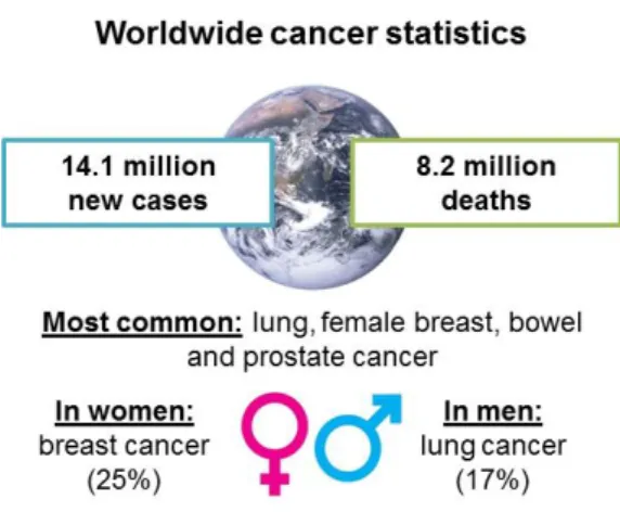
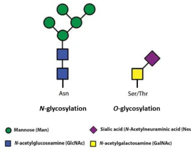
Figure 1.1 – Worldwide cancer statistics: number of new cases and deaths in 2012. Most common cancers (general), and top cancers affecting woman and men. Adapted from [90,91]. ... 1 Figure 1.2 – Schematic representation of the hallmarks of cancer. Adapted from [6]. ... 2 Figure 1.3 – Schematic representation of a N-glycosylated glycoprotein (left) and of a O-glycosylated
glycoprotein (right). Adapted from [92]. ... 3 Figure 1.4 – Structure of N-acetylneuraminic acid (Neu5Ac) [19]. ... 4
Figure 1.5 – Schematic representation of the biosynthesis of Thomsen-Friedenreich antigens. Adapted from [15,17,61]. ... 6 Figure 1.6 – Schematic representation of an immunoglobulin structure (IgG). Adapted from [61]. Abbreviations: C (constant region), V (variable region), L (light chain), H (heavy chain), VL (variable region of the light chain), VH (variable region of one heavy chain), CDRs (complementary-determining regions), Fab (fragment antigen binding), Fc (fragment cristallizable). ... 11 Figure 1.7 – Schematic representation of hybridoma technology [61]. ... 13 Figure 1.8 – Representation of the work-plan proposed for this thesis. ... 16
Chapter 2
Figure 2.1 – Schematic representation of the conditions of the co-culture experiments performed. ... 25
Chapter 3
Figure 3.1 - Phenotypical analysis performed, by flow cytometry, on the MDA-MB-231 STn cells. On all histograms, the yy axis represents the number of cells and the xx axis represents the fluorescence
intensity of each fluorophore used. On figures A-D, the black line represents the negative control (unstained cells) and the grey line represents the staining of anti-CD80-PE (A), anti-CD45-PE (B), anti-CD86-FITC (C) and anti-HLA-DR-APC (D). On figure E, the negative control (cells stained only with the secondary antibody) is represented by the grey filled histogram, as well as the staining of human IgG1κ isotype control in cells treated with sialidase (grey line) and in cells non-treated with sialidase (black line). ... 34 Figure 3.2 – Assessment, by flow cytometry, of the binding of the supernatant from the hybridoma clone 2C5 to the MDA-MB-231 STn cells. On the histograms, the yy axis represents the number of
cells and the xx axis represents the fluorescence intensity of FITC. The grey filled histogram
represents the negative control (cells stained only with the secondary antibody), the black line represents the binding to cells non-treated with sialidase and the grey line represents the binding to cells treated with sialidase. A) Positive control staining with an anti-STn antibody (3F1). B) Staining obtained when using the supernatant of the hybridoma clone 2C5. ... 36 Figure 3.3 – Assessment, by flow cytometry, of the binding of the supernatants from the hybridoma clones 2C5/1D6 and 2C5/2C4 to the MDA-MB-231 STn cells. On the histograms, the yy axis
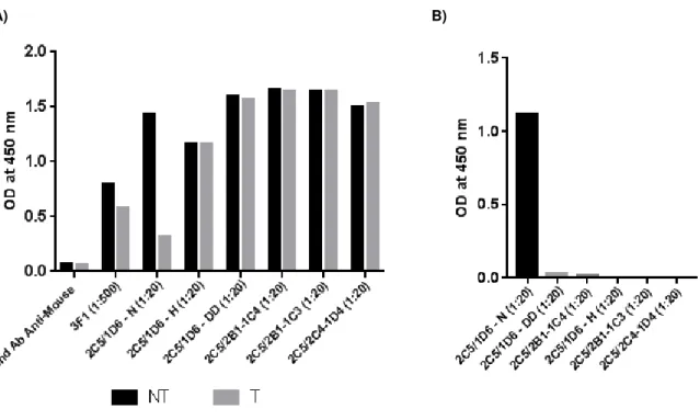
xxii filled histogram represents the negative control (cells stained only with the secondary antibody), the black line represents the binding to cells non-treated with sialidase and the grey line represents the binding to cells treated with sialidase. A) Positive control staining with an anti-STn antibody (3F1). B) Staining obtained when using the supernatant from the hybridoma clone 2C5/1D6). C) Staining obtained when using the supernatant from the hybridoma clone 2C5/2C4). ... 37 Figure 3.4 - ELISA results obtained for the clones resulting from the second limiting cloning of the 2C5/1D6 hybridoma clone. A) ODs at 450 nm obtained for all the clones, for the negative control (BSM with secondary antibody) and for the STn-positive control (3F1 bound to BSM). The ODs obtained for non-treated BSM are represented in black and the ODs obtained for sialidase-treated BSM are represented in grey. B) Method used to select the best clones: difference of the ODs obtained for non-treated and sialidase non-treated BSM for all the clones, ordered from highest (down) to lowest (up). ... 38 Figure 3.5 - ELISA results obtained for the six selected clones. A) ODs at 450 nm obtained for all the clones, for the negative control (secondary antibody) and for the STn-positive control (anti-STn mAb 3F1). The ODs obtained for non-treated BSM are represented in black and the ODs obtained for sialidase-treated BSM are represented in grey. B) Difference of the ODs obtained for non-treated and sialidase treated BSM for all the clones, ordered from highest to lowest. ... 39
Figure 3.6 - Assessment, by flow cytometry, of the binding of the supernatant from the final hybridoma clone 2 (FHC2) to the MDA-MB-231 STn cells. The yy axis represents the number of cells
and the xx axis represents the fluorescence intensity of FITC. The grey filled histogram represents the
negative control (cells stained only with the secondary antibody), the black line represents the binding to cells non-treated with sialidase and the grey line represents the binding to cells treated with sialidase. A) Positive control staining with an anti-STn antibody (B72.3). B) Staining obtained when using the supernatant from the hybridoma clone FHC2. ... 40 Figure 3.7- Assessment, by flow cytometry, of the binding of the supernatant from the final hybridoma clone 1 (FHC1) after culture in serum-free medium to the MDA-MB-231 STn cells. On the histograms, the yy axis represents the number of cells and the xx axis represents the fluorescence intensity of
FITC. The grey filled histogram represents the negative control (cells stained only with the secondary antibody), the black line represents the binding to cells non-treated with sialidase and the grey line represents the binding to cells treated with sialidase. A) Positive control staining with an anti-STn antibody (B72.3). B) Staining obtained when using the supernatant from the hybridoma clone FHC1.41 Figure 3.8 - Isotyping of the final hybridoma clone 1 supernatant by capture ELISA. An IgG1 anti-STn antibody (B72.3) was used as control; a purified IgM antibody was used as positive control; serum-free culture medium was used as negative control. ... 42 Figure 3.9 - Titration, by flow cytometry, of the humanized anti-STn mAbs against MDA-MB-231 STn cells. A) On the yy axis is represented the number of cells and on the xx axis is represented the
xxiii Figure 3.10 - Titration, by flow cytometry, of the humanized anti-STn mAbs against MDA-MB-231 STn cells. On the yy axis is represented the number of cells and on the xx axis is represented the
fluorescence intensity associated with FITC. The red line represents the negative control (cells stained only with the secondary antibody), and the mAbs are represented by the blue (mAb1), the orange (mAb2) and the black (mAb3) lines. The concentrations tested are indicated on the upper right corner of the histogram representations. ... 45 Figure 3.11 - Titration, by flow cytometry, of the humanized anti-STn mAb against MDA-MB-231 STn cells. On the yy axis is represented the number of cells and on the xx axis is represented the
fluorescence intensity associated with FITC. The grey filled histogram represents the negative control (cells stained only with the secondary antibody), and the staining observed with mAb2 is represented by the black line. Two of the five concentrations tested are indicated on the upper right corner of the histogram representations. ... 46 Figure 3.12 – Results, obtained by flow cytometry, for the co-culture assay without anti-STn blocking mAb (n=1). A) Percentage of dendritic cells that adhered to cancer cells. B) MFI values obtained for staining with anti-CD80 antibody. C) MFI values obtained for staining with anti-CD86 antibody. D) MFI values obtained for staining with anti-HLA-DR antibody. ... 48 Figure 3.13 – Results, obtained by flow cytometry, for the co-culture assay with anti-STn blocking mAb2, at a concentration of 0.1 μg/ml (n=2). The error bars represent the standard deviation from the average values. A) Percentage of dendritic cells that adhered to cancer cells. B) MFI values obtained for staining with anti-CD80 antibody. C) MFI values obtained for staining with anti-CD86 antibody. D) MFI values obtained for staining with anti-HLA-DR antibody. ... 50 Figure 3.14 – Ratio between the gene expression of moDCs incubated with MDA-MB-231 STn (cells only, and in the presence of isotype control and blocking mAb2), and the gene expression of moDCs incubated with MDA-MB-231 WT in the same conditions (n=1). A) IL6; B) IL10; C) IL12; D) TNF. ... 51
xxiv incubation of 6 hours. D) MFI values obtained for staining with anti-HLA-DR antibody, after incubation of 24 hours. E) Percentage of dendritic cells that adhered to cancer cells (6 and 24 hours). ... 56
Figure 3.18 – Ratio between the gene expression of moDCs incubated with MDA-MB-231 STn in the presence of blocking mAb2 and the gene expression of moDCs incubated with MDA-MB-231 STn in the presence of isotype control, after 6 (black bars) and 24h (grey bars) (n=3). The error bars represent the standard deviation from the average values. A) IL6; B) IL10; C) IL12; D) TNF. ... 58
Chapter 6
xxv
INDEX OF TABLES
Chapter 1
Table 1.1 – Human antibody isotype characteristics. Adapted from [53–55]. ... 12
Chapter 2
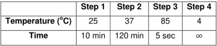
Table 2.1 – Program used in the cDNA synthesis reaction. ... 31 Table 2.2 – Reaction conditions used in the RT-PCR experiments. ... 31
Chapter 3
xxvii
LIST OF ABBREVIATIONS
Abbreviation Meaning
Ab(s) Antibody (Antibodies)
ADCC Antibody-dependent cell-mediated cytotoxicity Ag(s) Antigen(s)
APC Allophycocyanin APCs Antigen presenting cells
Asn Asparagine
BSA Bovine serum albumin BSM Bovine submaxillary mucin
C Constant region
CD Cluster of differentiation
CDC Complement-dependent cytotoxicity cDNA Complementary DNA
CDRs Complementary-determining regions CFG Consortium for Functional Glycomics CMP-sialic acid Cytidine monophosphate-sialic acid
CT Threshold cycle
DC(s) Dendritic cell(s)
DMEM Dulbecco’s Modified Eagle Medium DMSO Dimethyl sulfoxide
DNA Deoxyribonucleic acid
dNTP Deoxynucleoside triphosphate EDTA Ethylenediaminetetraacetic acid ELISA Enzyme-Linked Immunosorbent Assay
ER Endoplasmic reticulum EV Empty vector
Fab Fragment antigen binding FBS Fetal bovine serum
Fc Fragment cristallizable FHC Final hybridoma clone FITC Fluorescein isothiocyanate
fsc Forward-scatter Gal Galactose
GalNAc N-acetylgalactosamine
GAPDH Glyceraldehyde-3-phosphate dehydrogenase GlcNAc N-acetylglucosamine
GM-CSF Granulocyte macrophage colony-stimulating factor H Heavy chain
HAT Hypoxanthine-aminopterin-thymidine HBV Hepatitis B virus
HCV Hepatitis C virus
HGPRT Hypoxanthine-guanine-phosphoribosyltransferase HIV Human Immunodeficiency Virus
xxviii HTLV Human T-lymphotropic virus
IC Isotype control Ig(s) Immunoglobulin(s)
IL Interleukin
IPST Instituto Português do Sangue e da Transplantação
IUPAC International Union of Pure and Applied Chemistry L Light chain
LPS Lipopolysaccharide
mAb(s) Monoclonal antibody (antibodies) MFI Median intensity fluorescence MHC Major histocompatibility complex
MHC-I Major histocompatibility complex, class I MHC-II Major histocompatibility complex, class II moDCs Monocyte-derived dendritic cells
mRNA Messenger ribonucleic acid Neu Neuraminic acid
Neu5Ac N-acetylneuraminic acid
NK Natural killer OD Optical density
OSM Ovine submaxillary mucin
PAMPs Pathogen associated molecular patterns PBMCs Peripheral blood mononuclear cells
PBS Phosphate buffered saline PCR Polymerase chain reaction
PE Phycoerythrin PEG Polyethylene glycol
PRRs Pattern recognition receptors RNA Ribonucleic acid
RT Room temperature
RT-PCR Real-time polymerase chain reaction Ser Serine
ssc Side-scatter STn Sialyl-Tn
T or TF Thomsen–Friedenreich
TACA(s) Tumor-associated carbohydrate antigen(s) Tc Cytotoxic T cells (CD8+)
TE Trypsin-EDTA Th Helper T cells (CD4+) Thr Threonine
TMB 3,3',5,5'-tetramethylbenzidine TNF-α Tumor necrosis factor-alpha
UDP-GalNAc Uridine diphosphate N-acetylgalactosamine
V Variable region
VH Variable region (heavy chain)
VL Variable region (light chain)
1
1. INTRODUCTION
1.1. Cancer
Cancer is one of the leading causes of death worldwide (Figure 1.1), along with cardiovascular and neurodegenerative diseases, diabetes and chronic pulmonary diseases. The number of new cases is expected to increase by about 70% over the next twenty years. Thus, it is important that this disease can be correctly and early diagnosed and that effective treatments that allow its cure or that can considerably prolong the patient’s life are made available [1,2].
This disease results from the accumulation of somatic mutations in the progeny of normal cells, which generates abnormal cells with a selective growth advantage and with an upregulated and uncontrolled capacity of proliferation. Cancer cells can also undergo a process known as metastasizing, in which they gain the ability of invading adjacent normal tissues and organs and spread to distant sites throughout the body, which is the main cause of death related with this disease. These mutations are mostly related with deoxyribonucleic acid (DNA) repair genes, tumor suppressor genes and proto-oncogenes, arising from exposure to physical and chemical agents, from infection with viruses, from genetic predisposition and from behaviors related with known risk factors, like smoking [3,4]. The type of cell from which a tumor arises classifies it: tumors that arise from epithelial cells are named carcinomas, tumors with origin on connective tissues are named sarcomas, tumors that arise from cells of the immune system are called lymphomas and tumors generated from blood-forming cells are named leukemias. They can also be further classified according to their primary site [3].
Several aspects of the cell behavior distinguish cancer cells from their normal equivalents. These aspects, known as the hallmarks of cancer (Figure 1.2), are acquired functional abilities that allow cancer cells to survive, proliferate, and disseminate. Normal tissues ensure the homeostasis of cell growth and proliferation by controlling the production of growth signals. Cancer cells have the
2 capacity of sustaining chronic proliferation by deregulating those signals, an action that also has an influence on cell survival and energy metabolism. In addition, they have the ability to limit or evade apoptosis and express less surface adhesion molecules, which results in morphological and cytoskeletal modifications. Moreover, cancer cells secrete proteases, such as collagenase, that digest components of the extracellular matrix, allowing them to invade the adjacent normal tissues; they can secrete growth factors as well, thus stimulating angiogenesis (formation of new blood vessels), a process needed so that the proliferating tumor cells can obtain nutrients and oxygen. These properties of cancer cells are associated with their ability of migrating to adjacent tissues and to invade nearby blood and lymphatic vessels, which allows them to colonize distant sites and form metastasis [3–6].
1.2. Glycosylation
Glycosylation is an enzymatic process by which glycosidic linkages are created, allowing saccharides to be bound to other saccharides, lipids or proteins, and generating glycoconjugates. The diversity of glycans (term often applied to name the carbohydrate part of a glycoconjugate) derives from the ways in which monosaccharides can be linked together. Differences in monosaccharide composition, substitutions in the existing glycans, the presence or not of branching structures and the different linkages - to other carbohydrates, to proteins or to lipids – all contribute to an increasing diversity of glycans [7,8]. For this reason, a nomenclature strategy was adopted by the Nomenclature Committee of the Consortium for Functional Glycomics (CFG) [9]. This strategy provides a consistent system to represent glycan structures by both symbols and text (see Appendix I).
In eukaryotic cells, most of the secreted and cell-surface molecules originate in the endoplasmic reticulum (ER). They are then distributed from the trans-Golgi network to several
destinations and, in the process, proteins and lipids are modified by a series of glycosylation reactions. However, not all glycans are assembled within the ER-Golgi pathway, as some modifications occur in
3 the cytoplasm and others at the plasma membrane. Nucleotide sugars (activated forms of monosaccharides) are often transferred in glycosylation reactions, which are mediated by glycosyltransferases, to the glycan being synthetized [10]. The biological roles of glycans are broadly classified in four categories, all involving glycan-binding proteins: structural and modulatory roles, intrinsic recognition, extrinsic recognition and molecular mimicry of host glycans [11,12]. The two general classes of protein-bound glycans occur via linkages to oxygen (O-glycosylation) or nitrogen
(N-glycosylation). The process of N-glycosylation generates N-glycans through the binding of N
-acetylglucosamine (GlcNAc) residues to the amide groups of asparagine (Asn) side chains. The process of O-glycosylation generates O-glycans, in which N-acetylgalactosamine (GalNAc) residues
are linked to the oxygen atom of threonine (Thr) or serine (Ser) side chains (Figure 1.3) [7,8]. Because the work developed and presented in this thesis is more focused on O-glycans, more emphasis will be
given to these glycans throughout this chapter.
1.2.1. O-glycosylation
Glycoproteins are often heavily O-glycosylated, particularly serine- and threonine-rich mucins
that constitute mucous secretions and can also be found on the cell surface as transmembrane glycoproteins. The biosynthesis of O-glycan chains occurs in the Golgi apparatus and involves the
sequential action of different glycosyltransferases. Thus, the pattern of O-glycans that is expressed on
a certain cell depends on the level of expression, localization and substrate specificity of the glycosyltransferases. The structures generated by these enzymes can be further modified through processes such as sialylation, acetylation and fucosylation [8,13,14].The initial step of O-glycosylation
involves the transfer of a GalNAc residue from uridine diphosphate N-acetylgalactosamine
(UDP-GalNAc) to a threonine or to a serine in the amino acid chain, generating the Tn antigen (GalNAc-Ser/Thr), which is the simplest mucin O-glycan and will be mentioned later in this chapter. The chain is
then further elongated [15–17].
4 1.2.2. Sialic Acids
One of the modifications than can occur on glycan structures is sialylation, through the addition of monosaccharides, named sialic acids, which comprise a group of approximately 50 different chemical derivatives of neuraminic acid (Neu). In mammalian cells, the most common variant is the N-acetylneuraminic acid (Neu5Ac), with a N-acetyl group on carbon 5 (Figure 1.4) [18,19].
Sialic acids exhibit a significant diversity due to the different alpha linkages that can be formed. They are commonly found in the terminal positions of N- and O-glycans of glycoproteins and
are added by action of specific sialyltransferases that use cytidine monophosphate-sialic acid (CMP-sialic acid) as donors [18,19]. These monosaccharides have several biological roles, such as the stabilization of membranes and molecules and the modulation of interactions of the molecules and cells with the environment; they can also serve as a biological target, allowing its recognition by a receptor protein, or shield antigenic sites and receptors, protecting molecules from the action of proteases or glycosidases and weakening the immune-reactivity [19,20].
1.2.3. Sialyltransferases and Sialidases
Sialylation of glycoproteins is a modification that occurs in glycans and is associated to cancer. The level of cell surface sialylation is regulated by a diverse group of enzymes. This group includes the enzymes that control the synthesis and availability of the activated donor substrate (CMP-sialic acid); sialyltransferases, the enzymes that add the (CMP-sialic acids; and sialidases (or neuraminidases), the enzymes that remove the sialic acids. Most of the sialyltransferases are localized in the Golgi apparatus, since sialic acids are added during the biosynthesis of the glycoproteins, while sialidases are usually found in lysosomes or endosomes, for the removal of sialic acids during the degradation of glycoproteins [18].
Sialyltransferases are a family of enzymes that vary in tissue distribution and on the type of sialic acid linkage that they perform. Sialic acids can be added by ST3Gal-I sialyltransferase in an α2 -3 linkage to galactose (Gal); by ST6Gal-I and ST6Gal-II sialyltransferases in an α2-6 linkage to galactose or by ST6GalNAc sialyltransferases to N-acetylgalactosamine; or by the family of polysialyltransferases in an α2-8 linkage to other sialic acid. Aberrant expression of these enzymes, as well as of sialidases, is observed in cancer [18].
5 1.3. Glycosylation and Cancer: Tumor-Associated Carbohydrate Antigens
Glycosylation can be essential for the regulation of several physiopathological mechanisms. For this reason, variations in the resulting glycoconjugates can interfere with cancer cell processes, such as inflammation and immune surveillance, and with the tumor microenvironment, as they are associated with oncogenic transformation, cancer growth, progression and metastasis. Cancer cells exhibit a diverse series of differences on the levels and types of carbohydrate structures present on their surfaces. These carbohydrate structures allow them to be distinguished from normal cells and are known as tumor-associated carbohydrate antigens (TACAs). Alterations in the glycans present at the surface of the cell can result from abnormal expression and activity of glycosyltransferases or to their incorrect location in the Golgi apparatus [7,21].
The two main processes of tumor-associated carbohydrate alteration are the neo-synthesis and the incomplete synthesis. The first one is commonly observed in more advanced stages of cancer and results of a cancer-associated induction of genes that are related with the expression of carbohydrate determinants, which leads to the de novo expression of antigens in cancer tissue. The
second process is more commonly found in early stages of cancer and results of a deficiency on the normal synthesis of complex glycans that are usually expressed in normal epithelial cells, which leads to the generation of truncated structures in cancer tissues [7,22,23].
The most common alterations in glycosylation are fucosylation, sialylation, truncation of O
-glycans and branching of O- and N-linked glycans. Although all these alterations have been shown to
be related with cancer [24], sialylation and truncated O-glycans will be more explored in this thesis.
Truncation of O-glycans is commonly found in secreted and transmembrane glycoproteins. Sialylated
structures of Thomsen-Friedenreich antigens are examples of truncated glycans [25,26]. The increased expression of sialylated glycans promotes cell detachment from the original tumor site, by disrupting cell-cell adhesion processes, leading to metastasis, which is related with poor prognosis for the patients [7].
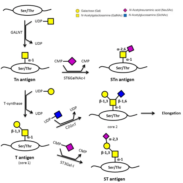
1.3.1. Thomsen–Friedenreich antigens
Thomsen–Friedenreich antigens are O-glycans found mainly on serine- and threonine-rich
mucins, thus being present in membrane glycoproteins. These antigens are a result of a defective elongation of the O-glycan on the first steps of mucin glycosylation [27]. The first Thomsen–
Friedenreich antigen described was called T (or TF) antigen, forms the core 1 structure of mucin O
-glycans and consists of the disaccharide composed by a galactose 1-3 linked to a N
-acetylgalactosamine, -linked to a serine or threonine residue (Gal1-3GalNAc1-Ser/Thr). The T antigen is the precursor of core 2 O-glycans, but if the cancer cells lose the ability to synthesize the
6 lost, its precursor, the Tn antigen, is exposed. This antigen consists of a single N
-acetyl-galactosamine -linked to serine or threonine residue (GalNAc1-Ser/Thr) [15,28]. The Tn antigen is not usually found on secreted or cell-surface proteins in normal tissues, but is found in most carcinomas, being also a pan-carcinoma antigen [30,31]. As with the T antigen, an increased expression of the Tn antigen is related with an enhancement of cancer invasiveness and cellular proliferation and because they are detected on early-stages of cancer development, they have potential for being studied and used as biomarkers [32]. Both T and Tn antigens can be further modified by, for instance, sialylation, which is how the sialyl-Tn (STn) antigen is generated (Figure 1.5).
7 1.3.2. Sialyl-Tn Antigen
The addition of a sialic acid residue to the carbon 6 of GalNAc (α2-6 linkage) on the Tn antigen originates the disaccharide known as sialyl-Tn (Neu5Ac2-6GalNAc1-Ser/Thr), also referred to as CD175s [15,17,33]. Similarly, STn is also a pan-carcinoma antigen: is generally not expressed in normal healthy tissues but is frequently detected in most carcinomas, such as pancreatic, ovarian and lung cancers [7,26,33]. Thus, the expression of this antigen is considered pathologic since it is not a normal biosynthetic precursor, as are the T and Tn antigens. In normal cells, the process of O
-glycosylation generates elongated and branched O-glycans that are often modified by sialylation.
However, the sialylation of the Tn antigen blocks the normal extension of the O-glycan chain, as the
STn structure cannot be further elongated, consequently blocking the synthesis of other glycosidic structures [15,18,26] (see Figure 1.5 in the previous section). Its overexpression results of mechanisms related to ST6GalNAc-I upregulation or the re-localization of this enzyme from the Golgi apparatus to the ER [7,15,25,34–37].
As previously mentioned, the expression of the STn antigen is correlated with increased tumor growth and with the disruption of cell-cell adhesion mechanisms, increasing the processes of migration ad invasion and, consequently, modulating a malignant phenotype and thus being associated with a poor prognosis in cancer patients [7,15,22,33,38,39].
Since STn is expressed early in the process of cancer development and is usually not found in healthy cells, this antigen is considered to be a helpful tumor marker in diagnosis [7,26,33]. Tumor markers can be detected in serum, since it is a simple, sensitive and non-invasive method of diagnosis. However, detection of STn in serum is linked to poor prognosis, as it is related with shedding of cells from tumors into the bloodstream. This is consequently correlated with increased tumor masses found in advanced cancers, thus making the STn antigen more useful as a prognosis than as a diagnosis marker [33].
1.4. Immunology and Immune system
Immunology studies the reactions to foreign substances, ranging from microbes to macromolecules, like proteins and carbohydrates, and the cellular and molecular events that occur after an organism encounters them, a process designated as immune response [40]. The immune system comprises different molecules, cell types, tissues and organs that protect the host by identifying potential threats and defending it against them. The process of host defense relies on the action and cooperation of two types of immunity: the innate immunity, which is not antigen-specific, and the adaptive immunity, which is antigen specific [40–42].
1.4.1. Innate immunity
8 complement system, cytokines and other inflammation mediators. The mechanisms on this type of immunity show a broad specificity and use a variety of pattern recognition receptors (PRRs) to recognize structures that are common in groups of related microbes, known as pathogen associated molecular patterns (PAMPs). One of these structures is, for instance, the lipopolysaccharide (LPS), a component of the external membrane of the cell wall of Gram-negative bacteria. The process of phagocytosis is one of the main mechanisms of innate immunity, enabling the destruction or neutralization of microbes. This process is also important in the activation of the adaptive immunity, as it will be mentioned further in this chapter [40,43].
1.4.2. Adaptive immunity
The adaptive immunity is the second line of defense of the immune system and is present only in vertebrate organisms. It provides a late (days) but specific response to an antigen, after innate signaling. This response is mediated by the action of T and B lymphocytes and can then generate immunological memory and increase in magnitude with repeated exposures to a particular antigen. The antigen can be recognized, in its native conformation, by the receptors of B lymphocytes, whereas the recognition by T lymphocytes occurs via the presentation of antigen-derived peptide fragments in the major histocompatibility complex (MHC) context. The recognition process will then induce the activation and differentiation of B cells, which will produce specific antibodies against the antigen, a mechanism known as humoral immunity; and of T cells, which will generate effector and regulator cells, a mechanism known as cell-mediated immunity. For the development of adaptive immunity it is required that the antigens are captured and presented to lymphocytes, a process performed by antigen presenting cells (APCs). Dendritic cells (DCs) are professional APCs and have a central role in antigen capture and in the induction of T lymphocyte responses against protein antigens, thus being considered an important association between the innate and adaptive immunities [40,43,44]. Due to the significance of these cells in the context of this thesis, they will be further explored in the next section.
1.4.3. Dendritic cells
9 contacted with antigens), instruct them by presenting the processed antigen-derived peptides and thus initiate the generation of a specific immunological response [40,45].
Immature DCs are highly endocytic and process endogenous and exogenous antigens, but have a low stimulatory capacity. The mechanisms involved in the uptake of antigens include phagocytosis, through membrane receptors, endocytosis, through Fc and C-type lectin receptors and pinocytosis [45]. Particularly, immature DCs can be obtained in vitro by culturing monocytes with
granulocyte macrophage colony-stimulating factor (GM-CSF) and interleukin-4 (IL-4) cytokines, thus promoting their differentiation into dendritic cells, being then usually known as monocyte-derived dendritic cells (moDCs) [46,47].
1.4.3.1. Antigen processing and maturation
The mechanism of antigen processing and presentation of antigen-derived peptides is mediated by MHC molecules. In humans, they are referred to as human leukocyte antigen (HLA). These molecules can be of two classes, I and II, designated then MHC-I and MHC-II, respectively. MHC-I is expressed in all nucleated cells and processes and presents endogenous antigens, whereas MHC-II processes and presents exogenous antigens, being expressed by APCs. The activation of T cells occurs through the binding of MHC molecules to the CD28 receptor that is present in those cells [48]. As previously mentioned, DCs possess the ability to capture and process antigens, producing antigenic peptides which will then bind to MHC-I and MHC-II molecules and that will be transported to the cell surface and presented for cellular recognition by T cells. The antigen processing by dendritic cells occurs mainly through two pathways: one cytosolic (endogenous) and one endocytic (exogenous), through which the antigenic peptides are bound to MHC-I and MHC-II molecules, respectively [45,48].
1.4.3.2. Antigen presentation through the cytosolic pathway: MHC-I
In this pathway, the intracellular antigens are bound to molecules of the MHC-I complex and later presented at the cell surface and recognized by CD8+ T cytotoxic (T
c) cells. Therefore, proteins of
endogenous origin, from the self or of pathogenic origin are ubiquitinated and degraded in peptides by the proteasome. The resulting peptides are then transported to the ER, where they are bound to the newly-synthetized MHC-I molecules. Then the MHC-I/peptide complexes are transported to the cytoplasmic membrane through the trans-Golgi network [45,48].
1.4.3.3. Antigen presentation through the endocytic pathway: MHC-II
In this pathway, the antigens that were captured from the extracellular environment by endocytosis, phagocytosis or pinocytosis are bound to molecules of the MHC-II complex and later presented at the cell surface and recognized by CD4+ T helper (T
10 1.4.3.4. Maturation of dendritic cells and antigen presentation – therapeutic potential
In immature DCs, the MHC-II molecules and degraded peptides accumulate in intracellular vesicles. The maturation process occurs upon the uptake of antigens, throughout the migration of DCs from peripheral tissues to lymphoid organs and is promoted by inflammatory mediators. During this process, dendritic cells undergo functional and phenotypical alterations. They are characterized by a reduction in the uptake machinery, losing the capacity to capture antigens, and acquire the ability to present the processed antigens to naïve T cells and stimulate them. After receiving an inducing stimulus, the pH in the endosomes of DCs decreases, easing the processing of exogenous antigens, which are then bound to MHC-II molecules and the MHC-II/peptide complexes migrate to the cell surface. Besides these functional alterations, mature dendritic cells suffer a reorganization of their cytoskeleton, increase the expression of adhesion molecules, chemokine receptors, I and MHC-II molecules, co-stimulatory molecules and pro-inflammatory cytokines. Additionally to the activation of T cells, DCs can also activate B cells, thus having a role in humoral immunity [45,46,48–51]. Since DCs have the potential to initiate both cellular and humoral adaptive responses, their use is currently considered in many anti-cancer therapies in order to improve the immune response against a certain antigen [49]. Immunotherapies regarding DCs often rely on the use of DCs obtained from the patient, which are loaded with cancer antigens ex vivo and injected back to the patient with the goal of
boosting his immune system to fight the cancer cells via expansion of T cells, eliciting a specific immune response towards the tumor [52].
1.4.4. Antibodies
11 The differences in the heavy chains of the constant regions of an antibody molecule determine their functional activity. Therefore, antibodies can be divided into five distinct classes or isotypes, named IgA, IgD, IgE, IgG and IgM (Table 1.1). The heavy chains are labelled by the letter of the Greek alphabet that corresponds to the isotype of the antibody: α, δ, ε,γ, andμ, respectively. Also, two types of light chains exist, called κ and λ, being classified by their carboxyl terminal constant regions. The antibody molecule has two κ light chains or two λ light chains [53,54].
Several antibody-mediated effector functions exist, depending on the isotype, besides their ability to specifically bind to target antigens with high affinity. These functions include the neutralization of microbes or toxic microbial products, the opsonization of antigens for enhanced phagocytosis and immediate hypersensitivity [53,56]. Antibodies can also mediate cell death processes by redirecting immune effector cells, a mechanism known as antibody-dependent cell-mediated cytotoxicity (ADCC), or by activating the complement system (known as complement-dependent cytotoxicity, CDC) and even through blocking actions on specific molecules and soluble mediators. The mechanisms of ADCC and CDC depend on the engagement of Fc receptors that are expressed on effector immune cells or of the Fc region of antibodies, respectively, in order to endorse targeted cell death. All of these characteristics are important mechanisms of action and contributed for the application of antibodies in many therapeutic fields, one of them being anti-cancer therapies [53,54,57,58].
Figure 1.6 – Schematic representation of an immunoglobulin structure (IgG). Adapted from [61].
Abbreviations: C (constant region), V (variable region), L (light chain), H (heavy chain), VL (variable region of the light chain),
VH (variable region of one heavy chain), CDRs (complementary-determining regions), Fab (fragment antigen binding), Fc
12 Table 1.1 – Human antibody isotype characteristics. Adapted from [53–55].
Isotype Subtypes Heavy chain Light chain Molecular weight (kDa) Serum half-life (days) General
structure Functions
IgA IgA1
IgA2
α1
α2 λ or κ 150-600 5-7
Monomer,
dimer, trimer Mucosal immunity
IgD None δ λ or κ 150 2-8 Monomer Naïve B cell antigen receptor
IgE None ε λ or κ 190 1-5 Monomer
Immediate hypersensitivity, protection against parasite worms IgG IgG1 IgG2 IgG3 IgG4 γ1 γ2 γ3 γ4
λ or κ 150 21-24 Monomer
Opsonization, complement activation, antibody-dependent cell-mediated cytotoxicity, neonatal immunity
IgM None μ λ or κ 750-900 5-10 Pentamer, Hexamer
Naïve B cell antigen receptor, complement
activation
1.4.4.1. Antibody production in vitro: Hybridoma Technology
13 hybridoma cells that have antibodies in their culture supernatants that show reactivity towards the antigen of interest) are then cloned and grown so that monoclonal antibodies can in that way be produced, continuously, in vitro (Figure 1.7)[60,61].
1.4.5. Anti-tumor immunity
An appropriate anti-tumor immune response involves the action of both cellular and humoral immune responses. Using the patient’s own immune system for the combat of cancer is becoming more common. This is mainly because the immune system has the potential to destroy specifically cancer cells without offering toxicity to normal tissues, while inducing long-term memory that will allow the prevention of cancer recurrence. Besides having this potential for cancer destruction, is it also known that cancer can suppress the anti-tumor immune responses, leading to cancer cells not being eliminated and continuing to proliferate. This immune suppression is associated with the aberrant expression of TACAs by the cancer cells [33,61,62].
Cytotoxic T cells and NK cells are activated in cellular immune responses in order to eliminate cancer cells by apoptosis. However, as previously mentioned, activation of cytotoxic T cells requires antigen-derived peptides to be presented via MHC-I, which can be a disadvantage on the development of a strong immune response against carbohydrate antigens. Humoral immune responses consist of tumor-specific antibodies, which can then assist on effector functions like antigen neutralization, phagocytosis, ADCC mediated by NK cells and complement-dependent cytotoxicity. Also, as opposite to T cells, B cells can directly recognize carbohydrate antigens through their
14 receptors, which enables their activation and antibody secretion, a process named T cell-independent B cell activation. T cell-dependent B cell activation, however, requires the involvement of antigen-specific T helper cells. These are activated by APCs via the presentation of antigen-derived peptides through MHC-II. The isotype switching from the initially produced IgM antibodies to high-affinity IgG antibodies is mediated through the secretion of cytokines by the helper T cells. This enables the generation of long-lasting anti-tumor humoral responses, as carbohydrate-specific plasma B cells differentiate into memory B cells and the IgG antibodies produced can bind to the target antigen on cancer cells, signaling them for destruction [33,38].
1.5. Anti-cancer immunotherapy
The main goal of anti-cancer immunotherapy is to neutralize or eliminate cancer cells and/or factors that enable the survival and proliferation of those cells, by using components or mechanisms of the patient’s immune system. The interest in this kind of therapy has increased in the last years, mainly due to its therapeutic potential: it relies on adaptive immunity for specific and effective anti-tumor responses and it can establish immunological memory, thus eventually being able to prevent the recurrence of the tumor. It is also an attractive strategy against cancer as it presents less side-effects and toxicity when compared to conventional therapies [33,63]. Examples of immunotherapies currently being applied against cancer are dendritic-cell based vaccines and monoclonal antibodies against TACAs.
Dendritic-cell based vaccines rely on the use of DCs as APCs and as the source of the tumor antigens to be presented to the immune system. Monocytes are collected from the patient and differentiated into DCs ex vivo, which are loaded with one or more tumor antigens, being then injected
back into the patient. Ideally, this vaccine initiates a local inflammatory response, which then results in the specific activation of T cells [63].
The use of monoclonal antibodies is usually combined with drugs in a mechanism of drug-delivery by which antibodies with specificity towards a certain antigen are conjugated to a drug and transport it to the tumor site, thus making the action of that drug more specific and localized. However, antibodies can also be used as therapy on their own, due to their effector characteristics (mentioned previously in section 1.4.4). Another goal towards the use of monoclonal antibodies is the reversion of the immunologic suppression or tolerance often caused by cancer cells, which prevents the activation of specific responses from T cells [63].
1.6. Introduction to the aim of the thesis
15 immune system in order to fight cancer cells and can not only eliminate cancer cells, but also provide immunological memory to prevent recurrence and metastasis. These therapies decrease the adverse effects often observed on conventional cancer therapies, offering in that way an advantage over those. Over the last decades the role of the immune system and its influence on cancer, as well as several cancer mechanisms, became better understood. In addition, new therapeutic and diagnostic targets were discovered, leading to an increased importance of the relevance of the field of glycobiology in cancer research [7]. In fact, cancer cells exhibit glycosylation-related alterations which are now considered interesting candidates for not only diagnostic but also as therapeutic targets. One example is the sialyl-Tn antigen, which became even more interesting due to its low or absent expression in normal cells, while being overexpressed on cancer tissues. Nonetheless, aberrantly expressed glycans may contribute for cancer cells to escape from immunological surveillance. For instance, it was shown that STn-expressing cancer cells have the ability to induce a tolerogenic phenotype on dendritic cells, impairing their maturation process [64]. These results have highlighted the importance of the STn antigen as a diagnostic marker and potential target for anti-cancer therapies. It also highlights the importance of developing new ways to block the tolerogenic immunomodulatory role of these cancer antigens.
Antibodies have been being broadly used as therapeutic strategies due to their high specificity and possibility of altering the immune system through recognition by immune cells. Additionally, there have been great improvements in the development and optimization of the methods involved in antibody production and engineering, which promoted the introduction of antibodies into clinics [65]. The group has shown that antibodies against STn may override the tolerogenic profile of dendritic cells [64]. Therefore, it was hypothesized that the development of targeted therapies based on antibodies against STn may offer effective means to improve the immune responses against cancer cells, thus being an attractive approach for immunotherapy.
16 Figure 1.8 – Representation of the work-plan proposed for this thesis.
17
2. MATERIALS AND METHODS
2.1 Cell culture
2.1.1 Culture of breast and bladder cancer cell lines
Several cancer cell lines were used during this thesis, specifically from breast and bladder cancer. The cell lines used were of three variants - the wild type (WT), the empty vector (EV) and the STn positive (STn). The breast cancer cell lines used were MDA-MB-231 WT, MDA-MB-231 STn, MCF-7 WT and MCF-7 STn and the bladder cancer cell lines used were MCR WT, MCR EV and MCR STn.
The cell line MDA-MB-231 was originally established from a pleural effusion of a 51 year old woman with breast adenocarcinoma [66]. The cell line MCF-7 was originally established from a pleural effusion of a 69 year old woman with breast adenocarcinoma [67]. Both of these cell lines were, previously, transfected with the plasmid pRc-CMV on which the complementary DNA (cDNA) of the gene that codes the enzyme ST6GalNAc-I had been inserted, generating the cell lines MDA-MB-231 STn and MCF-7 STn [36]. The cell line MCR was originally established from a subcutaneous metastatic lesion of a 51 year old man diagnosed with grade III transitional cell bladder carcinoma [68]. This cell line was previously transduced with a lentiviral vector, the pLenti6/V5 Directional TOPO in which the cDNA of the human gene that codes the enzyme ST6GalNAc-I had been inserted, generating the cell line MCR STn. Also, the same cell line was transduced with the same lentiviral vector without the cDNA insertion, generating the cell line MCR EV, which is used as a negative control of STn expression [35].
All the cancer cell lines used are adherent and were cultured on T25 and/or T75 culture flasks (Sarstedt) on an incubator (Panasonic) at 37oC, with a humidified atmosphere and 5% CO
2. Both bladder and breast cancer cells were cultivated in complete Dulbecco’s Modified Eagle Medium (DMEM) (Gibco) – see composition on Appendix II. All the variants of the MCR and MCF-7 cells were supplemented with 1 μl/ml of recombinant human insulin (Sigma) every two passages. The cells were detached from the culture flasks with Trypsin-EDTA (TE) (Gibco) when a confluence of 80-90% was observed, washed with phosphate buffered saline (PBS) (see composition on Appendix II) by centrifugation (Eppendorf) at 200xg for 5 minutes and subcultured according to the desired dilutions
for subsequent uses. Besides being subcultured, the cell lines were also stored at -80oC by resuspending the pellet in complete DMEM with 10% (v/v) of dimethyl sulfoxide (DMSO).
2.1.2 Culture of hybridoma cells
![Figure 1.4 – Structure of N-acetylneuraminic acid (Neu5Ac) [19].](https://thumb-eu.123doks.com/thumbv2/123dok_br/16502940.734115/34.892.326.561.282.387/figure-structure-n-acetylneuraminic-acid-neu-ac.webp)
![Figure 1.6 – Schematic representation of an immunoglobulin structure (IgG). Adapted from [61]](https://thumb-eu.123doks.com/thumbv2/123dok_br/16502940.734115/41.892.208.679.117.518/figure-schematic-representation-immunoglobulin-structure-igg-adapted.webp)