Catarina Ribeiro Correia
Degree in Cellular and Molecular Biology
Evaluation of pathogenic potential
of
Aeromonas spp.
strains
using
in vitro
methodologies
Dissertation to obtain a Master Degree in
Molecular Genetics and Biomedicine
Supervisor: Ana Luísa Ferreira Simplício, Ph.D IBET/ITQB-UNL
Co-Supervisor: Maria Teresa Crespo, Ph.D IBET/ITQB-UNL
Júri:
Presidente: Prof. Doutora Paula Maria Theriaga Mendes Bernardo Gonçalves Arguente: Doutora Teresa Maria Leitão Semedo-Lemsaddek
Vogal: Doutora Ana Luísa Ferreira Simplício
Evaluation of pathogenic potential of Aeromonas spp. strains using in vitro methodologies
Copyright Catarina Ribeiro Correia, FCT/UNL, UNL
ACKNOWLEDGMENTS
Gostaria de agradecer a todas as pessoas que, direta ou indiretamente, contribuíram para que este trabalho fosse possível, em particular à Doutora Ana Luísa Simplício, orientadora desta tese de Mestrado, um sincero agradecimento pela sua orientação, apoio, incentivo, paciência e disponibilidade total. À Doutora Maria Teresa Crespo, coorientadora desta tese de Mestrado, por me ter acolhido no seu grupo e por todos os ensinamentos e críticas.
A todas as pessoas do Instituto de Tecnologia Química e Biológica, que pertencem ao laboratório de Microbiologia de Ambientes Humanos, ao laboratório de Farmacocinética e Análise Biofarmacêutica, e ao laboratório de Nutracêuticos e Libertação Controlada por terem tornado o ambiente de trabalho impecável. À Doutora Joana Lamego pela disponibilidade e por ter sido a primeira pessoa a ensinar-me a trabalhar com culturas de células animais.
A todos os meus Amigos e Irmãos por terem sido um suporte fundamental, pela companhia e apoio que foram indispensáveis para tornar possível a concretização de mais uma etapa da minha vida. Por fim, mas também muito importante, à minha Mãe, pelo seu esforço e por sempre acreditar em mim.
Obrigada a todos!
ABSTRACT
Aims: To contribute to the evaluation of the pathogenic potential of Aeromonas, through the test of 24
Aeromonas spp. strains of Portuguese origin, for the adherence, invasion and cytotoxicity abilities in mammal cells.
Rationale: Studies on other enteropathogens indicate that a pathogen must be able to attach to host
target cells to cause gastrointestinal disease [Finlay and Falkow, 1997; Scoglio et al., 2001], via either toxin production or host cell invasion, or both [Knutton, et al. 1987].
Results: 19 (79%) and 12 (50%) strains were found to have the ability to adhere and invade
differentiated cells respectively while 22 (92%) and 13 (54%) strains had ability to adhere and invade undifferentiated Caco-2 cells. These results indicate that most Aeromonas spp. strains interact optimally with cultured human intestinal cells at cellular sites expressed in the brush border early in the differentiation process of Caco-2 cells. In 13 (54%) strains it was observed an aggregative adhesion pattern as observed in other enteropathogens, including all clinical strains. 6 (25%) isolates express both adherence and extracellular cytotoxicity, but preheating caused a decrease in the citotoxicity of the supernatants of 5 of these strains suggesting that the remainder clinical strain (A255) has the ability to produce extracellular heat-stable toxins. 17 (71%) isolates express cell contact dependent cytotoxicity, but only 13 of these strains were able to invade Caco-2 cells, indicating the presence of others mechanisms of cell lysis not yet determined.
Conclusions: Aeromonas spp. strains isolated from water, food and food processing surfaces
showed adhesive, invasive and cytotoxic patterns similar or larger than clinical strains, suggesting that environmental Aeromonas spp. stains have the potential to cause human illness and that food and water sources may act as dissemination vehicles of this human pathogen with implication in the public health in Portugal.
RESUMO
Objetivos: Contribuir para a avaliação do potencial patogénico das Aeromonas, através da análise de
24 estirpes de Aeromonas spp., com origem em Portugal, da capacidade de aderência, invasão e citotoxicidade em células mamíferas.
Base: Estudos sobre outros enteropatógenos indicam que um agente patogénico deve ser capaz de
aderir a célula alvo para causar doenças gastrointestinais [Finlay and Falkow, 1997; Scoglio et al., 2001], quer através da produção de toxinas ou quer pela invasão da célula hospedeira, ou por ambas [Knutton, et al. 1987].
Resultados: 19 (79%) e 12 (50%) estirpes apresentaram capacidade de aderir e invadir células
diferenciadas, respetivamente, enquanto 22 (92%) e 13 (54%) estirpes apresentaram capacidade de aderir e invadir células indiferenciadas Caco-2. Estes resultados indicam que a maioria das estirpes de Aeromonas spp. interagem otimamente com células intestinais humanas cultivadas em locais celulares expressas nas microvilosidades no início do processo de diferenciação das células Caco-2. Em 13 (54%) estirpes observou-se um padrão de adesão agregativa igual ao observado em outros enteropatogénicos, incluindo todas as estirpes clinicas. 6 (25%) estirpes expressam tanto aderência como citotoxicidade extracelular, mas o pré-aquecimento causou uma diminuição na citotoxicicidade dos sobrenadantes de 5 dessas estipes, o que sugere que a restante estirpe clinica (A255) têm a capacidade de produzir toxinas termorresistentes. 17 (71%) estirpes expressam citotoxicidade dependente de contacto celular, mas apenas 13 dessas estirpes foram capazes de invadir células Caco-2, indicando a presença de outros mecanismos de lise celular ainda não determinados.
Conclusão: As estirpes de Aeromonas spp. isoladas a partir de água, alimentos e superfícies de
processamento de alimentos apresentaram padrões de adesão, invasão e citotóxicos semelhantes ou maiores que as das estirpes com origens clínicas, sugerindo que as estirpes Aeromonas spp. com origens ambientais têm o potencial de causar doenças humanas e que alimentos e água podem podem atuar como veículo de difusão destes patogénicos humanos com implicação na saúde pública em Portugal.
CONTENTS
Chapter 1 Introduction ... 1
1.1. Taxonomy of genus Aeromonas ... 1
1.2. Occurrence ... 1
1.3. Health effect in humans ... 2
1.4. Health effect in animals ... 3
1.5. Virulence properties ... 3
1.5.1. Cell-associated virulence factors ... 3
1.5.2. Extracellular virulence factors ... 5
1.6. Caco-2 cell line as a model of intestinal barrier ... 7
Chapter 2 Objectives ... 9
Chapter 3 Materials and Methods ... 11
3.1. Aeromonas culture and growth study ... 11
3.2. Caco-2 cells culture and growth study ... 12
3.3. Bacterial adhesion and invasion evaluation on Caco-2 cell line ... 13
3.3.1. Adherence and invasion quantitative assays ... 13
3.3.2. Adherence patterns assays ... 14
3.4. Bacterial cytotoxicity evaluation on Caco-2 cell line ... 14
3.4.1. Cytotoxicity induced by culture supernatants assays ... 14
3.4.2. Cytotoxicity induced by cell-contact assays ... 15
Chapter 4 Results and Discussion ... 17
4.1. CFU determination by optical density ... 17
4.2. Aeromonas adhesion and invasion evaluated on Caco-2 cell line ... 17
4.2.1. Aeromonas spp. adherence activity ... 17
4.2.2. Aeromonas spp. adherence patterns ... 22
4.2.3. Aeromonas spp. invasion activity ... 24
4.3. Aeromonas cytotoxicity evaluation on Caco-2 cell line ... 26
4.3.1. Aeromonas spp. cell-contact cytotoxic activity ... 26
4.3.2. Aeromonas spp. extracellular cytotoxic activity ... 29
Chapter 5 Conclusions ... 33
Chapter 7 Future Work ... 35
Chapter 8 References ... 37
LIST OF TABLES
Table 4.1. Page 18 Adhesion abilities of Aeromonas isolates to undifferentiated (4-6 days old) and differentiated (19-21 days old) Caco-2 cells, as described in Chapter 3.3.1.
Table 4.2. Page 20 Distribution of adherent Aeromonas isolates by source and ability levels.
Table 4.3. Page 21 Comparison of the phenotype and genotype characterization of
Aeromonas isolates carried out to date by Barroco (2013) and the phenotype characterization carried out in this study.
Table 4.4. Page 22 Distribution of adherence patterns of Aeromonas isolates by source.
Table 4.5. Page 25 Invasion abilities of Aeromonas isolates to undifferentiated (4-6 days old) and differentiated (19-21 days old) Caco-2 cells, as described in Chapter 3.3.1.
Table 4.6. Page 26 Distribution of invasive Aeromonas isolates by source and ability levels.
Table 4.7. Page 27 Cell-contact cytotoxicity ability of Aeromonas isolates to undifferentiated (4-6 days old) Caco-2 cells, as described in Chapter 3.4.2.
Table 4.8. Page 28 Distribution of cell-contact cytotoxic Aeromonas isolates by source and ability levels.
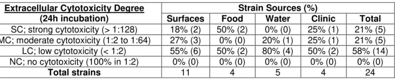
Table 4.9. Page 30 Extracellular cytotoxicity ability of Aeromonas isolates to undifferentiated (4-6 days old) Caco-2 cells, as described in Chapter 3.4.1.
Table 4.10. Page 31 Distribution of extracellular cytotoxic Aeromonas isolates by source and ability levels.
LIST OF FIGURES
Figure 4.1. Page 23 Optic microscopy observation showing the adherence ability and patterns of the Aeromonas isolates to undifferentiated (4-6 days old) Caco-2 cells, as described in Chapter 3.3.2.
Figure A1. ANNEX Calibration curve (A) and growth curve (B) of Escherichia coli K-12 C600 strain, performed as described in Chapter 3.1.
Figure A2. ANNEX Calibration curve (A) and growth curve (B) of Pseudomonas aeruginosa PAO1 strain, performed as described in Chapter 3.1.
Figure A3. ANNEX Average calibration curve (A) and growth curve (B) of Aeromona hydrophila subsp. hydrophila (DSM 30187t) strain and 5 Aeromonas isolates, performed as described in Chapter 3.1.
Figure A4. ANNEX Growth curves of Caco-2 cells line at passage 35, performed as described in Chapter 3.2.
Figure A5. ANNEX Adhesion and Invasion abilities of Aeromonas isolates to undifferentiated (4-6 days old) Caco-2 cells, as described in Chapter 3.3.1.
Figure A6. ANNEX Adhesion and Invasion abilities of Aeromonas isolates to differentiated (19-21 days old) Caco-2 cells, as described in Chapter 3.3.1.
Figure A7. ANNEX Cell-Contact cytotoxicity ability of Aeromonas isolates to undifferentiated (4-6 days old) Caco-2 cells at MOI 100:1, as described in Chapter 3.4.2.
Figure A8. ANNEX Cell-Contact cytotoxicity ability of Aeromonas isolates to undifferentiated (4-6 days old) Caco-2 cells at MOI 10:1, as described in Chapter 3.4.2.
Figure A9. ANNEX Extracellular cytotoxicity ability of Aeromonas isolates to undifferentiated (4-6 days old) Caco-2 cells at 4 hours of incubation, as described in Chapter 3.4.1.
Figure A10. ANNEX Extracellular cytotoxicity ability of Aeromonas isolates to undifferentiated (4-6 days old) Caco-2 cells at 24 hours of incubation, as described in Chapter 3.4.1.
Chapter 1
Introduction
1.1. Taxonomy of genus Aeromonas
The genus Aeromonas belongs to a single family of bacteria, Aeromonodaceae, of the order Aeromonadales. They consist of straight, coccobacillary to bacillary gram-negative bacteria with rounded ends measuring 0.3-1.0 x 1.0-3.5 µm [Martin-Carnahan and Joseph, 2005]. They occur
singly, in pairs, and rarely as short chains. Aeromonas spp. are facultative anaerobic, catalase positive, oxidase positive, chemoorganotrophic bacteria that exhibit both oxidative and fermentative metabolism of carbohydrates. Aeromonas spp. are waterborne and grow optimally within 22-35ºC, but some species can growth within 0-45ºC [Mateos et al., 1993], they tolerate a pH range from 4.5 to 9 and have a optimally growth at sodium chloride concentrations between of 0-4% [Isonhood and Drake, 2002].
Aeromonas spp. can be divided in two major groups, according to different phenotypical characteristics. The strains that grow at 35ºC to 37ºC and are motile by polar flagella, belong to mesophilic group and are commonly responsible for numerous human infections, being subdivided into Aeromonas hydrophila, Aeromonas caviae and Aeromonas sobria. The psychrophilic group comprises non-motile strains that grow better between 22ºC and 28ºC are responsible for causing fish infection, and are designated by Aeromonas salmonicida [Janda and Abbott, 1998].
Currently there are thirty one recognized Aeromonas species at list of prokaryotic names with standing in nomenclature: genus Aeromonas [Euzéby, 2013].
1.2. Occurrence
Aeromonas species are autochthonous (natural inhabitants) to the aqueous environment and due to their high adaptation capacity, are widely distributed in nature occurring in many different habitats, namely freshwater, marine and estuarine waters, sewages and ground waters [Havelaar et al., 1992; Holmes et al., 1995; Nielsen et al., 2001; Rahman et al., 2007 in USEPA, 2006] where they may be pathogenic for poikilotherms (cold-blooded animals) [Janda and Abbott, 1998].
USEPA, 2006] and in the U.S. Aeromonas hydrophila is listed by EPA (Protection Agency’s) on the Candidate Contaminant List (CCL) since 1998 [USEPA, 1998].
Isonhood and Drake (2002) reviewed Aeromonas spp. in foods. While Aeromonas spp. have been isolated from fish, shellfish, meats (chicken, beef, lamb, pork, etc.), vegetables, dairy products (cheese, raw milk, etc.) and ready to eat foods, few foodborne outbreaks have been reported and most resulted from ingestion of fish or shellfish. However a growing body of epidemiological evidence supports the possibility of Aeromonas causing foodborne gastroenteritis. While plethora of putative virulence factors has been postulated and demonstrated in food isolates, the exact role and mechanism of Aeromonas in causing diarrheal illness has not been elucidated. Evidence suggests that a high infective dose is necessary to produce gastrointestinal disease in a susceptible host, however, the fact that Aeromonas may survive and grow at refrigerator temperatures raises the concern that a reservoir of bacteria that may be created and able to achieve an infective dose when foods are mishandled.
As mentioned before, Aeromonas form biofilms on surfaces (including within water distributing systems) and this may pose a threat of contamination in food processing. Bal’a et al. (1998) found that heat and chlorine were effective against biofilms on stainless steel surfaces, but older biofilms were more resistant recently established biofilm to heat. An eight-day old biofilm was destroyed by heating to 60ºC and by exposure to 75 mg/L chlorine for 1 minute.
1.3. Health effect in humans
Figueras (2005) reviewed human infections caused by Aeromonas spp. They have multiple virulence factors that are typically associated with gastrointestinal diseases in other pathogenic bacteria, but while the production of extra-intestinal disease in humans is undisputable, the role of Aeromonas spp. as gastroenteritis agents is controversial, and has been based on extrapolation from anecdotal case reports, case-control studies, and a handful of outbreaks epidemiologically associated with food or water ingestion. The direct relationship between the presence of most of these virulence factors and gastrointestinal disease has not been proven. No epidemiological studies have indisputably linked Aeromonas with outbreaks of diarrheal disease. The association is strongest in children under the age of 2 years, adults over 50 and the immunocompromised, but a high number of asymptomatic persons carry Aeromonas in their gastrointestinal tract.
Fecal isolation rates of Aeromonas in asymptomatic persons, in developed countries, range between 0% to 4.0% [Millership et al., 1983; Agger et al., 1985; Svenungsson et al., 2000 in USEPA, 2006]; while in symptomatic persons it ranges between 0.8 to 7.4% [Agger et al., 1985; Moyer, 1987; Albert et al., 2000 in USEPA, 2006]. In lower developed regions like in Southeast Asia, asymptomatic carriage rates are as high as 27.5%, while in patients with diarrhea rates have been reported as high as 34% [Pazzaglia et al., 1990 in USEPA, 2006].
Aeromonas. hydrophila, Aeromonas caviae, Aeromonas veronii biovar Sobria, Aeromonas veronii biovar Veronii, Aeromonas jandaei, Aeromonas trota and Aeromonas schubertii are also known to be pathogenic to humans. They cause a variety of extra-intestinal infections such as wound infection, memingitis, osteomyelitis, septic arthritis, endocarditis, peritonitis, eye and urinary tract infections. Those extra-intestinal infections are usually serious and potentially life-threatening and for a successful recovery is necessary an aggressive antibiotic therapy after an antimicrobial susceptibility test, because multi-drug resistance is common among Aeromonas.
1.4. Health effect in animals
Gosling (1996) reviewed animal infections caused by Aeromonas spp. Diseases caused by Aeromonas represent a significant source of loss to the aquaculture industry. The disease mechanisms of Aeromonas that cause animal disease are essentially the same ones that produce disease in humans.
1.5. Virulence properties
Janda and Abbott (2010) reviewed the virulence factors produced by Aeromonas spp. and the pathogenicity of Aeromonas is complex and multifactorial and incompletely understood despite decades of intense investigation. Although an established animal model that faithfully reproduces the syndrome associated with Aeromonas does not exist, testing with isogenic mutants in animal cells show that many virulence factors produced by Aeromonas are associated with its pathogenesis. Factors contributing to virulence are present in two forms, cell-associated structures and extracellular products.
1.5.1. Cell-associated virulence factors
Structural factors have been discovered in Aeromonas ssp. that promote their attachment (pili, flagella) and colonization (adhesins, outer membrane proteins (OMPs)). Other factors protect Aeromonas from host response (S-layer, lipopolysaccharide (LPS), capsule) and are responsible for delivering virulence factors directly into the host cell (Type II and Type III secretion systems) [USEPA, 2006].
A vital step for the enteropathogens to initiate infection is through adherence to host cells, allowing localization and subsequent colonization of the appropriate target tissues [Finlay and Falkow, 1997; Scoglio et al., 2001]. It is essential to cause gastrointestinal disease, via either toxin production or host cell invasion, or both [Knutton et al., 1987].
Flagella have been referred to have a crucial role in adhesion, biofilm formation, and colonization of several other pathogenic bacteria, such as Pseudomonas aeruginosa [Stanley, 1983 in USEPA, 2006], Salmonella enterica [Ciacci-Woolwine et al., 1998 in USEPA, 2006], Escherichia coli [Pratt and Kotler, 1998 in USEPA, 2006], Helicobacter pylori [Eaton et al., 1996 in USEPA, 2006] and Vibrio cholerae [Gardel and Mekalanos, 1996 in USEPA, 2006].
Aeromona hydrophila and they do not seem to share structural genes or regulatory [McCarter, 2004 in Merino et al., 2006]. Polar flagella and lateral flagella were described by Rabaan et al. (2001) and Kirov et al. (2002). Kirov (2003) reviewed the expression of lateral flagella and their multi-functional role in pathogenesis.
The swimming motility has been linked to a single polar unsheathed flagellum in all mesophilic Aeromonas, which is expressed constitutively and highly regulated by a number of environmental factors [Merino et al., 2006]. They respond to sensory stimuli (chemotaxis), allowing locomotion toward new substrates and, therefore, confer an adaptive advantage in the colonization of different environments [Merino et al., 2006]. The swarming motility has been linked to many unsheathed peritrichous lateral flagella in 50 to 60% of Aeromonas [Kirov et al., 2004], which are expressed when grown in matrices that do not allow motility by a single polar flagellum, like viscous environments or over surfaces and solid media [Kirov et al., 2003; Naharro et al., 2011].
In this bacterial genus both types of flagella function as adhesins enabling adherence of human cell lines by most Aeromonas isolates, observed in experimental tests with Caco-2, HEp-2 e Henle 407 cells [Kirov et al., 2004; Sen and Lye, 2007]. Strains that loose polar flagella are virtually nonadherent to cell lines, while strains lacking lateral flagella have a reduced capacity for cell binding. Flagellar mutants were shown to have decreased binding capacity by more than 80% [Kirov et al., 2004]. In addition, the flagella have been described as colonization factors and biofilm formation in different surfaces [Kirov et al., 2004].
Several studies have shown a relationship between the adhesive patterns and the increased potential for virulence of bacteria [Parsot, 2005; Mitache et al., 2009; Mora et al., 2009]. Because they are well studied, adhesive patterns obtained with Escherichia coli serves as a model for studies of infection of other bacteria in eukaryotic cells. Diffuse adhesion (DA), can be observed in Escherichia coli as well as localized (LA) and aggregative (AA) with "stacked-brick" appearance. It is believed that the latter is related to the higher bacterial virulence, in contrast to other patterns which are seen in less virulent bacteria [Challapalli et al., 1988; Singh et al, 1992; Parras et al., 1993].
Several studies suggest that attachment to host cells is mediated by pili, extracellular filamentous appendages, and were described as potential colonization factors in Aeromonas hydrophila and Aeromonas veronii biovar Sobria [Hokama and Iwanaga, 1991 in USEPA, 2006]. Kirov (1993) reported that pili were important adhesive factors for mucosal surface attachment and described filamentous and nonfilamentous adhesins. Two morphotypes of pili have been observed in Aeromonas spp., short rigid pili (S/R type) similar to those of Escherichia coli Type I and Pap pili [Ho et al., 1992 in USEPA, 2006] and long wavy flexible pili (L/W type) belong to a class of Type IV bundle-forming pili (Bfp) [Kirov and Sanderson, 1996 in USEPA, 2006]. Removal of pili or neutralization of attachment sites by homologous antibody treatment limits or defeats adherence properties in cell culture systems in Aeromonas [Iwanaga and Hokama, 1992 in USEPA, 2006].
electron microscopy (TEM) did not demonstrate fimbrial structures on cell surfaces of highly-adherent Aeromonas caviae strains. These data suggest that OMPs mediate adherence in Aeromonas caviae instead of pili. Some OMP have hemagglutination activity, while other OMPs are thought to have pore-forming capability.
One of the possible mechanisms involved in the Aeromonas pathogenesis is associated with the production of type III (T3SS) or injectisome secretion system, which was reviewed by Coburn (2007). A broad clinical spectrum of diseases have been referred as being caused by T3SS containing pathogens, for example, infections with enteropathogenic Escherichia coli, Shigella, Salmonella and Yersinia species which result in serious intestinal diseases.
There is high structural similarity between the bacterial flagellum and the T3SS, many structural proteins are clearly homologous. This system is composed of several rings, the basal area is anchored in the inner membrane forming a canal that crosses the outer membrane and projected outwards, this way allows adherence to cell membranes and injection of bacterial toxins (effector proteins) directly in the cytosol of host cells [Alberts et al., 2002]. Some of these effector proteins have multiple biological functions, such as changing the cytoskeleton to facilitate the invasion or activation of intracellular signaling cascades within the host cells, eventually causing lysis of host epithelial cells and contributing to the degradation of tissues host [Alberts et al., 2002; Sha et al., 2005; Krzymińska
et al., 2012].
Several studies have shown invasive ability of Aeromonas to equal that of Campylobacter [Nishikawa et al., 1994; Shaw et al., 1995 in USEPA, 2006], while intracellular bacteria have been demonstrated by electron microscopy, no gene or product has been identified specifically with invasion [USEPA, 2006].
1.5.2. Extracellular virulence factors
Extracellular factors have been discovered in Aeromonas ssp. that promote their ability to obtain nutrients that allow the proliferation and dissemination of the microorganism in the host, such as enterotoxins, proteases, phospholipases and hemolysins associated [Khajanchi et al. 2010].
DNases can act as food enzymes used to obtain phosphorus and nitrogen [Pemberton et al., 1997] and provide a barrier to the entry of foreign DNA in the host cell, playing an important role in microbial defense mechanisms [Kamble and Deshmukh, 2012], but can also act as virulence factors due to the ability to degrade extracellular fibers released by neutrophils, preventing phagocytosis [Brinkmann et al., 2004].
gcat, pla, and apl-1 genes encode phospholipases often associated with intestinal lesions, while the lipA and lipH3 genes encode lipases without phospholipid action [Chuang et al., 1997].
Elastases have elastolytic and caseinolytic activities. In the Aeromonas genus, the caseinolytic activity is mainly a result of the AhyA serine protease, ahyA gene product which a weak elastolytic activity, and it has been associated with the acceleration of the maturity of the AhyB elastase, extracellular metalloprotease encoded by the ahyB gene, that the elastolytic activity has been associated [Cascón et al., 2000a, 2000b]. The role of AhyB elastase in Aeromonas spp. is not yet completely elucidated, but revealed a 52% aminoacid identity when compared to the sequence of the LasB elastase identified in Pseudomonas aeruginosa, which can degrade many components of the immune system, including chemokines and cytokines [Horvat et al., 1989; Kevin et al., 2003 in Kuang et al., 2011] and antimicrobial peptides [Schad et al., 1987 in Kuang et al., 2011].
Enterotoxins produced by Aeromonas spp. include into two categories, cytotoxic and cytotonic [Krzymińska et al., 2003; Von Gravaenitz, 2007]. Chopra and Houston (1999) reviewed enterotoxins of
Aeromonas spp. associated with production of gastrointestinal disease.
The cytotoxic enterotoxins may cause extensive damage to the epithelia, include heat-labile and stable compounds, with hemolytic and cytotoxic activities, like pore-forming toxin aerolysin and different α- and β-hemolysins [Galindo et al., 2006; Von Gravaenitz, 2007], while progress is being
made in understanding aerolysin activity, the actual mechanism is complex and incompletely understood. The cytotoxin Act, which is structural and functional closely related to the cytotoxin Aerolisina [Martin-Carnahan e Joseph, 2005], is consensually considered the most important virulence factor associated with the genus Aeromonas. It’s secreted by the type II secretion system (T2SS), expressing hemolytic and cytotoxic activity, which involves the formation of pores in the target cell membrane and consequently occurs the influx of water, resulting in cell lysis [Khajanchi et al., 2010]. The cytotoxin HlyA is a non-channel forming β-hemolisina similar to the Vibrio cholerae hemolysin, widespread in the genus Aeromonas and is virtually ubiquitous in Aeromonas hydrophila [Naharro et al., 2011].
The cytotonic enterotoxins, on the other hand, cause increase in the level of cAMP in intestinal epithelial cells, like cholera toxin [Galindo et al. 2006], resulting in fluid secretion from intestinal cells, not causing the degeneration of crypts and villi of the small intestine [Sha et al, 2002]. The cytotonic Ast and Alt cause fluid accumulation in ligated ileal loops in animal models and probably have an undescribed role in causing diarrhea in humans [Sha et al., 2002]. Laohachai et al. (2003) reviewed the role of bacterial toxins that induce changes in membrane transport leading to diarrheal disease.
1.6. Caco-2 cell line as a model of intestinal barrier
Several infection in vitro studies in animal cell lines support the ability of these bacteria to cause cellular damage, in addition to its ability to adhere and to invade cells [Schiavano et al., 1998; Martins et al., 2002; Balaji et al, 2004].
Caco-2 cells, which are derived from a human colon adenocarcinoma, have gained great attention in recent years as an in vitro model of the intestinal epithelium. In culture Caco-2 cells spontaneously differentiate and organize as a polarized monolayer with tight junctions and microvilli, mimicking the in vivo enterocyte, expressing relatively high levels of digestive brush border enzymes and display other morphological, structural and functional properties similar to intestinal enterocytes [Pinto et al., 1983 in Delie and Rubas, 1997]. Due to these characteristics, the Caco-2 cell culture is now widely used as a tissue model for studying adhesion and invasion of probiotic bacteria or entericpathogens [Delie and Rubas, 1997]. For example, Panigrahi et al. (1990) showed that the adhesion of non-01 Vibrio cholerae to Caco-2 cells correlated with human intestinal colonization and disease.
Traditional Caco-2 cell culture requires a 21-day period to attain a differentiated monolayer. During the growing phase Caco-2 cells remain undifferentiated and immediately after they reach the status of confluence, cells start the differentiation program that will be finished 18-21 days later [Delie and Rubas, 1997]. According to the morphological and functional grade of differentiation they can be divided into three subgroups:
1) cells homogeneously undifferentiated (sub confluent population); 2) cells heterogeneously polarized and differentiated (intermediate phase); 3) cells homogeneously polarized and differentiated (>15 days after seeding).
Chapter 2
Objectives
In the last decade Aeromonas have been referred as an emergent pathogen of gastrointestinal disease in humans and normally associated to ingestion of contaminated water or food [Figueras, 2005]. However the presence of virulence genes does not correlate with expression of gene products or with manifestations of disease in animals or humans [Bondi et al., 2000] and therefore the precise combination of virulence factors that invariably confer virulence on a particular strain has not been determined.
A vital step for the enteropathogens to initiate infection is through adherence to host cells, allowing localization and subsequent colonization of the appropriate target tissues [Finlay and Falkow, 1997; Scoglio et al., 2001]. Colonization is therefore essential to cause gastrointestinal disease, via either toxin production or host cell invasion, or both [Knutton, et al. 1987]. Knowledge of these may help to identify strains which pose a public health risk.
The Caco-2 cells are derived from a human carcinoma of the colon, they exhibit structural and differentiation patterns characteristic of mature enterocytes and are being increasingly used as a substitute for human intestinal cells to study the adhesion of enteric pathogens [Pinto et al., 1983; Russel and Blake, 1994 in Delie and Rubas, 1997].
The work reported here was therefore conducted on those cells as a preliminary investigation to determine, if Aeromonas present in several human environments, from food industry (slaughterhouse, cheese factory and supermarkets) to water treatment facilities (EPAL), in Portugal, may pose a threat to public health.
24 strains were chosen as representatives of the diversity present in a set of Aeromonas isolates obtained by the team of Doctor Teresa Semedo-Lemsaddek at the Faculty of Veterinary Medicine, University of Lisbon, according to the results of clusters diversity, presence of virulence factors and resistance to antibiotics.
The specific aims that were pursuit were:
To implement assays for adherence, invasion and cytotoxicity for bacteria in mammal cells, particularly the Caco-2 cell line;
To evaluate eventual cytotoxicity of bacterial culture medium to the intestinal Caco-2 cell line, in undifferentiated cells (UC);
To characterize the ability of the different bacterial strains to adhere and to invade the intestinal Caco-2 cell line, in both undifferentiated (UC) and differentiated cells (DC); To compare the phenotype profile determined in this study with the genetic profile
determined by Barroco (2013).
Chapter 3
Materials and Methods
3.1. Aeromonas culture and growth study
24 previously isolated Aeromonas spp. strains, identified by phenotypic and genomic typing, were used in this study and their source and characterization carried out to date by Barroco (2013) are listed in Table 1 in Annex. The strains were stored at -20ºC in brain heart infusion broth (BHI, AES Laboratories) containing 45% (v/v) glycerol (Hi-media Laboratories).
Escherichia coli K-12 C600 (No. 426, DSMZ; Braunschweig, Germany), a non-pathogenic strain, and Pseudomonas aeruginosa PAO1 (No. 19880, DSMZ; Braunschweig, Germany), an adherent, invasive and cell-contact cytotoxic strain, were included as negative and positive controls, respectively. Aeromona hydrophila subsp. hydrophila (No. 30187t, DSMZ; Braunschweig, Germany), an adherent, invasive and extracellular cytotoxic strain, was included as a reference of the Aeromonas genus.
Optical density was used to estimate colony forming units (CFU) in a bacterial suspension which was possible after taking some basic precautions to control the following: type of media, growth phase of microorganisms, nature and condition of the equipment [Scott, 2011]. Therefore the spectrophotometer (Ultraspec 20100 pro) used was calibrated for the linear range of absorption vs CFU relevant values, using the bacterial suspension under the specific conditions of the future assays.
Bacterial strains were subcultured on BHI agar (BHI broth with 15 g/L agar, Scharlau) plates and incubated at 30ºC overnight, then passed to new plates of BHI agar and incubated at 37ºC overnight. The bacterial were then washed by harvesting and suspending in BHI broth, centrifuged at 10.000xg for 10 minutes and resuspended in fresh BHI broth.
Bacterial suspensions were adjusted to 0.400 optical density at 600 nm wave-length (OD600) and twofold serial dilutions were performed in BHI broth (0.400 to 0.005). Spectrophotometer readings of the dilutions, stored on ice, were taken in triplicate before performing serial dilutions of the bacterial suspension in peptone water (Merck Millipore) and plated by the pour plate method, which consists in suspending 1 mL of the dilutions in a Petri-dish using molten Luria-Bertani agar (LBA, Bioreagentes Fisher) cooled to approximately 48°C (just above the point of solidification to minimize heat-induced cell death), after the medium solidifies the plates were inverted and incubated for 24-48 hours at 37ºC.
Each dilution was plated in duplicate and the viable counting was performed in the dilutions that have between 30 and 300 colonies. The results were expressed in colony forming unit (CFU) per mL by using the following formula:
Average of colonies x (1 / dilution factor) x (1 / dilution volume)
The calibration curves obtained with this method are represented in Figure A1A, 2.A and 3.A in Annex.
The growth curves obtained with this method are represented in Figure 1.B, 2.B and 3.B in Annex.
3.2. Caco-2 cells culture and growth study
Human colon carcinoma Caco-2 cells were purchased from Deutsche Sammlung von Mikroorganismen und Zellkulturen (DSMZ, Braunschweig, Germany). They were stored at -80ºC in fetal bovine serum (FBS, Gibco) containing 5% (v/v) dimethyl sulfoxide (DMSO, Sigma) at a concentration of 2x10⁶ cells/mL in passage 30. The cells were routinely grown in 75 cm2 plastic tissue culture flasks (Falcon) containing Dulbecco’s modified Eagle minimum essential medium (DMEM, Gibco) supplemented with 10% (v/v) FBS and 1% (v/v) nonessential amino acids (NAAS, Gibco) at 37ºC in a humidified atmosphere of 95% (v/v) air and 5% (v/v) CO2. The culture medium was changed every 48 hours and passed before reaching confluence (once a week).
For the cytotoxicity, adhesion and invasion assays, the cells were expanded in 175 cm2 plastic tissue culture flasks (Nunc) and when reaching 70-90% confluence, the cells were trypsinized with 0.25% (v/v) trypsin (Gibco) for 5 minutes and adjusted to a concentration of 2x10⁵ cells/mL in culture medium. Depending on the assay, 4.8 mL, 1 mL or 0.16 mL of cell suspension was dispensed into each well of a 6, 24 or 96-well tissue culture plate (Falcon), respectively, to attain 1x10⁵ cells/cm2 and incubated at 37ºC in 5% (v/v) CO2 to obtain, 3 days later, undifferentiated semi-monolayers (± 80% confluent), or 4 days later, confluent undifferentiated cells (UC) monolayers and, after 18 days or more, differentiated cells (DC) monolayers. The culture medium was changed every 48 hours. The Caco-2 cells used for those assays were at passage numbers between 35 and 45.
The existence of Caco-2 cell lines maintained in different laboratories and/or of different clonal origin, in addition to the effects of different culture protocols may result in variations in growth rate of the cell line. For this reason a growth study of Caco-2 cell line was undertaken in the in-house conditions of the assays to be performed in order to estimate growth kinetics and cell concentration.
Semi-confluent cell monolayers were trypsinized and adjusted to a concentration of 1x10⁵ cells/mL (5x10⁴ cells/cm2) in culture medium and then 1 mL cell suspension was dispensed into each 2 cm2 well (2x10⁴ cells/cm2) of a 24-well tissue culture plate was visually evaluated in an inverted microscope (OlymPus CKX41) every day of incubation. The cells of 3 wells were trypsinized and counted using a Fuchs-Rosenthal Counting Chamber (EMS #63512-10) and the cell viability was evaluated by diluting the cell suspension in Dulbecco's phosphate-buffered saline (DPBS, Gibco) containing 0.1% (v/v) trypan blue (Gibco) exclusion dye to establish the growth curves.
The results were expressed in cells per cm2 by using the following formula:
(Average of viable cells x (1 / dilution factor) x (1 / volume per quadrant)) / surface growth
3.3. Bacterial adhesion and invasion evaluation on Caco-2 cell line
3.3.1. Adherence and invasion quantitative assays
Bacterial strains were subcultured on BHI agar plates and incubated at 30ºC overnight, then passed to new plates of BHI agar and further incubated at 37ºC overnight. The bacterial were then washed as described previously. Bacterial suspensions were adjusted to approximately 5x10⁷ CFU/mL by measuring 0.115 OD600 (0.080 and 0.085 OD600 for K-12 and PAO1 strains, respectively), according to the calibration curves previously established (Figures 1, 2 and 3 in Annex), and kept on ice until use; the number of bacteria of each strain was confirmed by CFU determination immediately after use as described previously.
In the adherence assay, differentiated and undifferentiated Caco-2 cells monolayers plated in 24-well plates and containing 400 µL of fresh culture medium, were incubated at 37ºC in 5% CO2 for 90 minutes with 100 µL of bacterial suspension to give a MOI of 10 bacteria / Caco-2 cell (approximately
5x10⁵ epithelial cells per mL to 5x10⁶ bacteria per mL, according to the calibration curve presented in Figure 4 in Annex. To remove non-adherent bacteria, the monolayers were washed gently three times with 1 mL of DPBS and then the cells were lysed by adding 0.5 mL of DMEM containing 2% (v/v) Triton X-100 (Sigma-Aldrich) for 30 minutes at 37ºC. To ensure full lysis and release of all the bacteria, including those eventually internalized, the lysate was pipetted up and down several times, pointing the tip of the pipette directly at the surface of the well,
For the invasion assay, performed similarly and in the same plate as the adhesion test, after the period of infection, the cultures were replaced by 1 mL of a 300 µg/mL Gentamicin solution and
incubated for 1 hour at 37ºC to kill extracellular bacteria. To remove the antibiotic, the monolayers were washed gently 2 times with 1 mL of DPBS and then the intracellular bacteria were released by lysing the cells as described above.
Lastly, 0.5 mL of cold DMEM was added to 0.5 mL of each lysate, while keeping them on ice, for the determination of the number of adherent and invasive bacteria by CFU as described previously. The results are expressed in percentage of bacteria recovered in comparison to other wells where the monolayers were infected under the same conditions but untreated, the culture was kept on ice rather than discarded after the period of infection and the cell lysate was subsequently added. This step is performed because the bacteria in the inoculum can sometimes grow much faster than the bacteria in the presence of cells, skewing the size of the inoculum by comparison with the real total number of bacteria in wells with cells. The percentage of adherent/invasive bacteria can then be calculated by dividing the number of CFU of adherent/invasive bacteria by the addition of the number of CFU of adherent/invasive and non- adherent/non-invasive bacteria. All tests were performed in duplicate and in two separate experiments for each isolate. The variation is expressed as propagated error.
3.3.2. Adherence patterns assays
In this adhesion assays were used monolayers of cells plated on sterilized glass coverslips (20x20 mm, Marienfeld) in 6-well plates with 80% of confluence (3 days old cells) and containing 1 mL of fresh culture medium which were then incubated at 37ºC in 5 % CO2 for 90 minutes with 2 mL of bacterial suspension to give a MOI of 100 bacteria / Caco-2 cell. To remove non-adherent bacteria, the monolayers were washed gently 3 times with 2 mL of DPBS and then the remaining adherent bacteria and monolayers were fixed with 2 mL of 99.8% (v/v) methanol (J. T. Backer) for 5 minutes. Methanol was removed by washing the cells with 2 mL of PBS (Calbiochem) and then the cells were stained for 45 minutes with 2 mL of 10% (v/v) Giemsa stain (Sigma) prepared in Giemsa buffer (Sigma). The coverslips were removed from wells, washed in distilled water, air dried, mounted on glass slides and examined by oil immersion under a light microscope (Zeiss Imager A2 with Zeiss AxioCam MRm) at x1000 magnification.
Isolates which show a uniform distribution on the cell surface, were characterized as having a diffuse adhesion (DA) pattern; those which show a localized distribution, were characterized as having a localized adhesion (LA) pattern; and those which show a "stacked-brick" appearance on the cell surface, were characterized as having a aggregative adhesion (AA) pattern.
3.4. Bacterial cytotoxicity evaluation on Caco-2 cell line
In vitro cytotoxicity assays may make use of various cell lines and the quantitative assessment of cytotoxicity generally relies on the visual counting of cells [Balaji et al., 2004; Ghatak et al., 2006; Castilho et al., 2009], leading to low reproducibility of these approaches and consequently conferring significant limitations. Previous studies suggest that the MTS assay in vitro cytotoxicity assay combines all features of a good measurement system in terms of ease of use, precision, rapid [Berg et al., 1994], as well as sensitive and specific indication of toxicity whose performance is very competitive to other toxicological test systems [Gregor et al., 1997]. The MTS assay is based on the conversion of MTS, 3-(4,5-dimethylthiazol-2-yl)-5-(3-carboxymethoxyphenyl)-2-(4-sulfophenyl)-2H-tetrazolium, inner salt into a colored, aqueous soluble formazan product by mitochondrial activity of viable cells at 37°C. The amount of formazan produced by dehydrogenase enzymes is directly proportional to the number of living cells in culture and can be measured at 490 nm [Barltrop et al., 1991].
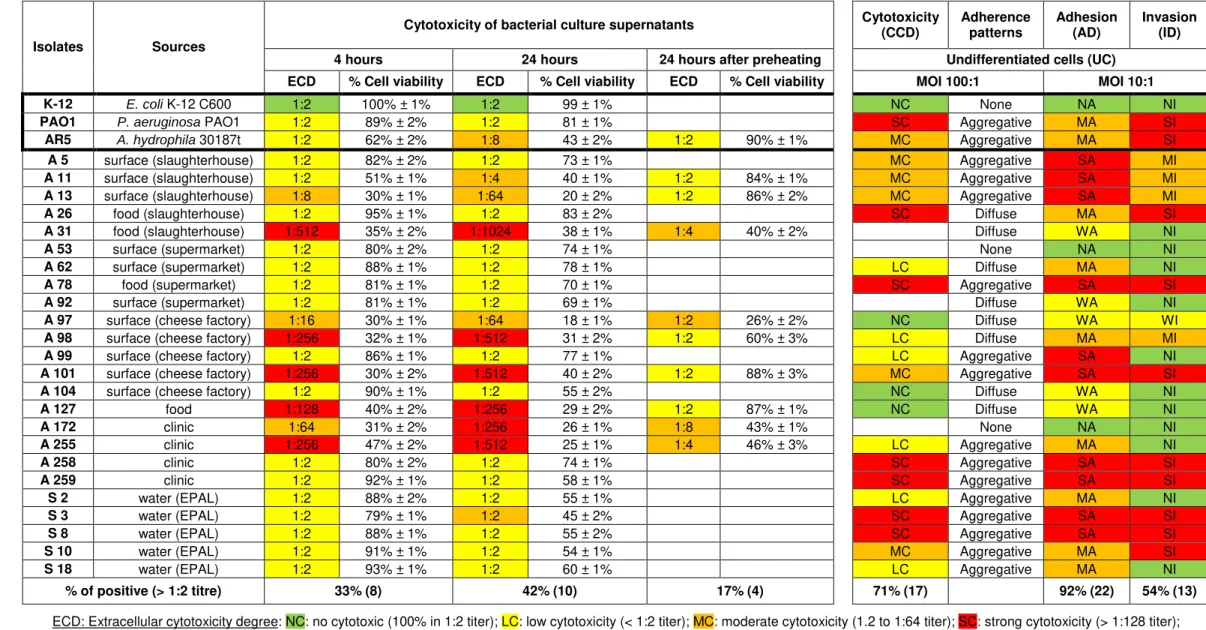
3.4.1. Cytotoxicity induced by culture supernatants assays
Undifferentiated Caco-2 cells monolayers, grown in 96-well plates, were incubated at 37ºC in 5% CO2 for 4 and 24 hours with 100 µL of twofold serial dilutions in culture medium with only 0,5% FBS (1:2 to
1:2048 v/v) of CFS of Aeromonas spp. strains, non-pathogenic E. coli K-12 and P. aeruginosa PAO1. Morphological changes were evaluated in an inverted microscope over the first 1-4 hours period. Afterwards the monolayers were washed gently with 200 µL DPBS and the viability of the cells was
assessed by MTS assay. For this assay, 100 µL of DMEM containing 2% (v/v) of the colorimetric reagent MTS (CellTiter 96® AQueous One Solution Cell Proliferation Assay, Sigma) were added in each well and incubated for 1-4 hours at 37ºC. The medium with the colorimetric reagent was transferred to a new 96-well plate and were perform measurements of the absorbance at 490 nm in a BioTekTM Power Wave XS microplate reader. For the isolates that shown to be positive a sample of the same supernatant was pre-heated to 65ºC for 20 minutes in a water bath thermostated and analyzed again.
The results are expressed in percentage of viable cells, using the following formula:
((OD cell treatment – OD negative control) / (OD positive control – OD negative control)) x 100,
where the negative control (total cellular damage) is culture medium alone and the positive control (no cellular damage) are cells that receive BHI broth instead of CFS. Assays were performed in duplicate and in two separate experiments for each isolate. The variation is expressed as propagated error. Cytotoxic titre was considered as the reciprocal of the highest dilution of the culture filtrate that caused destruction of 50% of the Caco-2 cells. The CFS preparations that induced cytopathic effect above to 1:2 dilution in 50% or more cells were recorded as cytotoxic positive isolates.
3.4.2. Cytotoxicity induced by cell-contact assays
Bacterial strains were subcultured on BHI agar plates and incubated at 30ºC overnight, then passed to new BHI agar plates and incubated again at 37ºC overnight. The bacterial were then washed by harvesting and suspending in BHI broth, centrifuged at 10.000xg for 10 minutes and resuspended in fresh BHI broth. Bacterial suspensions were adjusted to approximately 5x10⁷ CFU/mL by measuring 0.115 OD600 (0.080 and 0.085 OD600 for K-12 and PAO1 strains, respectively), according to the calibration curves previously established (Figures 1, 2 and 3 in Annex) and kept on ice until use; the number of bacteria of each strain was confirmed by CFU determination immediately after use as described previously.
Undifferentiated Caco-2 cells monolayers grown in 96-well plates containing 100 µL of fresh culture
medium were incubated at 37ºC in 5% CO2 for 90 minutes with 160 µL of bacterial suspension to give a multiplicity of infection (MOI) of 100 bacteria / Caco-2 cell, meaning that 1x10⁵ epithelial cells per mL were incubated with approximately 1x10⁷ bacteria per mL, according to the calibration curve presented in Figure 4 in Annex) and with 160 µL of bacterial suspension 1:10 diluted in BHI broth to give a MOI
of 10:1. Next, the bacteria were removed and replaced by 200 µL DMEM containing 300 µg/ml
To test the importance of bacteria-host cell contact in cytotoxicity, co-cultures of the bacteria and Caco-2 cells at the same time and under the same conditions were performed but by using cells cultivated on 96-well plates with 0.45 µm pore size transwell inserts (Corming). Caco-2 cells were cultured in the lower chamber and the bacteria cells were added in the upper chamber, preventing bacterial contact with Caco-2 cells.
Extracellular cytotoxicity activity was determined using transwells, while cell-contact cytotoxicity activity, after the subtraction of the extracellular cytotoxicity was determined in the plain wells, after the subtraction of the extracellular cytotoxicity activity.
The results are expressed in percentage of cell damage, which is the reverse percentage of the viable cells and was calculated by using the following formula:
(1 – ((OD cell treatment – OD negative control ) / (OD positive control – OD negative control ))) x 100,
Chapter 4
Results and Discussion
4.1. CFU determination by optical density
The species of each isolate had not yet been clearly established at the time of the experiments (ongoing work at the laboratory of Doctor Teresa Semedo-Lemsaddek at the Faculty of Veterinary Medicine, University of Lisbon), therefore, the Aeromona hydrophila subsp. hydrophila (DSM 30187t) strain and 5 isolates was used to obtain an average calibration curve to estimate the concentration of the remaining isolates, but it must be stressed that this calibration must be done for all isolates, or per species, if it were needed a more accurate determination for future assays.
4.2. Aeromonas adhesion and invasion evaluated on Caco-2 cell line
The observed percentage of bacteria recovered is often very dependent on the experimental set-up (and, to a lesser extent, on the analyst), particularly the MOI and the number of washes [Letourneau, 2011]. A method for determining adherence and cytotoxic activity of Aeromonas has not been standardized, and cautious comparisons of between published reports are advised.
Bacterial resistance or sensitivity in the presence of Triton X-100 and gentamicin for each strain was demonstrated in control experiments with equivalents numbers of bacteria and under the same assay conditions (concentration, temperature and duration). Cell viability was determined by visual observation of growth / no growth after inoculation in BHI broth. Complete lysis of monolayer Caco-2 cells in 2% Triton X-100 after 30 minutes incubation was confirmed by microscopy.
The CFU determination at the start (to determine the initial MOI) and at the end (to determine the total number of bacteria) of each assay proved that, after the 90 minutes infection period, bacterial multiplication, that is required for the adhesion process, occurred in all the strains.
4.2.1. Aeromonas spp. adherence activity
The clinical relevance of in vitro adhesion is sometimes contested, firstly because bacterial interaction with the intestinal mucosa is complex, and secondly because it cannot be assumed that tissue culture cells derived by cell transformation possess the same surface receptors for bacterial adherence as those found on human intestinal cells in vivo [Freter and Jones, 1983 in Delie and Rubas, 1997]. However, in several cases a correlation between in vitro adhesion and in vivo infectivity has been demonstrated [Mathewson et al., 1985; Kelly et al., 1993 in Delie and Rubas, 1997].
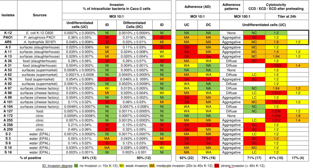
Table 4.1. Adhesion abilities of Aeromonas isolates to undifferentiated (4-6 days old) and differentiated (19-21 days old) Caco-2 cells, as described in Chapter 3.3.1.
The results are average ± propagated error.
Isolates Sources
Adherence
% of bacteria associated with Caco-2 cells Invasion (ID)
Adherence patterns
Cytotoxicity
CCD / ECD / ECD after preheating
MOI 10:1 MOI 10:1 MOI 100:1 Titer at 24h
Undifferentiated
cells (UC) AD
Differentiated
cells (DC) AD UC DC Undifferentiated cells (UC)
K-12 E. coli K-12 C600 0.32% ± 0.03% NA 0.3% ± 0.1% NA NI NI None NC 1:2
PAO1 P. aeruginosa PAO1 6% ± 1% MA 8.9% ± 0.9% MA SI SI Aggregative SC 1:2
AR5 A. hydrophila 30187t 9% ± 1% MA 5% ± 1% WA SI MI Aggregative MC 1:8 1:2
A 5 surfaces (slaughterhouse) 16% ± 4% SA 7% ± 2% MA MI SI Aggregative MC 1:2
A 11 surfaces (slaughterhouse) 16% ± 3% SA 11% ± 3% MA MI WI Aggregative MC 1:4 1:2
A 13 surfaces (slaughterhouse) 92% ± 14% SA 69% ± 14% SA MI WI Aggregative MC 1:64 1:2
A 26 food (slaughterhouse) 7% ± 2% MA 3% ± 1% WA SI SI Diffuse SC 1:2
A 31 food (slaughterhouse) 1.7% ± 0.3% WA 1.1% ± 0.2% NA NI NI Diffuse 1:1024 1:4
A 53 surfaces (supermarket) 0.9% ± 0.2% NA 0.9% ± 0.1% NA NI NI None 1:2
A 62 surfaces (supermarket) 4% ± 1% MA 3% ± 1% WA NI NI Diffuse LC 1:2
A 78 food (supermarket) 48% ± 7% SA 24% ± 4% SA SI MI Aggregative SC 1:2
A 92 surfaces (supermarket) 1.6% ± 0.3% WA 1.3% ± 0.2% NA NI NI Diffuse 1:2
A 97 surfaces (cheese factory) 2.5% ± 0.4% WA 1.8% ± 0.7% NA WI NI Diffuse NC 1:64 1:2
A 98 surfaces (cheese factory) 4.5% ± 0.8% MA 3.5% ± 0.7% WA MI WI Diffuse LC 1:512 1:2
A 99 surfaces (cheese factory) 14% ± 4% SA 7% ± 2% MA NI NI Aggregative LC 1:2
A 101 surfaces (cheese factory) 14% ± 3% SA 7% ± 3% MA SI MI Aggregative MC 1:512 1:2
A 104 surfaces (cheese factory) 2.5% ± 0.4% WA 2.1% ± 0.3% WA NI NI Diffuse NC 1:2
A 127 food 2.9% ± 0.7% WA 3% ± 1% WA NI NI Diffuse NC 1:256 1:2
A 172 clinic 1.0% ± 0.1% NA 0.95% ± 0.09% NA NI NI None 1:256 1:8
A 255 clinic 10% ± 4% MA 6.7% ± 0.7% MA NI NI Aggregative LC 1:512 1:4
A 258 clinic 34% ± 6% SA 17% ± 6% SA SI SI Aggregative SC 1:2
A 259 clinic 15% ± 2% SA 8% ± 1% MA SI SI Aggregative SC 1:2
S 2 water (EPAL) 15% ± 5% MA 6.7% ± 0.3% MA NI NI Aggregative LC 1:2
S 3 water (EPAL) 75% ± 13% SA 36% ± 8% SA SI MI Aggregative SC 1:2
S 8 water (EPAL) 23% ± 4% SA 16% ± 2% SA SI SI Aggregative SC 1:2
S 10 water (EPAL) 11% ± 2% MA 7% ± 2% MA SI WI Aggregative MC 1:2
S 18 water (EPAL) 5% ± 2% MA 2.3% ± 0.6% WA NI NI Aggregative LC 1:2
% of positive 92% (22) 79% (19) 54% (13) 50% (12) 71% (17) 41% (10) 17% (4)
AD: Adhesion degree: NA: no adhesion (< 3x K-12); WA: weak adhesion (3x to10x K-12); MA: moderate adhesion (10x to 30x K-12); SA: strong adhesion (> 30x K-12); ID: Invasion degree: NI: no Invasion (< 10x K-12); WI: weak invasion; MA: moderate invasion (20x to 40x K-12); SA: strong invasion (> 40x K-12);
CCD: Cell-contact cytotoxic degree: NC: no cytotoxic; LC: low cytotoxicity (< 25%); MC: moderate cytotoxicity (25 to 45%); SC: strong cytotoxicity (> 45%);
The adherent activity in 71% (17) and 54% (13) of the strains revealed adherence values equal to or greater than the adherence values of the pathogenic PAO1 strain to UC and to DC, respectively (Table 4.1). The highest adherent activity ranged between 14% ± 4%to 92% ± 14% and 16% ± 2% to 69% ± 14%, in 42% (10) and 21% (5) of the strains and only one of them is from clinical origin, where the pathogenic PAO1 strain only rated 6% ± 1% and 8,9% ± 0,9% in UD and DC, respectively (Table 4.1). In this study was observed that adherence of Aeromonas spp. strains were significantly higher in UC than in DC and that 3 weak adherent isolates to UC were not considered adherent to DC.
In Caco-2 cells, the time-course of the differentiation process, with UC exponentially dividing cells which differentiate when the cells stop dividing, closely mimics the situation found in the small intestine [Zweibaum et al., 1991]. A major tropism for UC or DC is related to the species of microorganism, for example, Salmonella typhimurium and enteropathogenic Escherichia coli were found to have a more efficient adherence to brush borders of DC, while Yersinia pseudotuberculosis and Listeria monocytogenes presented an optimum adherence to the borders of UC [Coconnier et al., 1993]. A decrease of adherence with the stage of differentiation of the cells was observed in all adherent Aeromonas strains and it is proportional; strains expressing very high levels of adherence in UC had consistently high levels of adherence in DC, and conversely, the adherence values of the PAO1 strain increased with the age of the cells.
These results indicate that Aeromonas interacts optimally with cultured human intestinal cells at cellular sites expressed in the brush border early in the differentiation process. These results also indicates a difference between the adhesion mechanism of the Aeromonas strains and the one belongs to the Pseudomonas aeruginosa PAO1 strain, whose adherence were significantly higher in DC than in UC, interacting optimally at cellular sites expressed late in the differentiation process,
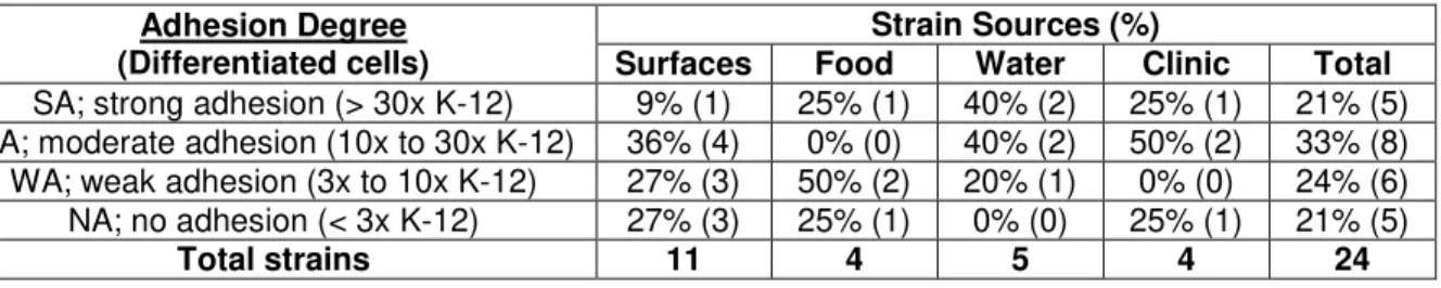
All strains originated from water samples are adherent. 80% (4/5) have moderate to high levels of adhesion (Table 4.2), which may be related to a need of these strains have to form biofilms in their niche. This was similar to what was observed in the clinical strains that showed 75% (3/4) with the same levels of adhesion (Table 4.2). This similarity supports the thesis that the major cause of gastrointestinal infections by Aeromonas spp.is from ingesting infected water [Statner and George, 1987; Holmberg et al., 1986], especially considering that only 25% (1/4) of the strains originated from food samples and 45% (5/11) of the strains originated from food processing surfaces have shown the same levels of adhesion (Table 4.2).
Table 4.2. Distribution of adherent Aeromonas isolates by source and level ability levels.
Adhesion Degree (Differentiated cells)
Strain Sources (%)
Surfaces Food Water Clinic Total
SA; strong adhesion (> 30x K-12) 9% (1) 25% (1) 40% (2) 25% (1) 21% (5) MA; moderate adhesion (10x to 30x K-12) 36% (4) 0% (0) 40% (2) 50% (2) 33% (8) WA; weak adhesion (3x to 10x K-12) 27% (3) 50% (2) 20% (1) 0% (0) 24% (6) NA; no adhesion (< 3x K-12) 27% (3) 25% (1) 0% (0) 25% (1) 21% (5)
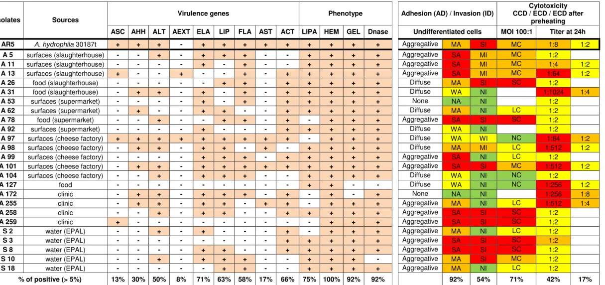
Barroco (2013) determined the presence of flaA/flaB genes in the strains used in this study (Table 4.3), they encode two subunits of flagellin that compose the complex filament of the polar flagellum, that allow the colonization of different niches, including the colonization of host tissues [Kirov, 2003]. Its expression is highly regulated by a number of environmental factors, but the molecular inhibition mechanism is not known [Merino et al., 2006].
The presence of flaA/flaB genes was detected in the genome of 2 strains, A53 and A172, considered nonadherent and in other 2 strains, A31 and A97, which expressed low adherence in UC and were considered nonadherent in DC. These results indicate the existence of a mechanism or genetic flaw in these strains that is preventing the polar flagellum to be expressed, at assay conditions, and the absence on the genome or no expression of other structures (e.g.: lateral flagella or pilli) and proteins (e.g.: OMPs) that can act as adhesins, allowing or facilitating adhesion to human epithelial cells in vitro.
The Aeromona hydrophila insertional flaH, flaJ, mutant and flaA/flaB double mutant resulted in the complete loss of motility, showing lateral flagella, absence of polar flagella, and a dramatic reduction in adhesion to HEp-2 cells and in ability to form biofilms [Canals et al., 2006].
Table 4.3. Comparison of the phenotype and genotype characterization of Aeromonas isolates carried out to date by Barroco (2013) and the phenotype characterization carried out in this study.
Isolates Sources Virulence genes Phenotype Adhesion (AD) / Invasion (ID)
Cytotoxicity CCD / ECD / ECD after
preheating ASC AHH ALT AEXT ELA LIP FLA AST ACT LIPA HEM GEL Dnase Undifferentiated cells MOI 100:1 Titer at 24h
AR5 A. hydrophila 30187t + + + - + + + + + + + + + Aggregative MA SI MC 1:8 1:2
A 5 surfaces (slaughterhouse) - - + - + + + - - + + + + Aggregative SA MI MC 1:2
A 11 surfaces (slaughterhouse) - - - - + - + - - + + + + Aggregative SA MI MC 1:4 1:2
A 13 surfaces (slaughterhouse) + - - + - - + - + + + + + Aggregative SA MI MC 1:64 1:2
A 26 food (slaughterhouse) - - - - - + + - + + + + + Diffuse MA SI SC 1:2
A 31 food (slaughterhouse) - + + - + - + - + + + + + Diffuse WA NI 1:1024 1:4
A 53 surfaces (supermarket) - - - - + - + - + + + + + None NA NI 1:2
A 62 surfaces (supermarket) - + - - + + - - + + + + + Diffuse MA NI LC 1:2
A 78 food (supermarket) - - + - - + + - + - + + + Aggregative SA SI SC 1:2
A 92 surfaces (supermarket) - - - - + - - - + + + + + Diffuse WA NI 1:2
A 97 surfaces (cheese factory) + + + + + + + + + - + + + Diffuse WA WI NC 1:64 1:2
A 98 surfaces (cheese factory) - + + - + + - + - + + + + Diffuse MA MI LC 1:512 1:2
A 99 surfaces (cheese factory) - - - - + + + - + + + + + Aggregative SA NI LC 1:2
A 101 surfaces (cheese factory) - + + - + + + + + + + + + Aggregative SA SI MC 1:512 1:2
A 104 surfaces (cheese factory) - - + - + + + - - + + + + Diffuse WA NI NC 1:2
A 127 food - - - - - - - - - + + - - Diffuse WA NI NC 1:256 1:2
A 172 clinic - + + - + + + - + - + - + None NA NI 1:256 1:8
A 255 clinic - + + - + + - + + - + + + Aggregative MA NI LC 1:512 1:4
A 258 clinic - - + - + + - - + + + + + Aggregative SA SI SC 1:2
A 259 clinic + - - - - - - - - - + + + Aggregative SA SI SC 1:2
S 2 water (EPAL) - - + - + - - - + - + + + Aggregative MA NI LC 1:2
S 3 water (EPAL) - - - - - - - - + + + + + Aggregative SA SI SC 1:2
S 8 water (EPAL) - - - - + + - - + + + + + Aggregative SA SI SC 1:2
S 10 water (EPAL) - - + - + + + - - + + + - Aggregative MA SI MC 1:2
S 18 water (EPAL) - - - - - + + - - + + + + Aggregative MA NI LC 1:2
% of positive (> 5%) 13% 30% 50% 8% 71% 63% 58% 17% 66% 75% 100% 92% 92% 92% 54% 71% 42% 17% Virulence genes: ASC (TTSS structural protein - ascV); AHH (β-hemolisina - hlyA); ALT (Aeromona heat-labile cytotonic enterotoxin- alt); AEXT (TTSS effector protein - ADP-Ribosyltransferase
- aexT); ELA (elastase - ahyB); LIP (lipases and phospholipases - pla/lip/lipH3/alp-1); FLA (flagellin A and B - flaA/B); AST (Aeromonas heat-stable cytotonic enterotoxina - ast); ACT (Aeromonas cytotoxic enterotoxina - act). Phenotype: LIPA (lipase); HEM (hemolysin); GEL (gelatinase); DNases.
ECD: Extracellular cytotoxicity degree: NC: no cytotoxic (100% in 1:2 titer); LC: low cytotoxicity (< 1:2 titer); MC: moderate cytotoxicity (1.2 to 1:64 titer); SC: strong cytotoxicity (> 1:128 titer); CCD: Cell-contact cytotoxic degree: NC: no cytotoxic; LC: low cytotoxicity (< 25%); MC: moderate cytotoxicity (25 to 45%); SC: strong cytotoxicity (> 45%);